
Международный эндокринологический журнал Том 22, №3, 2026
Вернуться к номеру
Гормональні основи патологічного перебігу першого триместру вагітності у жінок з аденоміозом
Авторы: Семенина Г.Б. (1), Мандзій І.М. (2), Дорошенко-Кравчик М.В. (1), Угрин О.М. (3), Коритко О.О. (1)
(1) - ДНП «Львівський національний медичний університет імені Данила Галицького», м. Львів, Україна
(2) - КП «Хмельницький міський перинатальний центр», м. Хмельницький, Україна
(3) - Львівська медична академія імені Андрея Крупинського, м. Львів, Україна
Рубрики: Эндокринология
Разделы: Клинические исследования
Версия для печати
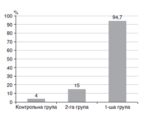
Актуальність. Аденоміозом називають внутрішній ендометріоз матки, при якому ендометріальні залози і строма глибоко і хаотично інфільтрують міометрій. Аденоміоз є поширеним захворюванням з недостатньо вивченим патогенезом та патофізіологією, частота якого сягає від 10 до 57 %. Факторами ризику вважають пологи, наявність в анамнезі процедур дилатації, кюретажу та інших операцій на матці, які можуть порушувати ендометріально-міометральний інтерфейс та сприяти інвазії, імплантації, вбудовуванню та утворенню ендометріальних колоній у стінці міометрія, збільшуючи розвиток аденоміозу. Перебіг вагітності у жінок з аденоміозом є нез’ясованим. Мета роботи: дослідити зміни гормонального балансу під час вагітності у жінок з аденоміозом та їх вплив на спектр ускладнень першого триместру вагітності. Матеріали та методи. Під спостереженням перебували 117 вагітних жінок. До 1-ї групи увійшли 27 жінок з аденоміозом, вагітність у них велась згідно з чинними нормативними документами, до 2-ї — 40 жінок, які отримали запропоноване нами лікування аденоміозу та пройшли прегравідарну підготовку, контрольну групу становили 50 вагітних здорових жінок. Вагітним проводили гормональні обстеження: визначення естрадіолу, естріолу, прогестерону, сексстероїдзв’язуючого глобуліну, пролактину, кортизолу та катехоламінів. Статистична обробка отриманих даних проводилася за допомогою стандартного пакета програм Statistica for Windows 13.0. Результати. У жінок 1-ї групи рівні естрадіолу, прогестерону, сексстероїдзв’язуючого глобуліну (р < 0,05) й естріолу (р < 0,01) були значно зниженими порівняно зі здоровими вагітними, у жінок 2-ї групи вміст цих гормонів не відрізнявся від контрольної групи (р > 0,05). У жінок з аденоміозом виявлено підвищений вміст стресових гормонів відносно контрольної групи: у 1-й групі це пролактин (р < 0,01) і кортизол (р < 0,05), у 2-й групі це кортизол (p < 0,05). У жінок 1-ї групи рівні адреналіну та норадреналіну є підвищеними порівняно з показниками контрольної (p < 0,01) та 2-ї (p < 0,05) групи. Вміст дофаміну є вдвічі нижчим за показник контрольної (p < 0,01) та в 1,9 раза за показник 2-ї (p < 0,05) групи. Отже, вагітність на тлі аденоміозу перебігає за умов біохімічно підтвердженого стресового гомеостазу, що разом з гормональними порушеннями отримало клінічну реалізацію у вигляді низки ускладнень перебігу першого триместру вагітності. Вагітність у жінок з аденоміозом, які не отримували прегравідарної підготовки, супроводжується невиношуванням майже у 90 % випадків, що вірогідно частіше порівняно з жінками 2-ї групи (p < 0,01), а також у 4 рази частіше, ніж у жінок із патологією плацентації. Висновки. Жінок з аденоміозом слід відносити до групи високого акушерського ризику. У жінок з аденоміозом у першому триместрі вагітності виявлено зниження вмісту естрогенів і прогестерону (p < 0,05), підвищений вміст стресових гормонів пролактину (р < 0,01) і кортизолу (р < 0,05). Виявлена кореляція між вмістом стресових гормонів у сироватці крові вагітних з аденоміозом та невиношуванням: адреналіну (r = +71, p < 0,01), дофаміну (r = –0,63, p < 0,05), кортизолу (r = +0,57, p < 0,05). Проведення прегравідарної підготовки у жінок з аденоміозом значно зменшує частоту невиношування вагітності та патологію плацентації.
Background. Adenomyosis is internal endometriosis of the uterus, in which endometrial glands and stroma deeply and chaotically infiltrate the myometrium. Adenomyosis is a common disease with poorly understood pathogenesis and pathophysiology, its incidence ranges from 10 to 57 %. Risk factors include childbirth, a history of dilation, curettage, and other uterine surgeries that can disrupt the endometrial-myometrial interface and promote invasion, implantation, embedding, and formation of endometrial colonies in the myometrial wall, increasing the development of adenomyosis. The course of pregnancy in women with adenomyosis is unclear. The purpose was to investigate changes in hormonal balance during pregnancy in patients with adenomyosis and their impact on the spectrum of complications in the first trimester of pregnancy. Materials and methods. One hundred and seventeen pregnant women were under observation. The first group included 27 patients with adenomyosis, whose pregnancy was followed up in accordance with current regulatory documents, the second group consisted of 40 women who received the adenomyosis treatment we proposed and underwent pre-pregnancy training, and the control group included 50 healthy pregnant women. Participants underwent hormonal examinations: determination of estradiol, estriol, progesterone, sex hormone-binding globulin, prolactin, cortisol, and catecholamines. Statistical processing of the obtained data was carried out using the standard Statistica for Windows 13.0 software package. Results. In group I, the content of estradiol, progesterone, sex hormone-binding globulin (p < 0.05) and estriol (p < 0.01) was significantly reduced compared to healthy pregnant women. In group II, the level of these hormones did not differ from that of the control group (p > 0.05). Women with adenomyosis showed an increased content of stress hormones compared to the control group: in group I, it was prolactin (p < 0.01) and cortisol (p < 0.05), in group II, it was cortisol (p < 0.05). In group I, the level of adrenaline and noradrenaline is higher than that of the control group (p < 0.01) and group II (p < 0.05). The dopamine content is halved compared to the controls (p < 0.01) and 1.9 times lower than in group II (p < 0.05). Therefore, pregnancy with adenomyosis occurs under conditions of biochemically confirmed stress homeostasis, which, together with hormonal disorders, manifests clinically in the form of complications during the first trimester of pregnancy. Pregnancy in women with adenomyosis who did not have pre-pregnancy training is accompanied by miscarriage in almost 90 % of cases, unlike women in group II (p < 0.01), and 4 times more often than in patients with placentation pathology. Conclusions. Patients with adenomyosis should be considered at high obstetric risk. In women with adenomyosis, a decrease in estrogen and progesterone (p < 0.05), an increase in stress hormones such as prolactin (p < 0.01) and cortisol (p < 0.05) was detected in the first trimester of pregnancy. A correlation was found between the content of stress hormones in the blood serum of pregnant women with adenomyosis and miscarriage: adrenaline (r = +71, p < 0.01), dopamine (r = –0.63, p < 0.05), cortisol (r = +0.57, p < 0.05). Pre-pregnancy training in women with adenomyosis significantly reduces the incidence of miscarriage and placentation pathology.
вагітність; аденоміоз; гормони; невиношування вагітності; патологія плацентації
pregnancy; adenomyosis; hormones; miscarriage; placentation pathology
Для ознакомления с полным содержанием статьи необходимо оформить подписку на журнал.
- Guo S.W. The pathogenesis of adenomyosis vis-à-vis endometriosis. J Clin Med. 2020 Feb 10;9(2):485. doi: 10.3390/jcm9020485.
- Semenyna H.B., Mandziy I.M., Popovych A.I., Korytko O.O., Doroshenko-Kravchyk M.V., et al. The state of hormonal balance in women with adenomyosis according to the phases of the menstrual cycle and its dependence on the stage of the disease. Int J End. 2025;21(5):537-543. doi: 10.22141/2224-0721.21.5.2025.1604.
- Guo S.W., Mao X., Ma Q., Liu X. Dysmenorrhea and its severity are associated with increased uterine contractility and overexpression of oxytocin receptor (OTR) in women with symptomatic adenomyosis. Fertil Steril. 2013;99:231-240. doi: 10.1016/j.fertnster.2012.08.038.
- Leyendecker G., Bilgicyildirim A., Inacker M., Stalf T., Huppert P., Mall G., et al. Adenomyosis and endometriosis. Re-visiting their association and further insights into the mechanisms of auto-traumatisation. An MRI study. Arch Gynecol Obs. 2015;291:917-932. doi: 10.1007/s00404-014-3437-8.
- Brunes M., Wennmo-Zuk K., Amark H., Nordborg J.W., Forslund M., Jokubkiene L., et al. Pregnancy in women with advanced endometriosis and adenomiosis: possible complications and the role of surgery. Reprod Biol Endocrinol. 2025 Nov 26;23(1):158. doi: 10.1186/s12958-025-01492-y.
- Elizur S.E., Mostafa J., Berkowitz E., Orvieto R. Endometriosis and infertility: pathophysiology, treatment strategies, and reproductive outcomes. Arch Gynecol Obstet. 2025 Oct;312(4):1037-1048. doi: 10.1007/s00404-025-08124-1.
- Alson S., Henic E., Hansson S.R., Sladkevicius P. Correlation of adenomiosis features to live birth rates after the first IVF/ICSI treatment when using the revised morphological uterus sonogra–phic assessment group definitions. Acta Obstet Gynecol Scand. 2024 Dec;103(12):2540-2553. doi: 10.1111/aogs.14986.
- Tsikouras P., Kritsotaki N., Nikolettos K., et al. The impact of adenomyosis on pregnancy. Biomedicines. 2024 Aug 22;12(8):1925. doi: 10.3390/biomedicines12081925.
- Hirashima H., Ohkuchi A., Usui R., Kijima S., Matsubara S. Magnetic resonance imaging of degeneration of uterine adenomyosis during pregnancy and post-partum period. J Obstet Gynaecol Res. 2018;44:1169-1173. doi: 10.1111/jog.13629.
- Pados G., Gordts S., Sorrentino F., Nisolle M., Nappi L., Daniilidis A. Adenomyosis and infertility: A literature review. Medicina. 2023;59:1551. doi: 10.3390/medicina59091551.
- Pokharel K., Peippo J., Weldenegodguad M., Honkatukia M., Li M.-H., Kantanen J. Gene expression profiling of corpus luteum reveals important insights about early pregnancy in domestic sheep. Genes. 2020;11:415. doi: 10.3390/genes11040415.
- Zhang X., Bao P., Ye N., Zhou X., Zhang Y., Liang C., et al. Identification of the key genes associated with the yak hair follicle cycle. Genes. 2021;13:32. doi: 10.3390/genes13010032.
- Zippl A.L., Kyvelidou C., Frank M., Gapp E. et al. The impact of GnRH agonists on endometrial immune cells in patients with adenomyosis: a prospective cohort study. BMC Med. 2025 Jun 9:23(1):338. doi: 10.1186/s12916-025-04162-3.
- Matsuzaki S., Ueda Y., Nagase Y., Matsuzaki S., Kakuda M., Sakaguchi H., et al. Placenta accrete spectrum disorder complicated with endometriosis: systematic review and meta-analysis. Biomedicines. 2022 Feb 6;10(2):390. doi: 10.3390/biomedicines10020390.
- Shi J., Xu Q., Yu S., Zhang T. Perturbations of the endome–trial immune microenvironment in endometriosis and adenomyosis: their impact on reproduction and pregnancy. Semin Immunopathol. 2025 Feb 18;47(1):16. doi: 10.1007/s00281-025-01040-1.
- Salmeri N., Farina A., Candiani M., Dolci C. Endometriosis and impaired placentation: A prospective cohort study comparing uterine arteries Doppler pulsatility index in pregnancies of patients with and without moderate-severe disease. Diagnostics (Basel). 2022 Apr 19;12(5):1024. doi: 10.3390/diagnostics12051024.
- Berlanda N., Alio W., Angioni S., et al. Impact of endome–triosis on obstetric outcome after natural conception: a multicenter Italian study. Arch Gynecol Obstet. 2022 Jan;305(1):149-157. doi: 10.1007/s00404-021-06243-z.
- de Rozario T., Jochum F., Schwaab T., Garbin O., Roy C., Host A. Adenomyosis and obstetric complications: A retrospective case-control study. Euro J Obstet Gynecol Reprod Biol. 2024;292:120-124. doi: 10.1016/j.ejogrb.2023.11.011.
- Hashimoto A., Iriyama T., Sayama S., Okamura A., Kato K., Fujii T., et al. Differences in the incidence of obstetric complications depending on the extern and location of adenomyosis lesions. J Matern Fetal Neonatal Med. 2023;36:2226789. doi: 10.1080/14767058.2023.2226789.
- Shi J., Dai Y., Zhang J., Li X., Jia S., Leng J. Pregnancy outcomes in women with infertility and coexisting endometriosis and adenomyosis after laparoscopic surgery: a long-term reproductive follow-up study. BMC Pregnancy Childbirth. 2021 May 18:21(1):383. doi: 10.1186/s12884-021-03851-0.
- Jan N., Yuan X., Huang S., Jie H., Wang J., Yuan Y. Ova–rian endometrioma increases the embrio aneuploidia rate: an analysis of 7092 biopsied blastocysts from fertile monogenetic disease carries. BMC Womens Health. 2023 May 9;23(1):244. doi: 10.1186/s12905-023-02406-Z.
- Stanecova V., Woodman R., Tremellen K. The rate of euploid miscarriage is increased in the setting of adenomyosis. Hum Reprod Open. 2019 Jan 29;2019(1):hoy026. doi: 10.1093/hropen/hoy026.
- Ticconi C., Nicastri E., D’Ippolito S., Chiaramonte C., Pie–tropolli A., Scambia G., Di Simone N. Diagnostic factors for recurrent pregnancy loss: an expanded workup. Arch Gynecol Obstet. 2023 Mar 25;308(1):127-142. doi: 10.1007/s00404-023-07001-z.
- Vercellini P., Vigano P., Bandini V., Buggio L., Berlanda N., Somigliana E. Association of endometriosis and adenomyosis with pregnancy and infertility. Fertil Steril. 2023 May;119:727-740. doi: 10.1016/j.fertnster.2023.03.018.
- Buggio L., Dridi D., Barbara G. Adenomyosis: Impact on fertility and obstetric outcomes. Reprod Sci. 2021;28:3081-3084. doi: 10.1007/s43032-021-00679-z.
- Cozzolino M., Tartaglia S., Pellegrini L., Troiano G., Rizzo G., Petraglia F. The effect of uterine adenomyosis on IVF outcomes: A systematic review and meta-analysis. Reprod Sci. 2022;29:3177-3193. doi: 10.1007/s43032-021-00818-6.
- Bourdon M., Mimouni A., Maignien C., Casalechi M., Vigano P., Bordonne C., et al. Reduced live birth rates following ART in adenomyosis patients: a matched control study. Hum. Reprod. 2025 May;40(5):855-864. doi: 10.1093/humrep/deaf052.
- Latif S., Kastora S., Al Wattar B.H., Yasmin E., Saridogan E., Mavrelos D. The effectiveness of prolonged downregulation with gonadotrophin-releasing hormone analogue (GnRHa) treatment in women with adenomyosis undergoing IVF/ICSI: A systematic review and meta-analysis. Eur J Obstet Gynecol Reprod Boil. 2024 Oct;301:87-94. doi: 10.1016/j.ejogrb.2024.07.063.
- Vaquero E., Selntigia A., Chiaramonte B., Soreca G., Rizzo G. Type and location of adenomyosis in women with reccurent pregnancy loss: A transvaginal ultrasonographic assessment. Reprod Sci. 2024 Aug;31(8):2447-2457. doi: 10.1007/s43032-024-01541-8.
- Harmsen M.J., Trommelen L.M., de Leeuw R.A., Tellum T., Juffermans L.J.M., Griffioen A.W., et al. Multidisciplinary view on uterine junctional zone in uteri affected by adenomyosis: explaining discrepancies between MRI and transvaginal ultrasound images on a microscopic level. Ultrasound Obstet Gynecol. 2022 Nov 12;62(1):42-60. doi: 10.1002/uog.26117.