
Архив офтальмологии Украины Том 14, №1, 2026
Вернуться к номеру
Ефективність диференційованого підходу до вітреоретинальної хірургії наскрізних розривів макули
Авторы: Путієнко О.О., Руднєв М.О., Денисюк Л.І., Лисенко М.Г.
Національний університет охорони здоров’я України імені П.Л. Шупика, м. Київ, Україна
Рубрики: Офтальмология
Разделы: Клинические исследования
Версия для печати
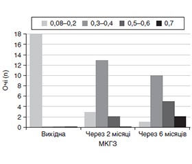
Актуальність. Сучасні можливості вітреоретинальної хірургії дозволяють досягти закриття наскрізного розриву макули у 90–100 % випадків залежно від розміру розриву. Видалення внутрішньої пограничної мембрани (ВПМ) призводить до витончення шару нервових волокон та інших ускладнень з боку сітківки і пов’язане зі зниженням зорових функцій. Тому розробка нового методу хірургічного лікування наскрізних розривів макули, який спрямований на максимальне збереження ВПМ, є актуальною. Мета: розробити диференційований підхід до методики вітреоретинальної хірургії наскрізних розривів макули з максимальним збереженням ВПМ. Матеріали та методи. Під спостереженням перебувало 45 хворих (50 очей) із наскрізними розривами макули з діаметром основи (ДО) розриву від 200 до 1000 мкм. Хворі були розподілені на три групи залежно від ДО розриву за даними оптичної когерентної томографії (ОКТ): I група — 10 хворих (10 очей) з ДО від 200 до 350 мкм, II група — 20 хворих (22 ока) з ДО > 350–600 мкм, III група — 15 хворих (18 очей) з ДО > 600–1000 мкм. Вітреоретинальне втручання виконували за стандартною методикою, у I групі хворих ВПМ не видаляли, у II групі видаляли ВПМ протяжністю до 1/4 відстані від фовеа до диска зорового нерва (ДЗН) (0,75 діаметра ДЗН) і в III групі — до середини відстані від фовеа до ДЗН (1,5 діаметра ДЗН). В усіх випадках для тампонади вітреальної порожнини застосовували 20% газоповітряну суміш перфторпропану. Результати. У I групі як через 2 місяці, так і через 6 місяців після операції закриття розриву було відмічене у всіх випадках, відновлення зовнішньої пограничної мембрани спостерігалось на 8 очах (80 %) через 2 місяці та у 100 % через 6 місяців. Гострота зору (ГЗ) значуще поліпшилась порівняно із вихідною в обох термінах спостереження. У II групі закриття розриву також було відмічено на всіх очах з відновленням зовнішньої пограничної мембрани в 16 випадках (72,7 %) через 2 місяці та в 90,9 % через 6 місяців, зі значущим поліпшенням ГЗ в обох термінах спостереження. У III групі розрив закрився на всіх 18 очах, а відновлення зовнішньої пограничної мембрани відбулося тільки в 10 випадках (55,6 %) в 2 місяці і в 77,8 % через 6 місяців зі значущим поліпшенням ГЗ. Висновки. Розроблений диференційований підхід дозволив у 100 % випадків повністю зберегти ВПМ при розривах з діаметром основи до 350 мкм, максимально зменшити її видалення у випадках розривів від 350 до 600 мкм та мінімізувати її видалення при розривах більше 600 мкм. Це супроводжується відновленням зовнішньої пограничної мембрани в 100 % випадків при розривах до 350 мкм, в 90,9 % випадків з діаметром від 350 до 600 мкм та 77,8 % випадків з діаметром розриву вище 600 мкм у термін 6 місяців спостереження.
Background. Current advances in vitreoretinal surgery enable to close full-thickness macular holes in 90–100 % of cases, depending on their size. Removal of the internal limiting membrane (ILM) leads to the thinning of the nerve fibre layer and other complications in the retina and is associated with a decline in visual function. Therefore, the development of a new method for the surgical treatment of full-thickness macular holes, aimed at maximising preservation of the ILM, is of great relevance. The purpose of the study was to develop a differentiated approach to the technique of vitreoretinal surgery for full-thickness macular holes with maximum preservation of the ILM. Materials and methods. The study included 45 patients (50 eyes) with full-thickness macular holes with a base diameter (BD) ranging from 200 to 1000 µm. Patients were divided into 3 groups depending on the BD of the hole as determined by optical coherence tomography: group I included 10 patients (10 eyes) with a BD of 200 to 350 µm, group II consisted of 20 participants (22 eyes) with a BD of > 350–600 µm, and group III included 15 patients (18 eyes) with a BD of > 600–1000 µm. Vitreoretinal surgery was performed using the standard technique; in group I, the ILM was not removed; in group II, the ILM was removed over a length of up to one-fourth of the distance from the fovea to the optic disc (0.75 diameter of the optic disc), and in group III, up to the midpoint of the distance from the fovea to the optic disc (1.5 diameter of the optic disc). In all cases, a 20% perfluoropropane gas-air mixture was used for vitreous cavity tamponade. Results. In group I, closure of the hole was observed in all cases both 2 and 6 months after surgery; restoration of the external limiting membrane was observed in 8 eyes (80 %) at 2 months and in 100 % of eyes at 6 months. Visual acuity (VA) improved significantly compared to baseline at both follow-up time points. In group II, closure of the hole was also observed in all eyes, with restoration of the external limiting membrane in 16 cases (72.7 %) at 2 months and in 90.9 % at 6 months, with a significant improvement in VA at both follow-up time points. In group III, the hole closed in all 18 eyes, whilst restoration of the external limiting membrane occurred in only 10 cases (55.6 %) at 2 months and in 77.8 % at 6 months, with a significant improvement in VA. Conclusions. The developed differentiated approach allowed the ILM to be fully preserved in 100 % of cases in holes with a BD of up to 350 µm, to minimise its removal as much as possible in cases of holes ranging from 350 to 600 µm, and to reduce its removal to a minimum in holes exceeding 600 µm. This is accompanied by the restoration of the external limiting membrane in 100 % of cases in holes of up to 350 µm, in 90.9 % of cases with a diameter of 350 to 600 µm, and in 77.8 % of cases with a hole diameter greater than 600 µm within a 6-month follow-up period.
наскрізні макулярні розриви; внутрішня погранична мембрана; вітреоретинальна хірургія; оптична когерентна томографія; гострота зору
full-thickness macular holes; internal limiting membrane; vitreoretinal surgery; optical coherence tomography; visual acuity
Для ознакомления с полным содержанием статьи необходимо оформить подписку на журнал.
- McCannel CA, Ensminger JL, Diehl NN, Hodge DN. Population‑based incidence of macular holes. Ophthalmology. 2009 Jul;116(7):1366-9. doi: 10.1016/j.ophtha.2009.01.052.
- Marten la Cour M, Friis J. Macular holes: Classification, epidemiology, natural history and treatment. Acta Ophthalmol Scand. 2002 Dec; 80(6):579-87. doi: 10.1034/j.1600-0420.2002.800605.
- Madi HA, Masri I., Steel DH. Optimal management of idiopathic macular holes. Clinical Ophthalmology. 2016 Jan 13;10:97-116. doi: 10.2147/OPTH.S96090.
- Elhusseiny AM, Schwartz SG, Flynn HW Jr, Smiddy WE. Long-Term Outcomes after Macular Hole Surgery. Ophthalmol Retina. 2020 Apr;4(4):369-376. doi: 10.1016/j.oret.2019.09.015.
- Xin Ye, Jiahao Xu, Shucheng He, Jun Wang, Jinglei Yang, Jiwei Tao, et al. Quantitative evaluation of dissociated optic nerve fibre layer (DONFL) following idiopathic macular hole surgery. Eye (2023) 37:1451-1457. doi: 10.1038/s41433-022-02150-y.
- Spaide FR. Dissociated optic nerve fiber layer appearance after internal limiting membrane removal is inner retinal dimpling. Retina. 2012 Oct;32(9):1719-26. doi: 10.1097/IAE.0b013e3182671191.
- Nukada K, Hangai M, Ooto S, Yoshikawa M, Yoshimura N. Tomographic features of macula after successful macular hole surgery. Invest Ophthalmol Vis Sci. 2013 Apr 1;54(4):2417-28. doi: 10.1167/iovs.12-10838.
- Clark A, Balducci N, Pichi F, Veronese C, Morara M, Torrazza C, Ciardella AP. Swelling of the arcuate nerve fiber layer after internal limiting membrane peeling. Retina. 2012 Sep;32(8):1608-13. doi: 10.1097/IAE.0b013e3182437e86.
- Demirel S, Abdullayev A, Yanık Ö, Batıoğlu F, Özmert E. Evaluation of Ganglion Cell-Inner Plexiform Layer Thickness after Vitreoretinal Surgery with Internal Limiting Membrane Peeling in Cases with Idiopathic Macular Hole. Turk J Ophthalmol. 2017;47:138-143. doi: 10.4274/tjo.34545.
- Kim YJ, Jo J, Lee JY, Yoon YH, Kim JG. Macular capillary plexuses after macular hole surgery: an optical coherence tomography angiography study. Br J Ophthalmol. 2018 Jul;102(7):966-970. doi: 10.1136/bjophthalmol-2017-311132.
- Yoshikawa M, Murakami T, Nishijima K, Uji A, Ogino K, Horii T, et al. Macular migration toward the optic disc after inner limiting membrane peeling for diabetic macular edema. Invest Ophthalmol Vis Sci. 2013 Jan;54(1):629-35. doi: 10.1167/iovs.12-10907.
- Kelly NE, Wendel RT. Vitreous surgery for idiopathic macular holes. Results of a pilot study. Arch Ophthalmol. 1991 May;109(5):654-9. doi: 0.1001/archopht.1991. 01080050068031.
- Kitao M, Wakabayashi T, Nishida K, Sakaguchi H, Nishida K. Long-term reconstruction of foveal microstructure and visual acuity after idiopathic macular hole repair: three-year follow-up study. Br J Ophthalmol. 2019 Feb;103(2):238-244. doi: 10.1136/bjophthalmol-2017-311689.
- Eckardt C, Eckardt U, Groos S, Luciano L, Reale E. [Removal of the internal limiting membrane in macular holes. Clinical and morphological findings]. Ophthalmologe. 1997 Aug;94(8):545-51. doi: 10.1007/s003470050156.
- Brooks HL Jr. Macular hole surgery with and without internal limiting membrane peeling. Ophthalmology. 2000 Oct;107(10):1939-48. doi: 10.1016/s0161-6420(00)00331-6.
- Mester V, Kuhn F. Internal limiting membrane removal in the management of full-thickness macular holes. Am J Ophthalmol. 2000 Jun;129(6):769-77. doi: 10.1016/s0002-9394(00)00358-5.
- Foulquier S, Glacet-Bernard A, Sterkers M, Soubrane G, Coscas G. [Study of internal limiting membrane peeling in stage-3 and -4 idiopathic macular hole surgery]. J Fr Ophtalmol. 2002 Dec;25(10):1026-31.
- Bae K, Kang SW, Kim JH, Kim SJ, Kim JM, Yoon JM. Extent of Internal Limiting Membrane Peeling and its Impact on Macular Hole Surgery Outcomes: A Randomized Trial. Am J Ophthalmol. 2016 Sep;169:179-88. doi: 10.1016/j.ajo.2016.06.041.
- Yao Y, Qu J, Dong C, Li X, Liang J, Yin H, et al. The impact of extent of internal limiting membrane peeling on anatomical outcomes of macular hole surgery: results of a 54-week randomized clinical trial. Acta Ophthalmol. 2019 May;97(3):303-12. doi: 10.1111/aos.13853.
- Goker YS, Koc M, Yuksel K, Yazici AT, Demir A, Gunes H, et al. Relationship between Peeled Internal Limiting Membrane Area and Anatomic Outcomes following Macular Hole Surgery: A Quantitative Analysis. J Ophthalmol. 2016;2016:5641273. doi: 10.1155/2016/5641273.
- Gass JD. Reappraisal of biomicroscopic classification of stages of development of a macular hole. Am J Ophthalmol. 1995 Jun;119(6):752-9. doi: 10.1016/s0002-9394(14)72781-3.
- Gotzaridis S, Liazos E, Petrou P, Georgalas I. Short-Acting Gas Tamponade with Strict Face-Down Posturing for the Treatment of Idiopathic Macular Hole. Semin Ophthalmol. 2017;32(5):597-601. doi: 10.3109/08820538.2015.1132333.
- Yao Y, Yan H, Qu J, Dong C, Liang J, Yin H, et al. Vitrectomy with sulfur hexafluoride versus air tamponade for idiopathic macular hole: a retrospective study BMC Ophthalmol. 2023 Jul 20;23(1):331. doi: 10.1186/s12886-023-03049-2.
- Shimada Y, Seno Y, Mizuguchi T, Tanikawa A, Horiguchi M. Patient adherence to the face-down positioning after macular hole surgery Clin. Ophthalmol. 2017 Jun 8;11:1099-1104. doi: 10.2147/OPTH.S135144.
- Vishal MY, Babu N, Kohli P, Rajendran A, Ramasamy K. Retrospective study of changes in ocular coherence tomography characteristics after failed macular hole surgery and outcomes of fluid‑gas exchange for persistent macular hole. Indian J Ophthalmol. 2018 Aug;66(8):1130-1135. doi: 10.4103/ijo.IJO_1119_17.
- Bodhankar PU, Joshi AK, Dronadula M, Patil AN. Postoperative microstructural re‑modelling and functional outcomes in idiopathic full thickness macular hole. Indian J Ophthalmol. 2022 Jun;70(6):2077-2083. doi: 10.4103/ijo.IJO_192_22.
- Lee MW, Kim TY, Song YY, Baek SK, Lee YH. Changes in each retinal layer and ellipsoid zone recovery after full thickness macular hole surgery. Sci Rep. 2021;11:11351. doi: 10.1038/s41598-021-90955-4.
- Houly JR, Veloso CE, Passos E, Nehemy MB. Quantitative analysis of external limiting membrane, ellipsoid zone and interdigitation zone defects in patients with macular holes. Graefes Arch Clin Exp Ophthalmol. 2017 Jul;255(7):1297-1306. doi: 10.1007/s00417-017-3636-7.