
Журнал "Гастроэнтерология" Том 60, №2, 2026
Вернуться к номеру
Епігенетичний антифібротичний ефект експресії довгих некодуючих РНК lncR-p21, MEG3, H19 на розвиток фіброзу при стеатотичній хворобі печінки, асоційованій із метаболічною дисфункцією (систематизований огляд та метааналіз)
Авторы: A.E. Abaturov, A.O. Nikulina
Dnipro State Medical University, Dnipro, Ukraine
Рубрики: Гастроэнтерология
Разделы: Справочник специалиста
Версия для печати
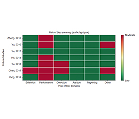
Актуальність. Стеатотична хвороба печінки, асоційована з метаболічною дисфункцією (metabolic dysfunction-associated steatotic liver disease — MASLD), є одним з найпоширеніших хронічних захворювань печінки в дітей та дорослих у всьому світі. Фіброз печінки є ключовим прогностичним фактором, що визначає прогресування захворювання до цирозу та гепатоцелюлярної карциноми. Довгі некодуючі РНК (lncRNAs) стали критичними епігенетичними регуляторами активації та фіброгенезу зірчастих клітин печінки (hepatic stellate cell — HSC). Мета: систематизовано оцінити епігенетичний антифібротичний вплив експресії довгих некодуючих РНК (lncR-p21, MEG3, H19) на розвиток і прогресування фіброзу печінки при MASLD/MASH з акцентом на ключові профібротичні сигнальні шляхи (TGF-β/SMAD, Hedgehog) і терапевтичні перспективи. Матеріали та методи. Цей систематизований огляд і метааналіз було проведено й представлено відповідно до заяви PRISMA 2020 (реєстрація PROSPERO CRD420250652980). Виконано комплексний пошук у базах PubMed, Cochrane Library, Scopus, Embase та Web of Science (2010–2026) з використанням попередньо визначених логічних рядків. Було включено оригінальні клінічні, тваринні й дослідження in vitro, які продемонстрували знижену експресію цільових антифіброзних lncRNAs та їх зв’язок зі збільшенням фіброзу або активацією HSC у встановлених моделях MASLD/MASH або людських когортах. Два незалежні рецензенти виконали відбір досліджень, вилучення даних та оцінку ризику систематичної помилки (SYRCLE для досліджень на тваринах, ROBINS-I для обсерваційних клінічних досліджень, модифікований OHAT для досліджень in vitro). Результати. Зі 106 визначених звітів у 53 розглядали експресію довгих некодуючих РНК (lncR-p21, MEG3, H19) як потенційні антифібротичні біомаркери; зрештою, 7 досліджень відповідали суворим критеріям включення. Знижена експресія lncR-p21, MEG3, H19 послідовно пов’язувалася з активацією HSC, епітеліально-мезенхімальним переходом й активацією основних профібротичних шляхів, що призводило до збільшення експресії α-SMA та COL1A1. Висновки. LncR-p21, MEG3, H19 відіграють значну епігенетичну роль у розвитку вираженого фіброзу печінки при MASLD/MASH. Зниження їх експресії призводить до дерепресії профібротичної сигналізації в ключових профібротичних сигнальних шляхах (TGF-β/SMAD, Hedgehog).
Background. Metabolic dysfunction-associated steatotic liver disease (MASLD) is one of the most common chronic liver disorders in children and adults worldwide. Liver fibrosis is the key prognostic factor determining disease progression to cirrhosis and hepatocellular carcinoma. Long non-coding RNAs (lncRNAs) have emerged as critical epigenetic regulators of hepatic stellate cell (HSC) activation and fibrogenesis. The purpose was to systematically evaluate the epigenetic impact of reduced expression of anti-fibrotic long non-coding RNAs (lncR-p21, MEG3, and H19) on the development and progression of liver fibrosis in MASLD/MASH, with emphasis on key profibrotic signaling pathways (TGF-β/SMAD, Hedgehog) and therapeutic perspectives. Materials and methods. This systematic review and meta-analysis was conducted and reported in accordance with the PRISMA 2020 statement (PROSPERO registration CRD420250652980). A comprehensive search of PubMed, Cochrane Library, Scopus, Embase, and Web of Science (2010–2026) was performed using predefined Boolean strings. Original clinical, animal, and in vitro studies that demonstrated reduced expression of the target anti-fibrotic lncRNAs and their association with increased fibrosis or HSC activation in established MASLD/MASH models or human cohorts were included. Two independent reviewers performed study selection, data extraction, and risk-of-bias assessment (SYRCLE for animal studies, ROBINS-I for observational clinical studies, modified OHAT for in vitro studies). Quantitative meta-analysis is planned when at least three comparable studies per lncRNAs are available. Results. Of 106 identified reports, 53 addressed lncRNAs (lncR-p21, MEG3, H19) expression as potential biomarkers; ultimately, 7 studies met strict inclusion criteria. Reduced expression of lncR-p21, MEG3, and H19 was consistently associated with HSC activation, epithelial-mesenchymal transition, and activation of the main profibrotic pathways, leading to increased α-SMA and COL1A1 expression. Conclusions. LncR-p21, MEG3, H19 play a significant epigenetic role in the development of advanced liver fibrosis in MASLD/MASH. A decrease in their expression leads to derepression of profibrotic signaling in key profibrotic signaling pathways (TGF-β/SMAD, Hedgehog).
фіброз печінки; стеатотична хвороба печінки, асоційована з метаболічною дисфункцією; довгі некодуючі РНК lncR-p21, MEG3, H19
liver fibrosis; metabolic dysfunction-associated steatotic liver disease; long noncoding RNAs lncR-p21, MEG3, H19
Для ознакомления с полным содержанием статьи необходимо оформить подписку на журнал.
- Guo X, Yin X, Liu Z, Wang J. Non-Alcoholic Fatty Liver Di–sease (NAFLD) Pathogenesis and Natural Products for Prevention and Treatment. Int J Mol Sci. 2022 Dec 7;23(24):15489. doi: 10.3390/ijms232415489.
- Younossi ZM, Kalligeros M, Henry L. Epidemiology of metabolic dysfunction-associated steatotic liver disease. Clin Mol Hepatol. 2025 Feb;31(Suppl):S32-S50. doi: 10.3350/cmh.2024.0431.
- Abaturov O, Nikulina A. Characterization of anti-steatogenic long noncoding RNAs and their epigenetic influence on the development of metabolic fatty liver disease — a systematic review. Eur J Clin Exp Med. 2025;23(3):767-775. doi: 10.15584/ejcem.2025.3.16.
- Spengler EK, Loomba R. Recommendations for Diagnosis, Referral for Liver Biopsy, and Treatment of Nonalcoholic Fatty Li–ver Disease and Nonalcoholic Steatohepatitis. Mayo Clin Proc. 2015 Sep;90(9):1233-46. doi: 10.1016/j.mayocp.2015.06.013.
- Li Y, Yang P, Ye J, Xu Q, Wu J, Wang Y. Updated mechanisms of MASLD pathogenesis. Lipids Health Dis. 2024 Apr 22;23(1):117. doi: 10.1186/s12944-024-02108-x.
- Oliveira LM, Teixeira FME, Sato MN. Impact of Retinoic Acid on Immune Cells and Inflammatory Diseases. Mediators Inflamm. 2018 Aug 9;2018:3067126. doi: 10.1155/2018/3067126.
- Haaker MW, Vaandrager AB, Helms JB. Retinoids in health and disease: A role for hepatic stellate cells in affecting retinoid levels. Biochim Biophys Acta Mol Cell Biol Lipids. 2020 Jun;1865(6):158674. doi: 10.1016/j.bbalip.2020.158674.
- Stroes AR, Vos M, Benninga MA, Koot BGP. Pediatric MASLD: current understanding and practical approach. Eur J Pediatr. 2024 Nov 19;184(1):29. doi: 10.1007/s00431-024-05848-1.
- Kamm DR, McCommis KS. Hepatic stellate cells in physiology and pathology. J Physiol. 2022 Apr;600(8):1825-1837. doi: 10.1113/JP281061.
- Wiering L, Subramanian P, Hammerich L. Hepatic Stellate Cells: Dictating Outcome in Nonalcoholic Fatty Liver Disease. Cell Mol Gastroenterol Hepatol. 2023;15(6):1277-1292. doi: 10.1016/j.jcmgh.2023.02.010.
- Bink DI, Pauli J, Maegdefessel L, Boon RA. Endothelial microRNAs and long noncoding RNAs in cardiovascular ageing. Atherosclerosis. 2023 Jun;374:99-106. doi: 10.1016/j.atherosclerosis.2023.03.019.
- Busscher D, Boon RA, Juni RP. The multifaceted actions of the lncRNA H19 in cardiovascular biology and diseases. Clin Sci (Lond). 2022 Aug 12;136(15):1157-1178. doi: 10.1042/CS20210994.
- Zeng Q, Liu CH, Wu D, Jiang W, Zhang N, Tang H. LncRNA and circRNA in Patients with Non-Alcoholic Fatty Liver Di–sease: A Systematic Review. Biomolecules. 2023 Mar 20;13(3):560. doi: 10.3390/biom13030560.
- Wu Z, Huang S, Zheng X, Gu S, Xu Q, et al. Regulatory long non-coding RNAs of hepatic stellate cells in liver fibrosis (Review). Exp Ther Med. 2021 Apr;21(4):351. doi: 10.3892/etm.2021.9782.
- Hanson A, Wilhelmsen D, DiStefano JK. The Role of Long Non-Coding RNAs (lncRNAs) in the Development and Progression of Fibrosis Associated with Nonalcoholic Fatty Liver Disease (NAFLD). Noncoding RNA. 2018 Aug 21;4(3):18. doi: 10.3390/ncrna4030018.
- Ganguly N, Chakrabarti S. Role of long non‑coding RNAs and related epigenetic mechanisms in liver fibrosis (Review). Int J Mol Med. 2021 Mar;47(3):04856. doi: 10.3892/ijmm.2021.4856.
- Jiang J, Gareev I, Ilyasova T, Shumadalova A, Du W, Yang B. The role of lncRNA-mediated ceRNA regulatory networks in liver fibrosis. Noncoding RNA Res. 2024 Jan 9;9(2):463-470. doi: 10.1016/j.ncrna.2024.01.001.
- Gerhard GS, Davis B, Wu X, Hanson A, Wilhelmsen D, et al. Differentially expressed mRNAs and lncRNAs shared between activated human hepatic stellate cells and NASH fibrosis. Biochem Biophys Rep. 2020 Mar 24;22:100753. doi: 10.1016/j.bbrep.2020.100753.
- DiStefano JK, Gerhard GS. Long Noncoding RNAs and Human Liver Disease. Annu Rev Pathol. 2022 Jan 24;17:1-21. doi: 10.1146/annurev-pathol-042320-115255.
- Li QY, Gong T, Huang YK, Kang L, Warner CA, et al. Role of noncoding RNAs in liver fibrosis. World J Gastroenterol. 2023 Mar 7;29(9):1446-1459. doi: 10.3748/wjg.v29.i9.1446.
- Page MJ, McKenzie JE, Bossuyt PM, Boutron I, Hoffmann TC, et al. The PRISMA 2020 statement: an updated guideline for reporting systematic reviews. BMJ. 2021 Mar 29;372:n71. doi: 10.1136/bmj.n71.
- Zheng J, Dong P, Mao Y, Chen S, Wu X, et al. lincRNA-p21 inhibits hepatic stellate cell activation and liver fibrogenesis via p21. FEBS J. 2015 Dec;282(24):4810-21. doi: 10.1111/febs.13544.
- Yu F, Lu Z, Chen B, Dong P, Zheng J. Identification of a No–vel lincRNA-p21-miR-181b-PTEN Signaling Cascade in Liver Fibrosis. Mediators Inflamm. 2016;2016:9856538. doi: 10.1155/2016/9856538.
- Yu F, Guo Y, Chen B, Shi L, Dong P, et al. LincRNA-p21 Inhibits the Wnt/β-Catenin Pathway in Activated Hepatic Stellate Cells via Sponging MicroRNA-17-5p. Cell Physiol Biochem. 2017;41(5):1970-1980. doi: 10.1159/000472410.
- He Y, Wu YT, Huang C, Meng XM, Ma TT, et al. Inhibitory effects of long noncoding RNA MEG3 on hepatic stellate cells activation and liver fibrogenesis. Biochim Biophys Acta. 2014 Nov;1842(11):2204-15. doi: 10.1016/j.bbadis.2014.08.015.
- Yu F, Geng W, Dong P, Huang Z, Zheng J. LncRNA-MEG3 inhibits activation of hepatic stellate cells through SMO protein and miR-212. Cell Death Dis. 2018 Oct 3;9(10):1014. doi: 10.1038/s41419-018-1068-x.
- Chen MJ, Wang XG, Sun ZX, Liu XC. Diagnostic value of LncRNA-MEG3 as a serum biomarker in patients with hepatitis B complicated with liver fibrosis. Eur Rev Med Pharmacol Sci. 2019 May;23(10):4360-4367. doi: 10.26355/eurrev_201905_17943.
- Yang JJ, She Q, Yang Y, Tao H, Li J. DNMT1 controls LncRNA H19/ERK signal pathway in hepatic stellate cell activation and fibrosis. Toxicol Lett. 2018 Oct 1;295:325-334. doi: 10.1016/j.toxlet.2018.07.013.
- Chaudhary R, Lal A. Long noncoding RNAs in the p53 network. Wiley Interdiscip Rev RNA. 2017 May;8(3):10.1002/wrna.1410. doi: 10.1002/wrna.1410.
- Murphy MR, Ramadei A, Doymaz A, Varriano S, Natelson D, et al. Long Non-Coding RNA Generated from CDKN1A Gene by Alternative Polyadenylation Regulates p21 Expression during DNA Damage Response. Nucleic Acids Res. 2023 Nov 27;51(21):11911-11926.
- Wang Z, Yang X, Gui S, Yang F, Cao Z, et al. The Roles and Mechanisms of lncRNAs in Liver Fibrosis. Front Pharmacol. 2021 Nov 24;12:779606. doi: 10.3389/fphar.2021.779606.
- Liu LH, Fang CK, Ge FC, Wang JN, Zhang XB, et al. JPHYD Inhibits miR-21-5p/Smad7-Mediated Epithelial-Mesenchymal Transition of Hepatocellular Carcinoma Cells. J Oncol. 2022 Apr 26;2022:7823433. doi: 10.1155/2022/7823433.
- Wang H, He F, Liang B, Jing Y, Zhang P, et al. p53-Dependent LincRNA-p21 Protects Against Proliferation and Anti-apoptosis of Vascular Smooth Muscle Cells in Atherosclerosis by Upregulating SIRT7 via MicroRNA-17-5p. J Cardiovasc Transl Res. 2021 Jun;14(3):426-440. doi: 10.1007/s12265-020-10074-9.
- Dimitrova N, Zamudio JR, Jong RM, Soukup D, Resnick R, et al. LincRNA-p21 activates p21 in cis to promote Polycomb target gene expression and to enforce the G1/S checkpoint. Mol Cell. 2014 Jun 5;54(5):777-90. doi: 10.1016/j.molcel.2014.04.025.
- Tu X, Zhang Y, Zheng X, Deng J, Li H, et al. TGF-β-induced hepatocyte lincRNA-p21 contributes to liver fibrosis in mice. Sci Rep. 2017 Jun 7;7(1):2957. doi: 10.1038/s41598-017-03175-0.
- Geng W, Zhou G, Zhao B, Xiao Q, Li C, et al. Liquiritigenin suppresses the activation of hepatic stellate cells via targeting miR-181b/PTEN axis. Phytomedicine. 2020 Jan;66:153108. doi: 10.1016/j.phymed.2019.153108.
- Zhu D, Shi C, Jiang Y, Zhu K, Wang X, Feng W. Cisatracu–rium inhibits the growth and induces apoptosis of ovarian cancer cells by promoting lincRNA-p21. Bioengineered. 2021 Dec;12(1):1505-1516. doi: 10.1080/21655979.2021.1916271.
- Wu YY, Wu S, Li XF, Luo S, Wang A, et al. LncRNA MEG3 reverses CCl4-induced liver fibrosis by targeting NLRC5. Eur J Pharmacol. 2021 Nov 15;911:174462. doi: 10.1016/j.ejphar.2021.174462.
- Mahpour A, Mullen AC. Our emerging understanding of the roles of long non-coding RNAs in normal liver function, disease, and malignancy. JHEP Rep. 2020 Sep 3;3(1):100177. doi: 10.1016/j.jhepr.2020.100177.
- Zhang L, Yang Z, Trottier J, Barbier O, Wang L. Long noncoding RNA MEG3 induces cholestatic liver injury by interaction with PTBP1 to facilitate ShpmRNA decay. Hepatology. 2017 Feb;65(2):604-615. doi: 10.1002/hep.28882.
- Schönke M, Rensen PCN. Mouse Models for the Study of Liver Fibrosis Regression In Vivo and Ex Vivo. J Clin Transl Hepatol. 2024 Nov 28;12(11):930-938. doi: 10.14218/JCTH.2024.00212.
- Takata A, Otsuka M, Kishikawa T, Yamagami M, Ishiba–shi R, et al. RASAL1 is a potent regulator of hepatic stellate cell activity and liver fibrosis. Oncotarget. 2017 May 4;8(39):64840-64852. doi: 10.18632/oncotarget.17609.
- Hsieh PF, Yu CC, Chu PM, Hsieh PL. Long Non-Co–ding RNA MEG3 in Cellular Stemness. Int J Mol Sci. 2021 May 19;22(10):5348. doi: 10.3390/ijms22105348.
- Abaturov O, Nikulina A. Epigenetic influence of long non-coding steatogenic RNAs on the development of metabolic dysfunction-associated steatotic liver disease. Int J Endocrinol (Ukraine). 2026;22(2):222-230. doi: 10.22141/2224-0721.22.2.2026.1699.
- Dong Z, Li S, Wang X, Si L, Ma R, et al. lncRNA GAS5 restrains CCl4-induced hepatic fibrosis by targeting miR-23a through the PTEN/PI3K/Akt signaling pathway. Am J Physiol Gastrointest Liver Physiol. 2019 Apr 1;316(4):G539-G550. doi: 10.1152/ajpgi.00249.2018.
- Montoya-Durango DE, Gobejishvili L. Long nonco–ding RNA H19 in liver development and disease. Cell Signal. 2026 Jun;142:112413. doi: 10.1016/j.cellsig.2026.112413.
- Sohn EJ, Oh SO. Mechanisms of Action and Biomarker Potential of ncRNAs in Hepatocellular Carcinoma and Liver Fibrosis: A Review. Anal Cell Pathol (Amst). 2026;2026(1):e2283163. doi: 10.1155/ancp/2283163.
- Yang JJ, Liu LP, Tao H, Hu W, Shi P, et al. MeCP2 silencing of LncRNA H19 controls hepatic stellate cell proliferation by targe–ting IGF1R. Toxicology. 2016 Jun 1;359–360:39-46. doi: 10.1016/j.tox.2016.06.016.
- Tzavlaki K, Moustakas A. TGF-β Signaling. Biomolecules. 2020 Mar 23;10(3):487. doi: 10.3390/biom10030487.
- Ahmed H, Umar MI, Imran S, Javaid F, Syed SK, et al. TGF-β1 signaling can worsen NAFLD with liver fibrosis backdrop. Exp Mol Pathol. 2022 Feb;124:104733. doi: 10.1016/j.yexmp.2021.104733.
- Massagué J, Sheppard D. TGF-β signaling in health and disease. Cell. 2023 Sep 14;186(19):4007-4037. doi: 10.1016/j.cell.2023.07.036.
- Gao L, Zhang Z, Zhang P, Yu M, Yang T. Role of canoni–cal Hedgehog signaling pathway in liver. Int J Biol Sci. 2018 Sep 7;14(12):1636-1644. doi: 10.7150/ijbs.28089.
- Ingham PW. Hedgehog signaling. Curr Top Dev Biol. 2022;149:1-58. doi: 10.1016/bs.ctdb.2022.04.003.