
Журнал "Гастроэнтерология" Том 60, №2, 2026
Вернуться к номеру
Особливості діагностики та можливості ендоскопічного лікування кавернозних гемангіом шийного відділу стравоходу: клінічний випадок і огляд літератури
Авторы: A.I. Stupnytskyy (1), N.D. Stupnytska (2), I.L. Korshovskyi (3), Ya.R. Kozak (2), I.T. Hrechyn (2), H.V. Hula (1, 2), I.B. Kozyak (1), Yu.B. Kuzminov (1, 4)
(1) - Medical Center “Horizons”, Lviv, Ukraine
(2) - Saint Paraskeva Medical Center, Lviv, Ukraine
(3) - Ternopil City Communal Hospital of Emergency Care, Ternopil, Ukraine
(4) - Danylo Halytsky Lviv National Medical University, Lviv, Ukraine
Рубрики: Гастроэнтерология
Разделы: Справочник специалиста
Версия для печати
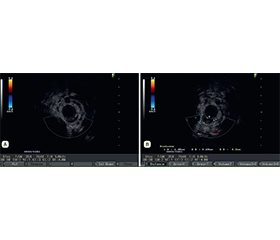
Актуальність. Гемангіома стравоходу (ГС) — доволі рідкісне доброякісне судинне новоутворення, яке становить менше 0,04 % усіх ендоскопічних знахідок. Через розташування й судинну природу ця патологія створює значні діагностичні труднощі та високий ризик небезпечної для життя кровотечі. Традиційно хірургічна резекція була основою лікування, але зараз парадигма змістилася в бік малоінвазивних ендоскопічних методів. Мета: підкреслити важливість мультимодального діагностичного підходу до диференціації рідкісних ГС від інших субепітеліальних уражень, продемонструвати доцільність, безпеку й ефективність ендоскопічної підслизової дисекції при лікуванні гігантської кавернозної гемангіоми у шийному відділі стравоходу, а також надати оновлений огляд сучасних діагностичних і терапевтичних стратегій на основі останніх клінічних даних. Клінічний випадок. Описано клінічний випадок ГС у 57-річної жінки, яка звернулася зі скаргами на прогресуючу дисфагію під час вживання рідини. При ендоскопічному дослідженні виявлено велике м’якоеластичне горбисте ціанотичне підслизове утворення по задній стінці шийного відділу стравоходу на відстані 16–21 см від різців (50 мм у довжину), яке заповнювало до 2/3 просвіту стравоходу, сплощувалося під тиском пристрою та відновлювало свою форму. Ендоскопічна ультрасонографія підтвердила наявність гетерогенного гіпоехогенного утворення в підслизовому шарі, без активного внутрішньоструктурного артеріального кровотоку. МСКТ-ангіографія виявила периферичні флеболіти, що додатково підтвердило діагноз кавернозної ГС. Було успішно проведено ендоскопічну підслизову дисекцію. Незважаючи на технічні труднощі, ураження було видалено en bloc без ускладнень. Гістопатологічне дослідження підтвердило кавернозну гемангіому (МКХ-О 9120/0). Пацієнтку було виписано в доброму стані на другий післяопераційний день. Висновки. Цей випадок ілюструє, що сучасна оперативна ендоскопія, зокрема ендоскопічна підслизова дисекція, є безпечною та ефективною органозберігаючою альтернативою радикальній хірургії при великих ГС, навіть у таких складних анатомічних ділянках, як шийний відділ стравоходу. Мультимодальна візуалізація (ендоскопічна ультрасонографія та МСКТ) має вирішальне значення для точного передопераційного планування.
Background. Esophageal hemangioma (EH) is an exceedingly rare benign vascular neoplasm, accounting for less than 0.04 % of all endoscopic findings. Due to location and vascular nature, it poses significant diagnostic challenges and a high risk of life-threatening hemorrhage. Traditionally, surgical resection was the mainstay of treatment, but the paradigm has shifted toward minimally invasive endoscopic techniques at present. The purpose of the study was to highlight the importance of a multimodal diagnostic approach in differentiating rare EH from other subepithelial lesions, to demonstrate the feasibility, safety, and effectiveness of endoscopic submucosal dissection (ESD) for the treatment of a giant cavernous EH in the cervical esophagus and to provide an updated review of current diagnostic and therapeutic strategies based on recent clinical evidence. Case presentation. We report a clinical case of a 57-year-old female presenting with progressive dysphagia when drinking liquids. Endoscopy revealed a large soft elastic tuberculate cyanotic subepithelial mass on the posterior wall of the cervical esophagus, extending from 16 to 21 cm from the incisors (50 mm in length), which filled up to 2/3 of the esophageal lumen, flattened under pressure with the device and restored its shape. Endoscopic ultrasonography (EUS) confirmed a heterogeneous hypoechoic lesion within the submucosal layer with no active intralesional arterial blood flow. MSCT-angiography identified peripheral phleboliths, further supporting the diagnosis of a cavernous EH. The patient successfully underwent ESD. Despite the technical complexity, the lesion was resected en bloc without complications. Histopathology confirmed a cavernous hemangioma (ICD-O 9120/0). The patient was discharged on the second postoperative day. Conclusions. This case illustrates that advanced operative endoscopy, specifically ESD, is a safe and effective organ-preserving alternative to radical surgery for large EH, even in challenging anatomical locations such as the cervical esophagus. Multimodal imaging (EUS and MSCT) is crucial for accurate preoperative planning.
гемангіома стравоходу; підслизове утворення; ендоскопічна підслизова дисекція; ендоскопічна ультрасонографія; шийний відділ стравоходу
esophageal hemangioma; subepithelial lesion; endoscopic submucosal dissection; endoscopic ultrasonography; cervical esophagus
Для ознакомления с полным содержанием статьи необходимо оформить подписку на журнал.
- Araki K, Ohno S, Egashira A, Saeki H, et al. Esophageal hemangioma: a case report and review of the literature. Hepatogastroenterology. 1999 Nov-Dec;46(30):3148-3154.
- Liu D, Yu XH, Zheng QF, Liu BR. A giant lumen-occluding hemangioma at the cervical esophagus resected by endoscopic submucosal dissection. Gastrointest Endosc. 2020 Apr;91(4):944-945. doi: 10.1016/j.gie.2019.11.001.
- Probst A, Schaller T, Weber T, Messmann H, Scheppach MW. Endoscopic submucosal dissection of a large cavernous hemangioma in the proximal esophagus. Endoscopy. 2025 Dec;57(S 01):E1123-E1124. doi: 10.1055/a-2707-3514.
- Chen Z, Wang L, Yu PJ. Early and accurate diagnosis and selection of appropriate treatment plans are crucial for patients with gastrointestinal hemangiomas. World J Gastrointest Surg. 2025 Feb 27;17(2):99432. doi: 10.4240/wjgs.v17.i2.99432.
- Wu C, Pang Y, Liu X, Wang L. Endoscopic submucosal dissection for oesophageal cavernous haemangioma accompanied by squamous epithelial dysplasia. Dig Liver Dis. 2024 Jan;56(1):211-213. doi: 10.1016/j.dld.2023.10.022.
- Chedgy FJ, Bhattacharyya R, Bhandari P. Endoscopic submucosal dissection for symptomatic esophageal cavernous hemangioma. Gastrointest Endosc. 2015 Apr;81(4):998. doi: 10.1016/j.gie.2014.10.023.
- Li W, Shao M, Hu S, Xie S, He B. The diagnostic value of endoscopic ultrasound for esophageal subepithelial lesions: A review. Medicine (Baltimore). 2024 Nov 15;103(46):e40419. doi: 10.1097/MD.0000000000040419.
- Yang G, Huang S, Liao S, Lv Y, Gong W, Qiu Q, Liu N. Efficacy of endoscopic submucosal tunnel dissection in the management of a large esophageal cavernous hemangioma. Endoscopy. 2024 Dec;56(S 01):E886-E887. doi: 10.1055/a-2421-6218.
- Feczko AF, Louie BE. Endoscopic Resection in the Esophagus. Thorac Surg Clin. 2018 Nov;28(4):481-497. doi: 10.1016/j.thorsurg.2018.07.006.
- Businello G, Dal Pozzo CA, Sbaraglia M, et al. Histopatholo–gical landscape of rare oesophageal neoplasms. World J Gastroenterol. 2020 Jul 21;26(27):3865-3888. doi: 10.3748/wjg.v26.i27.3865.
- Baek IH, Jeon JW, Shin HP, et al. Successful en bloc resection of an esophageal hemangioma by combined EBL & EMR: a case report and technical review. Transl Gastroenterol Hepatol. 2016 May 24;1:45. doi: 10.21037/tgh.2016.05.07.
- Pimentel-Nunes P, Libânio D, Bastiaansen BAJ, et al. Endoscopic submucosal dissection for superficial gastrointestinal lesions: European Society of Gastrointestinal Endoscopy (ESGE) Guideline — Update 2022. Endoscopy. 2022 Jun;54(6):591-622. doi: 10.1055/a-1811-7025.
- Libânio D, Pimentel-Nunes P, Dinis-Ribeiro M. Complications of endoscopic resection techniques for upper GI tract lesions. Best Pract Res Clin Gastroenterol. 2016 Oct;30(5):735-748. doi: 10.1016/j.bpg.2016.09.010.
- Cantero D, Yoshida T, Ito T, Suzumi M, Tada M, Okita K. Esophageal hemangioma: endoscopic diagnosis and treatment. Endo–scopy. 1994 Feb;26(2):250-3. doi: 10.1055/s-2007-1008954.
- Qu J, Wang Z, Zhang H, et al. How to update esophageal masses imaging using literature review (MRI and CT features). Insights Imaging. 2024 Jul 6;15(1):169. doi: 10.1186/s13244-024-01754-0.
- Santamaria-Barria JA, Banki F, Rajendran S, Floyd C. Hemangioma of the cervical esophagus: A rare case of dysphagia mimi–cking a large esophageal polyp on endoscopy. J Thorac Cardiovasc Surg. 2016 Sep;152(3):e59-61. doi: 10.1016/j.jtcvs.2016.04.050.
- Draganov PV, Wang AY, Othman MO, Fukami N. AGA Institute Clinical Practice Update: Endoscopic Submucosal Dissection in the United States. Clin Gastroenterol Hepatol. 2019 Jan;17(1):16-25.e1. doi: 10.1016/j.cgh.2018.07.041.
- Sogabe M, Taniki T, Fukui Y, et al. A patient with esophageal hemangioma treated by endoscopic mucosal resection: a case report and review of the literature. J Med Invest. 2006 Feb;53(1–2):177-82. doi: 10.2152/jmi.53.177.
- Hyun JH, Jeen YT, Chun HJ, et al. Endoscopic resection of submucosal tumor of the esophagus: results in 62 patients. Endoscopy. 1997 Mar;29(3):165-70. doi: 10.1055/s-2007-1004157.
- Kim SH, Lee JM, Han JK, et al. Three-dimensional MDCT imaging and CT esophagography for evaluation of esophageal tumors: preliminary study. Eur Radiol. 2006 Nov;16(11):2418-26. doi: 10.1007/s00330-006-0337-8.
- Liu J, Wang HH, Gao W, Chi Y, Tian Y. Cervical esopha–geal capillary hemangioma removed by combined and sequential endoscopic ligation and snare polypectomy. Endoscopy. 2008 Sep;40(Suppl 2):E179-80. doi: 10.1055/s-2007-995810.
- Yu Y, Shen B, Zhang C, Zhang J, Cao L, Lu P, Liu M. Successful en bloc resection of large esophageal hemangioma by endoscopic submucosal dissection: A case report. Medicine (Baltimore). 2020 Oct 23;99(43):e22821. doi: 10.1097/MD.0000000000022821.
- Kobara H, Mori H, Masaki T. Successful en bloc resection of an esophageal hemangioma by endoscopic submucosal dissection. Endo–scopy. 2012;44 Suppl 2 UCTN:E134-5. doi: 10.1055/s-0030-1256703.
- Wang HY, Bi JX, Guo SJ. Endoscopic submucosal dissection of a proximal esophageal hemangioma accompanied with gastrointestinal bleeding. Dig Endosc. 2020 Sep;32(6):990. doi: 10.1111/den.13721.
- Al Shamousi K, Al-Naamani Z, Abbas Q, Al-Busafi S, El Bingawi H. Treatment with Endoscopic Ultrasound (EUS)-Guided Transesophageal Coil Embolization Without Sclerotherapy: A Novel Therapy for Giant Distal Esophageal Hemangioma. Cureus. 2022 May 24;14(5):e25303. doi: 10.7759/cureus.25303.
- Zhongsheng L, Yan D, Ezzat R, et al. Endoscopic Submucosal Dissection: A Safe and Effective Alternative to Surgical Intervention for Esophageal Hemangioma. Surg Laparosc Endosc Percutan Tech. 2024 Apr 1;34(2):124-128. doi: 10.1097/SLE.0000000000001266.