
Журнал «Здоровье ребенка» Том 21, №3, 2026
Вернуться к номеру
Лінійний IgA-бульозний дерматоз: ретроспективна клінічна серія випадків із запропонованим діагностичним алгоритмом у дітей та дорослих
Авторы: Шостакович‑Корецька Л.Р. (1), Будаєва І.В. (1), Білоконь О.О. (1), Серневич О.В. (2)
(1) - Дніпровський державний медичний університет, м. Дніпро, Україна
(2) - Комунальне некомерційне товариство «Регіональний медичний центр родинного здоров’я» Дніпропетровської обласної ради, м. Дніпро, Україна
Рубрики: Педиатрия/Неонатология
Разделы: Клинические исследования
Версия для печати
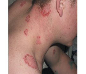
Актуальність. Лінійний IgA-бульозний дерматоз (ЛАБД) — рідкісне автоімунне субепідермальне везикуло-бульозне захворювання, що може виникати у дітей та дорослих і часто імітує інфекційні або алергічні дерматози. У клінічній практиці інфекціоніста ЛАБД нерідко потрапляє до диференційного ряду з вітряною віспою, рецидивуючим оперізуючим герпесом, медикаментозними токсикодерміями та іншими автоімунними бульозними дерматозами. Мета: на підставі аналізу власної серії клінічних випадків та актуальних доказових джерел сформувати практичний алгоритм диференційної діагностики інфекційних і автоімунних везикуло-бульозних захворювань з акцентом на доступні діагностичні кроки в умовах обмежених ресурсів. Матеріали та методи. Дослідження має дизайн ретроспективної клінічної серії випадків і включає 5 пацієнтів із клінічно встановленим діагнозом ЛАБД (2 дитини 3 і 5 років; 3 дорослих 19, 25, 27 років). Усім пацієнтам проводили клінічну оцінку, базові лабораторні дослідження; за показаннями — ПЛР-дослідження вмісту везикул на ДНК VZV та HSV-1/2 для виключення інфекційної природи хвороби. На основі аналізу літератури і власних даних узагальнені сучасні підходи до клінічної морфологічної та імунопатологічної верифікації ЛАБД (біопсія + ПІФ/НІФ, лінійний IgA). Звітність підготовлено відповідно до рекомендацій CARE 2013 та STROBE. Результати. У 4 з 5 хворих можливий тригер передував за 1–3 місяці (ГРВІ, лакунарний тонзиліт; прийом НПЗП/парацетамолу або ібупруфену). Для всіх пацієнтів була характерна відсутність гарячки та токсичного синдрому. Висипання поліморфні, з тенденцією до кільцеподібних/дугоподібних елементів («нитки перлів»), із помірним свербежем у 4/5 хворих. Ураження слизових не спостерігали, за винятком ерозивного хейліту у дитини 3 років. Запропоновано покроковий алгоритм діагностики з обов’язковим виключенням VZV/HSV-інфекцій перед призначенням медикаментозної імуносупресії та пріоритетом застосування прямої імунофлуоресценції як золотого стандарту діагностики. Висновки. ЛАБД є хворобою міждисциплінарного диференційного пошуку. В умовах обмеженої доступності імунопатологічної верифікації хвороби доцільно використовувати структурований алгоритм: 1) оцінка клінічної морфології висипань та можливих тригерів; 2) виключення герпесвірусних інфекцій (VZV/HSV) при бульозних висипаннях; 3) консультація дерматолога; 4) направлення на біопсію з ПІФ; 5) оцінка тяжкості; 6) індивідуалізований вибір терапії з урахуванням тяжкості та безпеки.
Background. Linear IgA bullous dermatosis (LABD) is a rare autoimmune subepidermal vesiculobullous disease affecting both children and adults and frequently mimicking infectious or allergic dermatoses. In clinical infectious disease practice, LABD may be misdiagnosed as varicella, recurrent herpes zoster, drug-induced toxicoderma, and other autoimmune bullous dermatoses. The purpose was to provide a pragmatic diagnostic algorithm for differentiating infectious and autoimmune vesiculobullous diseases based on an analysis of own clinical case series and up‑to‑date evidence, with an emphasis on accessible diagnostic steps under limited resources. Materials and methods. This study is a retrospective clinical case series including 5 patients with clinically established LABD (two children aged 3 and 5; three adults aged 19, 25, and 27 years). All participants underwent clinical examination, basic laboratory tests; if indicated, polymerase chain reaction of vesicle contents for varicella zoster virus (VZV) and herpes simplex virus (HSV) 1/2 DNA to exclude the infectious nature of the disease. Based on the analysis of the literature and our own data, modern approaches to clinical morphological and immunopathological verification of LABD (biopsy + direct (DIF)/indirect immunofluorescence, linear IgA) were summarized. The report was prepared in accordance with the CARE 2013 and STROBE guidelines. Results. In 4 of 5 cases, a potential trigger occurred 1–3 months before onset (acute respiratory viral infection or lacunar tonsillitis; nonsteroidal anti-inflammatory drugs/paracetamol use). Fever or systemic toxicity was absent in all patients. Lesions were polymorphic with annular/arciform clusters (“string of pearls”), with moderate pruritus in 4 of 5 cases. Mucosal involvement was not observed, except erosive cheilitis in one child. We propose a stepwise diagnostic algorithm prioritizing exclusion of VZV/HSV infection before prescription of immunosuppressive drugs and emphasizing DIF as the diagnostic gold standard. Conclusions. LABD remains a multidisciplinary diagnostic challenge. In conditions of limited availability of immunopathological verification of the disease, it is advisable to use a structured algorithm: 1) assessment of clinical morphology of rashes and possible triggers; 2) exclusion of herpesvirus infections (VZV/HSV) in bullous rashes; 3) consultation with a dermatologist; 4) referral for biopsy with DIF; 5) assessment of severity; 6) individualized selection of therapy taking into account severity and safety.
лінійний IgA-бульозний дерматоз; діагностичний алгоритм; ПЛР; VZV; HSV; пряма імунофлуоресценція
linear IgA bullous dermatosis; diagnostic algorithm; polymerase chain reaction; varicella zoster virus; herpes simplex virus; direct immunofluorescence