
Oral and General Health Том 7, №1, 2026
Вернуться к номеру
Ангуляція та тип ретенції верхньощелепних іклів: клініко-статистичний аналіз
Авторы: Якуш О.Г.
Національний університет охорони здоров’я імені П.Л. Шупика, м. Київ, Україна
Рубрики: Стоматология
Разделы: Клинические исследования
Версия для печати
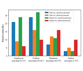
Актуальність. Вперше проведено системний аналіз ангуляції верхньощелепних ретинованих іклів з урахуванням типу ретенції та локалізації правий/лівий. Виявлено, що тип ретенції є прогностичним фактором ангуляції: двобічна ретенція асоціюється з більш вираженим відхиленням від вертикальної осі, що підвищує ризик ускладнень при прорізуванні. Крім того, результати дозволяють визначити клінічно значущі критерії для вибору тактики ортодонтичного втручання та прогнозування складності прорізування. Мета: оцінити взаємозв’язок між ангуляцією ретинованих верхньощелепних іклів та клінічними факторами (тип ретенції, тип прикусу, наявність скупченості зубів) у пацієнтів різного віку, а також визначити прогностичне значення цих параметрів для планування ортодонтичного втручання. Матеріали та методи. Проведено клініко-радіологічне обстеження 162 пацієнтів (204 ретиновані ікла) віком 15–45 років. Пацієнти були розподілені на підлітків (15–18 років), молодих дорослих (19–25 років) та дорослих (26–45 років). Використано конусно-променеву комп’ютерну томографію (КПКТ) для тривимірної оцінки положення ікла, ангуляції та взаєморозташування щодо сусідніх зубів. Ангуляцію класифіковано як нормальну (0–15°), помірну (16–45°), виражену (46–75°) та крайню (> 75°). Статистичний аналіз включав -тест для порівняння розподілу ангуляції між однобічною та двобічною ретенцією, а також між лівими та правими іклами. Результати. Однобічно ретиновані ікла переважно демонстрували нормальну та помірну ангуляцію (80,0 %), тоді як для двобічно ретинованих іклів частіше були характерні виражена та крайня ангуляція (51,2 %). Виявлені відмінності були статистично значущими ( = 12,58; p = 0,006). Ліві ікла з однобічною ретенцією частіше мали помірну ангуляцію, тоді як праві ікла показували більшу варіабельність кутів, включно з крайніми. Двобічно ретиновані ікла, особливо праві, мали високу частоту крайніх ангуляцій (> 75°). Аналіз за віковими групами показав, що відхилення ангуляції найбільш виражене у підлітків та молодих дорослих. Висновки. Тип ретенції верхньощелепних іклів є прогностичним фактором ангуляції. Двобічна ретенція асоціюється з більш вираженим відхиленням від вертикальної осі. Наявність скупченості зубів та дистальний тип прикусу (II клас за Енглем) вірогідно корелюють зі збільшенням кутів нахилу ретинованих іклів, що підвищує ризик ускладнень при прорізуванні. Раннє виявлення ангуляції за допомогою КПКТ дозволяє оптимізувати планування ортодонтичного лікування з урахуванням індивідуальних анатомо-клінічних особливостей пацієнта.
Background. For the first time, a systematic analysis of retained maxillary canine angulation was performed considering retention type and laterality (left/right). Retention type was identified as a prognostic factor for angulation: bilateral retention is associated with more pronounced deviation from the vertical axis, increasing the risk of eruption complications. Furthermore, the results provide clinically significant criteria for selecting orthodontic treatment strategies and predicting eruption complexity. The purpose was to assess the relationship between the angulation of retained maxillary canines and clinical factors (retention type, occlusion type, presence of dental crowding) in patients of different ages, as well as to determine the prognostic significance of these parameters for planning orthodontic intervention. Materials and methods. A clinical and radiological examination was conducted on 162 patients (204 retained canines) aged 15–45 years. They were divided into adolescents (15–18 years), young adults (19–25 years), and adults (26–45 years). Cone-beam computed tomography was used for three-dimensional evaluation of canine position, angulation, and relationship to adjacent teeth. Angulation was classified as normal (0–15°), moderate (16–45°), severe (46–75°), and extreme (> 75°). Statistical analysis included the -test to compare angulation distribution between unilateral and bilateral retention, as well as between left and right canines. Results. Unilateral retained canines predominantly showed normal and moderate angulation (80.0 %), whereas bilateral retained canines more frequently exhibited severe and extreme angulation (51.2 %), which was statistically significant ( = 12.58; p = 0.006). Left canines with unilateral retention more often had moderate angulation, while right canines showed greater variability, including extreme angles. Bilateral retained canines, especially right ones, had a high frequency of extreme angulation (> 75°). Age group analysis revealed that angulation deviation was most pronounced among adolescents and young adults. Conclusions. The type of maxillary canine retention is a prognostic factor for their angulation. Bilateral retention is associated with a more pronounced deviation from the vertical axis. The presence of dental crowding and a distal occlusion type (Angle’s class II) significantly correlate with increased angulation of retained canines, which raises the risk of eruption complications. Early detection of angulation using cone-beam computed tomography allows for the optimization of orthodontic treatment planning, taking into account the individual anatomical and clinical characteristics of the patient.
ангуляція іклів; ретенція іклів; однобічна та двобічна ретенція; конусно-променева комп’ютерна томографія; ортодонтичне планування; тривимірна діагностика
canine angulation; canine retention; unilateral and bilateral retention; cone-beam computed tomography; orthodontic treatment planning; three-dimensional diagnosis