
Международный эндокринологический журнал Том 22, №4, 2026
Вернуться к номеру
Підвищений базовий рівень фактора фон Віллебранда як предиктор тяжкості та неефективності лікування діабетичної ретинопатії
Авторы: A.V. Serdiuk (1), S.Yu. Mogilevskyy (2), M.S. Babenko (3), S.V. Ziablitsev (3)
(1) - Dnipro State Medical University, Dnipro, Ukraine
(2) - Shupyk National Healthcare University of Ukraine, Kyiv, Ukraine
(3) - Bogomolets National Medical University, Kyiv, Ukraine
Рубрики: Эндокринология
Разделы: Клинические исследования
Версия для печати
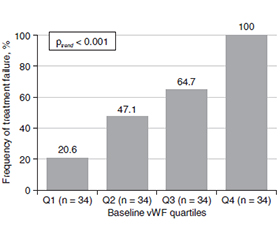
Актуальність. Пошук системних біомаркерів, здатних відображати активність діабетичної ретинопатії (ДР) і прогнозувати несприятливі результати лікування, є важливим клінічним завданням сучасної офтальмології. Особливу увагу привертає фактор фон Віллебранда (vWF), який є маркером ендотеліальної дисфункції. Відповідно до цього метою дослідження було оцінити звʼязок базового рівня vWF зі стадіями ДР та неефективністю лікування, визначеною за швидким прогресуванням ДР протягом 2 років, а також встановити його потенційну прогностичну цінність у пацієнтів із цукровим діабетом 2-го типу (ЦД2). Матеріали та методи. Проаналізовано дані 136 пацієнтів із ЦД2 та ДР. Стадію ДР класифікували за критеріями ВООЗ (непроліферативна, препроліферативна, проліферативна). Звʼязок vWF із прогресуванням хвороби оцінювали за допомогою порядкової та бінарної логістичної регресії з поправкою на клінічні конфаундери (вік, стать, стаж ЦД2, глікозильований гемоглобін (HbA1c), систолічний артеріальний тиск, вихідне офтальмологічне лікування). Прогностичну здатність визначали за допомогою ROC-аналізу із багаторазовою (50 повторів) 5-кратною перехресною валідацією. Результати. Неефективність лікування було зафіксовано у 58,1 % хворих. Рівень vWF послідовно зростав залежно від тяжкості ДР: 16,37 мкг/мл при непроліферативній, 30,66 мкг/мл при препроліферативній та 41,49 мкг/мл при проліферативній стадії (p < 0,001). За неефективності лікування рівень vWF був вірогідно вищим (34,26 мкг/мл), ніж за його ефективності (18,01 мкг/мл; p < 0,001). У скоригованих моделях підвищення vWF на 1 мкг/мл незалежно збільшувало шанси більш тяжкої стадії ДР (OR 1,23; p < 0,001) та неефективності лікування (OR 1,19; p < 0,001). Площа під ROC-кривою становила 0,846; поріг 29,37 мкг/мл забезпечив чутливість 64,6 % та специфічність 93,0 %. Висновки. Базовий рівень vWF має стійкий звʼязок із тяжкістю ДР та є клінічно релевантним предиктором щодо стратифікації ризику неефективності лікування в пацієнтів із ЦД2.
Background. The search for systemic biomarkers capable of reflecting the activity of diabetic retinopathy (DR) and predicting adverse treatment outcomes is an important clinical task of modern ophthalmology. Of particular interest is von Willebrand factor (vWF), which is a marker of endothelial dysfunction. Therefore, our aim was to evaluate the association of baseline vWF levels with the stage of DR and treatment failure, defined as rapid DR progression within 2 years, as well as to establish its potential prognostic utility in patients with type 2 diabetes mellitus (T2DM). Materials and methods. The study analyzed data from 136 T2DM patients with DR. DR stages were classified according to the WHO criteria as non-proliferative, pre-proliferative, and proliferative DR. The association of vWF with disease progression was assessed using ordinal and binary logistic regression models, adjusted for clinical confounders (age, sex, T2DM duration, glycated hemoglobin (HbA1c), systolic blood pressure, and baseline active ophthalmic treatment). Predictive utility was determined via ROC analysis with repeated stratified 5-fold cross-validation (50 repeats). Results. The presence of treatment failure was observed in 58.1 % of cases. vWF levels consistently increased with DR severity: 16.37 µg/mL in non-proliferative, 30.66 µg/mL in pre-proliferative, and 41.49 µg/mL in proliferative DR (p < 0.001). In treatment failure, vWF levels were significantly higher (34.26 µg/mL) compared to its absence (18.01 µg/mL; p < 0.001). In adjusted models, a 1 µg/mL increase in vWF independently increased the odds of a more severe DR stage (OR 1.23; p < 0.001) and treatment failure (OR 1.19; p < 0.001). The area under the ROC curve was 0.846; an optimal cut-off of 29.37 µg/mL yielded a sensitivity of 64.6 % and a specificity of 93.0 %. Conclusions. Baseline vWF levels demonstrate a consistent association with DR severity and represent a clinically relevant predictor for risk stratification of treatment failure in patients with T2DM.
діабетична ретинопатія; фактор фон Віллебранда; цукровий діабет 2-го типу; біомаркери; неефективність лікування
diabetic retinopathy; von Willebrand factor; diabetes mellitus type 2; biomarkers; treatment failure
Для ознакомления с полным содержанием статьи необходимо оформить подписку на журнал.
- Teo ZL, Tham YC, Yu M, Chee ML, Rim TH, Cheung N, et al. Global Prevalence of Diabetic Retinopathy and Projection of Burden through 2045: Systematic Review and Meta-analysis. Ophthalmology. 2021 Nov;128(11):1580-1591. doi: 10.1016/j.ophtha.2021.04.027.
- Flaxel CJ, Adelman RA, Bailey ST, Fawzi A, Lim JI, Vemu–lakonda GA, et al. Diabetic Retinopathy Preferred Practice Pattern®. Ophthalmology. 2020 Jan;127(1):P66-P145. doi: 10.1016/j.ophtha.2019.09.025.
- Wong TY, Cheung CM, Larsen M, Sharma S, Simó R. Diabetic retinopathy. Nat Rev Dis Primers. 2016 Mar 17;2:16012. doi: 10.1038/nrdp.2016.12.
- Forrester JV, Kuffova L, Delibegovic M. The Role of Inflammation in Diabetic Retinopathy. Front Immunol. 2020 Nov 6;11:583687. doi: 10.3389/fimmu.2020.583687.
- Miyamoto K, Khosrof S, Bursell SE, Rohan R, Murata T, Clermont AC, et al. Prevention of leukostasis and vascular leakage in streptozotocin-induced diabetic retinopathy via intercellular adhesion molecule-1 inhibition. Proc Natl Acad Sci USA. 1999 Sep 14;96(19):10836-41. doi: 10.1073/pnas.96.19.10836.
- Joussen AM, Murata T, Tsujikawa A, Kirchhof B, Bursell SE, Adamis AP. Leukocyte-mediated endothelial cell injury and death in the diabetic retina. Am J Pathol. 2001 Jan;158(1):147-52. doi: 10.1016/S0002-9440(10)63952-1.
- Vazzana N, Ranalli P, Cuccurullo C, Davì G. Diabetes mellitus and thrombosis. Thromb Res. 2012 Mar;129(3):371-7. doi: 10.1016/j.thromres.2011.11.052.
- Lenting PJ, Casari C, Christophe OD, Denis CV. Von Willebrand factor: the old, the new and the unknown. J Thromb Haemost. 2012 Dec;10(12):2428-37. doi: 10.1111/jth.12008.
- Kawecki C, Lenting PJ, Denis CV. Von Willebrand factor and inflammation. J Thromb Haemost. 2017 Jul;15(7):1285-1294. doi: 10.1111/jth.13696.
- Gragnano F, Sperlongano S, Golia E, Natale F, Bianchi R, Crisci M, et al. The Role of von Willebrand Factor in Vascular Inflammation: From Pathogenesis to Targeted Therapy. Mediators Inflamm. 2017;2017:5620314. doi: 10.1155/2017/5620314.
- Jenkins AJ, Joglekar MV, Hardikar AA, Keech AC, O’Neal DN, Januszewski AS. Biomarkers in Diabetic Retinopathy. Rev Diabet Stud. 2015;12(1–2):159-95. doi: 10.1900/RDS.2015.12.159.
- Spijkerman AM, Gall MA, Tarnow L, Twisk JW, Lauritzen E, Lund-Andersen H, et al. Endothelial dysfunction and low-grade inflammation and the progression of retinopathy in type 2 diabetes. Diabet Med. 2007 Sep;24(9):969-76. doi: 10.1111/j.1464-5491.2007.02217.x.
- Mogilevskyy SYu, Serdiuk AV, Ziablitsev SV. Endothelin-1 as a biomarker of diabetic retinopathy and a factor in its progression. Archives of Ukrainian Ophthalmology. 2025;13(2):107-114. doi: 10.22141/2309-8147.13.2.2025.411 (in Ukrainian).
- Muni RH, Kohly RP, Lee EQ, Manson JE, Semba RD, Schaum–berg DA. Prospective study of inflammatory biomarkers and risk of diabetic retinopathy in the diabetes control and complications trial. JAMA Ophthalmol. 2013 Apr;131(4):514-21. doi: 10.1001/jamaophthalmol.2013.2299.
- Rajab HA, Baker NL, Hunt KJ, Klein R, Cleary PA, Lachin J, et al.; DCCT/EDIC Group of Investigators. The predictive role of markers of inflammation and endothelial dysfunction on the course of diabetic retinopathy in type 1 diabetes. J Diabetes Complications. 2015 Jan-Feb;29(1):108-14. doi: 10.1016/j.jdiacomp.2014.08.004.
- Ekelund C, Dereke J, Nilsson C, Landin-Olsson M. Are solub–le E-selectin, ICAM-1, and VCAM-1 potential predictors for the development of diabetic retinopathy in young adults, 15–34 years of age? A Swedish prospective cohort study. PLoS One. 2024 Jun 6;19(6):e0304173. doi: 10.1371/journal.pone.0304173.
- World Health Organization. Prevention of blindness from dia–betes mellitus: report of a WHO consultation in Geneva, Switzerland, 9–11 November 2005. Geneva: World Health Organization; 2006.
- Kanda Y. Investigation of the freely available easy-to-use software EZR for medical statistics. Bone Marrow Transplant. 2013 Mar;48(3):452-8. doi: 10.1038/bmt.2012.244.
- Tan TE, Wong TY. Diabetic retinopathy: Looking forward to 2030. Front Endocrinol (Lausanne). 2023 Jan 9;13:1077669. doi: 10.3389/fendo.2022.1077669.
- Wang J, Song X, Xia Z, Feng S, Zhang H, Xu C, et al. Serum biomarkers for predicting microvascular complications of diabetes mellitus. Expert Rev Mol Diagn. 2024 Aug;24(8):703-713. doi: 10.1080/14737159.2024.2391021.
- Jia G, Bai H, Mather B, Hill MA, Jia G, Sowers JR. Diabetic Vasculopathy: Molecular Mechanisms and Clinical Insights. Int J Mol Sci. 2024 Jan 9;25(2):804. doi: 10.3390/ijms25020804.
- Wei H, Xiao X, Zeng S, Liu Y, Liu X, Zeng T, et al. Alterations in factors associated with diabetic retinopathy combined with thrombosis: A review. Medicine (Baltimore). 2023 Aug 4;102(31):e34373. doi: 10.1097/MD.0000000000034373.
- Kaur G, Harris NR. Endothelial glycocalyx in retina, hyperglycemia, and diabetic retinopathy. Am J Physiol Cell Physiol. 2023 May 1;324(5):C1061-C1077. doi: 10.1152/ajpcell.00188.2022.
- Kaštelan S, Orešković I, Bišćan F, Kaštelan H, Gverović Antunica A. Inflammatory and angiogenic biomarkers in diabetic reti–nopathy. Biochem Med (Zagreb). 2020 Oct 15;30(3):030502. doi: 10.11613/BM.2020.030502.
- Jonny, Violetta L, Kartasasmita AS, Supriyadi R, Rita C. Circulating Biomarkers to Predict Diabetic Retinopathy in Patients with Diabetic Kidney Disease. Vision (Basel). 2023 Apr 9;7(2):34. doi: 10.3390/vision7020034.
- Kassab HS, Morsy EY, Abdirahman AN, Amin NG. Study of the relation between plasma level of von Willebrand factor and diabetic retinopathy in type 2 diabetes. Int J Diabetes Dev Ctries. 2023;43:785-791. doi: 10.1007/s13410-022-01139-3.
- Siemianowicz K, Francuz T, Gminski J, Telega A, Syzdól M. Endothelium dysfunction markers in patients with diabetic retinopathy. Int J Mol Med. 2005 Mar;15(3):459-62.
- Kadıköylü G, Bolaman AZ, Sönmez HM, Oge M, Güney E, Akyol A, et al. Does von Willebrand Factor Have an Effect on the Occurrence of the Diabetic Complications? Turk J Haematol. 2002 Mar 5;19(1):31-7.
- Stehouwer CD, Zellenrath P, Polak BC, Baarsma GS, Nauta JJ, Donker AJ, et al. Von Willebrand factor and early diabetic reti–nopathy: no evidence for a relationship in patients with type 1 (insulin-dependent) diabetes mellitus and normal urinary albumin excretion. Diabetologia. 1992 Jun;35(6):555-9. doi: 10.1007/BF00400484.
- Feng D, Bursell SE, Clermont AC, Lipinska I, Aiello LP, Laffel L, et al. Von Willebrand factor and retinal circulation in early-stage retinopathy of type 1 diabetes. Diabetes Care. 2000 Nov;23(11):1694-8. doi: 10.2337/diacare.23.11.1694.
- Domingueti CP, Fuzatto JA, Fóscolo RB, Reis JS, Dusse LM, Carvalho MDG, et al. Association between von Willebrand factor, disintegrin and metalloproteinase with thrombospondin type 1 motif member 13, D-dimer and cystatin C levels with retinopathy in type 1 diabetes mellitus. Clin Chim Acta. 2016 Aug 1;459:1-4. doi: 10.1016/j.cca.2016.05.011.
- Oggianu L, Lancellotti S, Pitocco D, Zaccardi F, Rizzo P, Martini F, et al. The oxidative modification of von Willebrand factor is associated with thrombotic angiopathies in diabetes mellitus. PLoS One. 2013;8(1):e55396. doi: 10.1371/journal.pone.0055396.
- Boeri D, Cagliero E, Podestá F, Lorenzi M. Vascular wall von Willebrand factor in human diabetic retinopathy. Invest Ophthalmol Vis Sci. 1994 Feb;35(2):600-7.
- Peng X, Wang X, Fan M, Zhao J, Lin L, Liu J. Plasma levels of von Willebrand factor in type 2 diabetes patients with and without cardiovascular diseases: A meta-analysis. Diabetes Metab Res Rev. 2020 Jan;36(1):e3193. doi: 10.1002/dmrr.3193.
- Frankel DS, Meigs JB, Massaro JM, Wilson PW, O’Donnell CJ, D’Agostino RB, et al. Von Willebrand factor, type 2 diabetes mellitus, and risk of cardiovascular disease: the Framingham offspring study. Circulation. 2008 Dec 9;118(24):2533-9. doi: 10.1161/CIRCULATIONAHA.108.792986.
- Jager A, van Hinsbergh VW, Kostense PJ, Emeis JJ, Nijpels G, Dekker JM, et al. Prognostic implications of retinopathy and a high plasma von Willebrand factor concentration in type 2 diabe–tic subjects with microalbuminuria. Nephrol Dial Transplant. 2001 Mar;16(3):529-36. doi: 10.1093/ndt/16.3.529.
- Perais J, Agarwal R, Evans JR, Loveman E, Colquitt JL, Owens D, et al. Prognostic factors for the development and progression of proliferative diabetic retinopathy in people with diabetic retinopathy. Cochrane Database Syst Rev. 2023 Feb 22;2(2):CD013775. doi: 10.1002/14651858.CD013775.pub2.
- Serdiuk AV. Glycated hemoglobin as a prognostic factor for the progression of non-proliferative diabetic retinopathy in type 2 diabetes. Archives of Ukrainian Ophthalmology. 2024;12(2):26-30. doi: 10.22141/2309-8147.12.2.2024.377 (in Ukrainian).
- Abu El-Asrar AM, Nawaz MI, Ahmad A, Siddiquei M, Allegaert E, Adyns L, et al. ADAMTS13 Improves Endothelial Function and Reduces Inflammation in Diabetic Retinopathy. Cells. 2025 Jan 9;14(2):85. doi: 10.3390/cells14020085.
- Mogilevskyy SYu, Serdiuk AV, Serdiuk VN, Ziablitsev SV. Model for predicting the efficacy of treating diabetic retinopathy in type 2 diabetes on the basis of determination of markers of endothelial dysfunction. Journal of Ophthalmology (Ukraine). 2025;5(527):3-12. doi: 10.31288/oftalmolzh20256312.