
Інформація призначена тільки для фахівців сфери охорони здоров'я, осіб,
які мають вищу або середню спеціальну медичну освіту.

Підтвердіть, що Ви є фахівцем у сфері охорони здоров'я.

Журнал «Травма» Том 27, №3, 2026

Вернуться к номеру

Оцінка рівнів кальцію, фосфору, вітаміну D3 і прокальцитоніну в пацієнтів з остеопорозом та їх зв’язок зі статтю, віком й індексом маси тіла
Авторы: Ahmed Abduladheem (1), Riam Yousfe Muttair (2), Qammar Shaker Hmood (2), Rawa Abdulkareem Abd (2)
(1) - Basic Sciences Branch, College of Nursing, National University of Science and Technology, Thi-Qar, Iraq
(2) - College of Science, University of Thi-Qar, Thi-Qar, Iraq

Рубрики: Травматология и ортопедия

Разделы: Клинические исследования

Версия для печати


Резюме

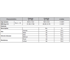
Актуальність. Зі старінням населення світу зростає важливість профілактики та лікування остеопоротичних переломів, спричинених ламкістю кісток. У цьому дослідженні пропонується комплексний аналіз частоти й розподілу переломів, спричинених ламкістю кісток, впливу медикаментозних втручань на їх виникнення, а також потенціалу інноваційних скринінгових програм для зменшення медичних і фінансових наслідків остеопоротичних переломів. Мета: оцінити рівні Ca, Pi, вітаміну D3 та прокальцитоніну (ПКТ) у пацієнтів з остеопорозом і їх зв’язок зі статтю, віком й індексом маси тіла. Матеріали та методи. У цьому дослідженні з січня до березня 2024 року взяли участь 160 осіб, включаючи 80 хворих із підтвердженим остеопорозом та 80 здорових людей контрольної групи. Вік пацієнтів з остеопорозом (58 жінок та 22 чоловіки) становив 50–75 років. Біохімічні параметри визначали за допомогою аналізатора Cobas е 402. Результати. Спостерігалося вірогідне збільшення віку й індексу маси тіла в осіб з остеопорозом порівняно з контрольною групою. Поширеність захворювання була вищою серед жінок, ніж серед чоловіків. Результати показали, що 82,5 % пацієнтів мали хронічні захворювання, як-от діабет, гіпертензія та серцево-судинна патологія. Щодо місця проживання, 70 % пацієнтів у цьому дослідженні були мешканцями міст. Рівень Ca, вітаміну D3 значно знизився, тоді як Pi — значно підвищився у хворих порівняно з контрольною групою. Водночас уміст ПКТ не зазнавав впливу патології. Щодо віку, було показано, що рівні як Ca, Pi, так і вітаміну D3 знижувалися зі збільшенням віку. Крім того, всі біохімічні параметри не залежали від статі пацієнтів. Висновки. Дослідження продемонструвало, що при остеопорозі спостерігаються значні порушення біохімічних маркерів, пов’язаних із кістковим метаболізмом, що характеризується зниженням рівня кальцію та вітаміну D3 у сироватці крові й підвищенням умісту фосфору порівняно зі здоровими особами контрольної групи. Ці зміни були більш вираженими у людей віком понад 60 років, що свідчить про вікове погіршення мінерального гомеостазу. Зв’язку ПКТ з остеопорозом не виявлено.

Background. As the global population continues to age, the significance of preventing and treating osteoporotic fragility fractures is growing. This study provides a comprehensive analysis of the occurrence and distribution of fragility fractures, the impact of medication interventions on this occurrence, and the potential of innovative screening programs to decrease the medical and financial impact of osteoporotic fractures. The purpose was to evaluate the levels of Ca, Pi, vitamin D3, and procalcitonin (PCT) in osteoporosis patients, and their association with sex, age, and body mass index. Materials and methods. The current study involved a total of 160 participants, comprising 80 patients with confirmed osteoporosis and 80 healthy controls during the period from January to March 2024. The age of patients suffering from osteoporosis (58 females and 22 males) was from 50 to 75 years. The biochemical parameters were evaluated by Cobas е 402. Results. There was a significant increase in both age and body mass index in osteoporosis patients compared to control group. The disease prevalence was higher in females than in males. Results showed that 82.5 % of patients had chronic diseases such as diabetes, hypertension, and cardiovascular pathology. Regarding residency, 70 % of patients in this study were from cities. The level of Ca, vitamin D3 decreased significantly, while the level of Pi increased significantly in patients compared to control group. On the other hand, the level of PCT was not affected by disease. In terms of age, the study revealed that the level of both Ca, Pi and vitamin D3 decreased with increasing age. In addition, all biochemical parameters were not affected by sex of patients. Conclusions. The study demonstrates that osteoporosis patients exhibit significant disturbances in bone-related biochemical markers, characterized by reduced serum calcium and vitamin D3 levels and elevated phosphorus compared to healthy controls. These alterations were more pronounced in individuals over 60 years of age, indicating an age-related decline in mineral homeostasis. PCT showed no association with osteoporosis.


Ключевые слова

остеопороз; прокальцитонін; Ca; Pi; віта-мін D3

osteoporosis; procalcitonin; Ca; Pi; vitamin D3


Для ознакомления с полным содержанием статьи необходимо оформить подписку на журнал.


Список литературы

1. Sobh MM, Abdalbary M, Elnagar S, Nagy E, Elshabrawy N, et al. Secondary osteoporosis and metabolic bone diseases. Journal of Clinical Medicine. 2022 Apr 24;11(9):2382. doi: 10.3390/jcm11092382.
2. Seeman E. Reduced bone formation and increased bone resorption: rational targets for the treatment of osteoporosis. Osteoporosis International. 2003 Mar;14:2-8. doi: 10.1007/s00198-002-1340-9.
3. Aibar-Almazn A, Voltes-Martnez A, Castellote-Caballero Y, Afanador-Restrepo DF, Carceln-Fraile MD, ––Lpez-Ruiz E. Current status of the diagnosis and management of osteoporosis. International Journal of Molecular Sciences. 2022 Aug 21;23(16):9465. doi: 10.3390/ijms23169465.
4. Ebeling PR, Nguyen HH, Aleksova J, Vincent AJ, Wong P, Milat F. Secondary osteoporosis. Endocrine Reviews. 2022 Apr 1;43(2):240-313. doi: 10.1210/endrev/bnab028.
5. Nappi RE, Cucinella L. Long-term consequences of menopause. Female Reproductive Dysfunction. 2020:1-3. doi: 10.1007/978-3-030-03594-5_17-1.
6. Shen Y, Huang X, Wu J, Lin X, Zhou X, et al. The global burden of osteoporosis, low bone mass, and its related fracture in 204 countries and territories, 1990–2019. Frontiers in Endocrinology. 2022 May 20;13:882241. doi: 10.3389/fendo.2022.882241.
7. Fernandez P, Pasqualini M, Locrelle H, Normand M, Bonneau C, et al. The effects of combined amplitude and high-frequency vibration on physically inactive osteopenic postmenopausal women. Front Physiol. 2022 Sep 7;13:952140. doi: 10.3389/fphys.2022.952140.
8. Hofbauer LC, Busse B, Eastell R, Ferrari S, Frost M, et al. Bone fragility in diabetes: novel concepts and clinical implications. The Lancet Diabetes & Endocrinology. 2022 Mar 1;10(3):207-20. doi: 10.1016/s2213-8587(21)00347-8.
9. Mostafa AM, Kyriacou H, Chimutengwende-Gordon M, Khan WS. An overview of the key principles and guidelines in the management of pelvic fractures. Journal of Perioperative Practice. 2021 Sep;31(9):341-8. doi: 10.1177/1750458920947358.
10. Srivastava RK, ed. Osteoporosis: Pathophysiology, Diagnosis, Management and Therapy. Books on Demand; 2024. Available from: https://www.intechopen.com/books/1003108.
11. Xiao PL, Cui AY, Hsu CJ, Peng R, Jiang N, et al. Global, regional prevalence, and risk factors of osteoporosis according to the World Health Organization diagnostic criteria: a syste–matic review and meta-analysis. Osteoporosis International. 2022 Oct;33(10):2137-53. doi: 10.1007/s00198-022-06454-3.
12. Kutsal FY, Ergani GO. Vertebral compression fractures: still an unpredictable aspect of osteoporosis. Turkish Journal of Medical Sciences. 2021;51(2):393-9. doi: 10.3906/sag-2005-315.
13. Ning HT, Du Y, Zhao LJ, Tian Q, Feng H, Deng HW. Racial and gender differences in the relationship between sarcopenia and bone mineral density among older adults. Osteoporosis International. 2021 May;32:841-51. doi: 10.1007/s00198-020-05744-y.
14. Xu G, Guo Y, Wang W, Yu WQ, Chen QM, Wang H. Zoledronic acid improves periodontal health, reduces serum inflammation, and enhances bone metabolism in postmenopausal osteoporosis complicated by periodontitis. Am J Transl Res. 2024 Nov 15;16(11):6519-6527. doi: 10.62347/GXEB5902.
15. Bouillon R, Antonio L, Olarte OR. Calcifediol (25OH vitamin D3) deficiency: A risk factor from early to old age. Nutrients. 2022 Mar 10;14(6):1168. doi: 10.3390/nu14061168.
16. Zubairi RB, Hasan SA, Okab HF. Effect of IL-17 and IL-23 Levels in Women with Polycystic Ovary Syndrome. Reproductive Health in Eastern Europe. 2025;15(1):43-50. doi: 10.34883/PI.2025.15.1.045.
17. Shaker JL, Deftos L. Calcium and phosphate homeostasis. Endotext. 2023 May 17. Available from: https://pubmed.ncbi.nlm.nih.gov/25905252/.
18. Leung EK. Parathyroid hormone. Advances in Clinical Chemistry. 2021 Jan 1;101:41-93. doi: 10.1016/bs.acc.2020.06.005.
19. Tsuprykov O, Elitok S, Buse C, Chu C, Krmer BK, Hocher B. Opposite correlation of 25-hydroxy-vitamin D- and 1,25-dihydroxy-vitamin D-metabolites with gestational age, bone- and lipid-biomarkers in pregnant women. Scientific Reports. 2021 Jan 21;11(1):1923. doi: 10.1038/s41598-021-81452-9.
20. Christakos S, Li S, DeLa Cruz J, Verlinden L, Carmeliet G. Vitamin D and bone. Bone Regulators and Osteoporosis Therapy. 2020:47-63. doi: 10.1007/164_2019_338.
21. Bellavia D, Costa V, De Luca A, Maglio M, Pagani S, et al. Vitamin D level between calcium-phosphorus homeostasis and immune system: new perspective in osteoporosis. Current Osteoporosis Reports. 2016 Oct 13:1-2. doi: 10.1007/s11914-016-0331-2.
22. Okab HF, Salih MB, Jarulla BA. Evaluation of CXCL 10 and IL-10 in COVID-19 pneumonia. Pneumologia. 2022;71(4). doi: 10.2478/pneum-2023-0043.
23. Burt LA, Billington EO, Rose MS, Raymond DA, Hanley DA, Boyd SK. Effect of high-dose vitamin D supplementation on volumetric bone density and bone strength: a randomized clinical trial. JAMA. 2019 Aug 27;322(8):736-45. doi: 10.1001/jama.2019.11889.
24. Siregar G, Jabbar MF, Effendi F, Alhair IH, Prabudi T, et al. Correlation between serum vitamin D levels and bone mass density evaluated by radiofrequency echographic multi-spectrometry technology (REMS) in menopausal women. Narra J. 2024;4(1):e452. doi: 10.52225/narra.v4i1.452.
25. Rahme M, Sharara SL, Baddoura R, Habib RH, Halaby G, et al. Impact of calcium and two doses of vitamin D on bone metabolism in the elderly: a randomized controlled trial. Journal of Bone and Mineral Research. 2017 Jul 1;32(7):1486-95. doi: 10.1002/jbmr.3122.
26. Rangarajan R, Mondal S, Thankachan P, Chakra–barti R, Kurpad AV. Assessing bone mineral changes in response to vitamin D supplementation using natural variability in stable isotopes of calcium in urine. Scientific Reports. 2018 Nov 13;8(1):16751. doi: 10.1038/s41598-018-34568-4.
27. Lerchbaum E, Trummer C, Theiler-Schwetz V, Kollmann M, Wlfler M, et al. Effects of vitamin D supplementation on bone turnover and bone mineral density in healthy men: a post-hoc analysis of a randomized controlled trial. Nutrients. 2019 Mar 29;11(4):731. doi: 10.3390/nu11040731.
28. Alonge TO, Adebusoye LA, Ogunbode AM, Olowookere OO, Ladipo MA, et al. Factors associated with osteoporosis among older patients at the Geriatric Centre in Nigeria: a cross-sectional study. South African Family Practice. 2017 May 1;59(3):87-93. doi: 10.1080/20786190.2016.1272248.
29. Thomas-John M, Codd MB, Manne S, Watts NB, Mongey AB. Risk factors for the development of osteoporosis and osteoporotic fractures among older men. The Journal of Rheumatology. 2009 Sep 1;36(9):1947-52. doi: 10.3899/jrheum.080527.
30. Sahni S, Mangano KM, McLean RR, Hannan MT, Kiel DP. Dietary approaches for bone health: lessons from the Framingham Osteoporosis Study. Current Osteoporosis Reports. 2015 Aug;13:245-55. doi: 10.1007/s11914-015-0272-1.
31. Hashmi FR, Elfandi KO. Heel ultrasound scan in detecting osteoporosis in low trauma fracture patients. Orthopedic Reviews. 2016 Jun 6;8(2). doi: 10.4081/or.2016.6357.
32. Mesinovic J, Mousa A, Wilson K, Scragg R, Plebanski M, et al. Effect of 16-weeks vitamin D replacement on calcium-phosphate homeostasis in overweight and obese adults. The Journal of Steroid Biochemistry and Molecular Biology. 2019 Feb 1;186:169-75. doi: 10.1016/j.jsbmb.2018.10.011.
33. Feng Y, Zhou M, Zhang Q, Liu H, Xu Y, et al. Synergistic effects of high dietary calcium and exogenous parathyroid hormone in promoting osteoblastic bone formation in mice. British Journal of Nutrition. 2015 Mar;113(6):909-22. doi: 10.1017/s0007114514004309.
34. Voulgaridou G, Papadopoulou SK, Detopoulou P, Tsoumana D, Giaginis C, et al. Vitamin D and calcium in osteoporosis, and the role of bone turnover markers: A narrative review of recent data from RCTs. Diseases. 2023 Feb 8;11(1):29. doi: 10.3390/diseases11010029.
35. Munshi R, Kochhar A, Garg V. Impact of dietary habits and physical activity on bone health among 40 to 60 year old females at risk of osteoporosis in India. Eco–logy of food and nutrition. 2015 Sep 3;54(5):470-92. doi: 10.1080/03670244.2015.1015120.
36. Ali NK. Estimation of some mineral (calcium, phosphorous, vitamin 25(OH)D and alkaline phosphatase) in osteoporosis patients in Kirkuk city. J Osteopor Phys Act. 2018;6(2):2-4. doi: 10.4172/2329-9509.1000215.
37. Hamdi RA. Evaluation of Serum Osteocalcin level in Iraqi Postmenopausal women with primary osteoporosis. Journal of the Faculty of Medicine Baghdad. 2013 Jul 1;55(2):166-9. doi: 10.32007/jfacmedbagdad.552649.
38. Al-Azzawie AF, Husain WN, Salih MH, Al-Assie AH. The relationship between some electrolyte levels and MTHFR C667T gene polymorphism in premenopausal and postmenopausal osteoporosis Iraqi women. EurAsian Journal of Biosciences. 2020 Jan 1;14(1). Available from: https://www.researchgate.net/publication/352439665.
39. Jayaram N, Bijoor AR, Rajagopalan N, Venkatesh T. The value of serum and urinary n-Telopeptide in the diagnosis of osteoporosis. Indian J Orthop. 2002;36(9). Available from: https://www.semanticscholar.org/paper.
40. Deng WM, Wei QS, Tan X, Shao Y, Chen XH, Sun WS. Relation of serum 25 hydroxyvitamin D levels to bone mineral density in southern Chinese postmenopausal women: A preliminary study. Indian Journal of Medical Research. 2015 Oct 1;142(4):430-7. doi: 10.4103/0971-5916.169206.
41. Hargrove L, Francis T, Francis H. Vitamin D and GI cancers: shedding some light on dark diseases. Annals of Translational Medicine. 2014 Jan;2(1). doi: 10.3978/j.issn.2305-5839.2013.03.04.
42. Chandy DD, Kare J, Singh SN, Agarwal A, Das V, et al. Effect of vitamin D supplementation, directly or via breast milk for term infants, on serum 25 hydroxyvitamin D and related biochemistry, and propensity to infection: a randomised placebo-controlled trial. British Journal of Nutrition. 2016 Jul;116(1):52-8. doi: 10.1017/s0007114516001756.
43. Labronici PJ, Blunck SS, Lana FR, Esteves BB, Franco JS, et al. Vitamin D and its relation to bone mineral density in postmenopause women. Revista Brasileira de Ortopedia (English Edition). 2013 May 1;48(3):228-35. doi: 10.1016/j.rboe.2012.07.003.

Вернуться к номеру