Резюме
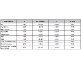
Актуальність. Грудопоперековий перехід — найчастіша локалізація травм хребта (близько половини всіх ушкоджень); значна їх частка супроводжується неврологічним дефіцитом, що суттєво впливає на функціональний результат і якість життя. Чинники регресу дефіциту вивчені недостатньо, а прогностична цінність морфометрії порівняно з вихідною тяжкістю лишається дискусійною. Мета: визначити незалежні чинники регресу дефіциту через 12 місяців після травми грудопоперекового переходу, передусім роль вихідного класу American Spinal Injury Association Impairment Scale (AIS) та морфометрії. Матеріали та методи. Обсерваційне дослідження 240 пацієнтів із вихідним дефіцитом за AIS A-D і 12-місячним катамнезом після травми грудопоперекового переходу (Th11-L2). Тип класифікували за AO Spine, статус — за ISNCSCI. Застосовано кореляцію Спірмена, точний критерій Фішера та логістичну регресію. Результати. Регрес щонайменше на один клас AIS відзначено у 122 (50,8 %) пацієнтів, на два класи й більше — у 10 (4,2 %; 5,8 % серед пацієнтів із вихідним класом A-C). Частота регресу ≥ 1 класу не залежала від типу ушкодження (p = 0,498), проте зростала зі зменшенням вихідної тяжкості дефіциту — від 41,8 % при класі A до 60,9 % при класі D ( = 0,165; p = 0,011). Значний регрес (≥ 2 класи) переважав при ушкодженнях типу C (20,7 проти 2,8 %; відношення шансів 9,0; p = 0,0019), що має пошуковий характер. Морфометричні параметри корелювали з вихідним класом AIS (кут Cobb = –0,48; задня дисторсія = –0,40; передня клиноподібна деформація = +0,36; усі p < 0,001), але не з величиною подальшого регресу; вплив задньої дисторсії зникав після врахування вихідного класу AIS. У багатофакторній моделі єдиним незалежним предиктором регресу була вихідна тяжкість дефіциту (відношення шансів 1,34; 95% довірчий інтервал 1,04–1,73; p = 0,026); тип ушкодження, вік, час від травми до оперативного втручання, рівень енергії травми та щільність кісткової тканини не продемонстрували незалежного впливу на ймовірність регресу. Аналіз чутливості підтвердив той самий напрям ефекту. Дискримінаційна здатність моделі була обмеженою (площа під ROC-кривою 0,612). Висновки. Неврологічне відновлення спостерігалося часто, але переважно в межах одного класу AIS. Основним незалежним чинником регресу є вихідна тяжкість дефіциту, тоді як морфометрія не продемонструвала переконливої додаткової прогностичної цінності понад вихідний клас AIS. Вища частка значного регресу при типі C потребує підтвердження.
Background. The thoracolumbar junction is the most common site of spinal trauma (about half of all spinal injuries), and accompanying neurological deficit strongly affects functional outcome and quality of life. Yet the factors governing recovery of this deficit remain poorly defined, and the prognostic value of morphometry relative to the initial neurological severity is debated. Objective: to determine independent predictors of deficit regression 12 months after thoracolumbar junction injury, focusing on the admission AIS grade and morphometry. Materials and methods. This observational study analysed 240 patients with a thoracolumbar junction injury (Th11-L2), a baseline deficit (AIS A-D), and 12-month follow-up. We classified injuries with the AO Spine system and neurological status with the ISNCSCI. The analysis used Spearman correlation, the Fisher exact test, and multivariable logistic regression. Results. At least one-grade improvement on AIS occurred in 122 (50.8 %) patients and improvement of two grades or more in 10 (4.2 %; 5.8 % among patients with a baseline A-C grade). The rate of ≥ 1 grade regression did not depend on injury type (p = 0.498) but rose as the baseline deficit became milder, from 41.8 % in grade A to 60.9 % in grade D ( = 0.165; p = 0.011). Substantial regression (two grades or more) predominated in type C injuries (20.7 versus 2.8 %; odds ratio 9.0; p = 0.0019); this finding is exploratory. Morphometric parameters correlated with the admission AIS grade (Cobb angle: = –0.48; posterior distraction: = –0.40; anterior wedging: = +0.36; all p < 0.001) but not with the magnitude of later regression; moreover, the effect of posterior distraction disappeared after adjustment for the admission AIS grade. In the multivariable model, the baseline deficit severity emerged as the only independent predictor of regression (odds ratio 1.34; 95 % confidence interval 1.04–1.73; p = 0.026); injury type, age, the injury-to-surgery interval, injury energy, and bone density did not have an independent effect on the likelihood of regression. A sensitivity analysis confirmed the same direction of effect. Consequently, the model showed limited discrimination (area under the ROC curve 0.612). Conclusions. Neurological recovery is common but rarely exceeds one AIS grade. Admission neurological severity is the main independent determinant of regression, whereas morphometry showed no convincing added prognostic value beyond the admission AIS grade. The higher proportion of substantial regression in type C warrants confirmation.
Список литературы
1. Zileli M, Sharif S, Fornari M. Incidence and Epidemio–logy of Thoracolumbar Spine Fractures: WFNS Spine Committee Re–commendations. Neurospine. 2021;18(4):704-712. doi: 10.14245/ns.2142418.209. PMID: 35000323.
2. Wang H, Zhang Y, Xiang Q, Wang X, Li C, et al. Epidemiology of traumatic spinal fractures: experience from medical university-affi–liated hospitals in Chongqing, China, 2001-2010. J Neurosurg Spine. 2012;17(5):459-468. doi: 10.3171/2012.8.SPINE111003. PMID: 22978439.
3. Bruno AG, Burkhart K, Allaire B, Anderson DE, Bouxsein ML. Spinal Loading Patterns From Biomechanical Modeling Explain the High Incidence of Vertebral Fractures in the Thoracolumbar Region. Journal of bone and mineral research: the official journal of the American Society for Bone and Mineral Research. 2017;32(6):1282-1290. doi: 10.1002/jbmr.3113. PMID: 28244135.
4. Suijkerbuijk M, Cranenbroek D, Van Ameijden SI, Van Egmond PW, Joosen MCW, De Jongh MAC, Geuze RE. Return to work following traumatic fractures of the thoracolumbar spine without spinal cord injury: a scoping review. Acta orthopaedica. 2026;97:83-90. doi: 10.2340/17453674.2026.45364. PMID: 41718639.
5. Gao S, Yuexin W, Daole H, Fang Z, Yun T, Yang L. Effect and prognosis of thoracolumbar fracture combined with incomplete spinal cord injury on male sexual function. Journal of orthopaedic surgery and research. 2025;20(1):176. doi: 10.1186/s13018-025-05587-5. PMID: 39972507.
6. Harrop JS, Hunt GE Jr., Vaccaro AR. Conus medullaris and cauda equina syndrome as a result of traumatic injuries: management principles. Neurosurg Focus. 2004;16(6):e4. doi: 10.3171/foc.2004.16.6.4. PMID: 15202874.
7. Bican O, Minagar A, Pruitt AA. The spinal cord: a review of functional neuroanatomy. Neurol Clin. 2013;31(1):1-18. doi: 10.1016/j.ncl.2012.09.009. PMID: 23186894.
8. Harrop JS, Naroji S, Maltenfort MG, Ratliff JK, Tjoumakaris SI, Frank B, et al. Neurologic improvement after thoracic, thoracolumbar, and lumbar spinal cord (conus medullaris) injuries. Spine (Phila Pa 1976). 2011;36(1):21-25. doi: 10.1097/BRS.0b013e3181fd6b36. PMID: 21192220.
9. Brouwers E, Meent HV, Curt A, Maier DD, Abel RF, Weidner N, et al. Recovery after traumatic thoracic- and lumbar spinal cord injury: the neurological level of injury matters. Spinal Cord. 2020;58(9):980-987. doi: 10.1038/s41393-020-0463-1. PMID: 32371940.
10. Kim NH, Lee HM, Chun IM. Neurologic injury and recovery in patients with burst fracture of the thoracolumbar spine. Spine (Phila Pa 1976). 1999;24(3):290-293; discussion 294. doi: 10.1097/00007632-199902010-00020. PMID: 10025025.
11. Khurana B, Prevedello LM, Bono CM, Lin E, McCormack ST, Jimale H, et al. CT for thoracic and lumbar spine fractures: Can CT findings accurately predict posterior ligament complex injury? Eur Spine J. 2018;27(12):3007-3015. doi: 10.1007/s00586-018-5712-z. PMID: 30076543.
12. McCormack T, Karaikovic E, Gaines RW. The load sharing classification of spine fractures. Spine (Phila Pa 1976). 1994;19(15):1741-1744. doi: 10.1097/00007632-199408000-00014. PMID: 7973969.
13. Costa F, Sharif S, Bajamal AH, Shaikh Y, Anania CD, Zileli M. Clinical and Radiological Factors Affecting Thoracolumbar Fractures Outcome: WFNS Spine Committee Recommendations. Neurospine. 2021;18(4):693-703. doi: 10.14245/ns.2142518.259. PMID: 35000322.
14. Vaccaro AR, Oner C, Kepler CK, Dvorak M, Schnake K, Bellabarba C, et al. AOSpine thoracolumbar spine injury classification system: fracture description, neurological status, and key modifiers. Spine (Phila Pa 1976). 2013;38(23):2028-2037. doi: 10.1097/BRS.0b013e3182a8a381. PMID: 23970107.
15. Magerl F, Aebi M, Gertzbein SD, Harms J, Nazarian S. A comprehensive classification of thoracic and lumbar injuries. Eur Spine J. 1994;3(4):184-201. doi: 10.1007/BF02221591. PMID: 7866834.
16. Rupp R, Biering-Srensen F, Burns SP, Graves DE, Guest J, Jones L, et al. International Standards for Neurological Classification of Spinal Cord Injury: Revised 2019. Top Spinal Cord Inj Rehabil. 2021;27(2):1-22. doi: 10.46292/sci2702-1. PMID: 34108832.
17. Kirshblum S, Botticello A, Benedetto J, Donovan J, Marino R, et al. A Comparison of Diagnostic Stability of the ASIA Impairment Scale Versus Frankel Classification Systems for Traumatic Spinal Cord Injury. Arch Phys Med Rehabil. 2020;101(9):1556-1562. doi: 10.1016/j.apmr.2020.05.016. PMID: 32531222.
18. Kaminski L, Cordemans V, Cernat E, M'Bra KI, Mac-Thiong JM. Functional Outcome Prediction after Traumatic Spinal Cord Injury Based on Acute Clinical Factors. J Neurotrauma. 2017;34(12):2027-2033. doi: 10.1089/neu.2016.4955. PMID: 28129730.
19. Dimar JR, Fisher C, Vaccaro AR, Okonkwo DO, Dvorak M, Fehlings M, et al. Predictors of complications after spinal stabilization of thoracolumbar spine injuries. J Trauma. 2010;69(6):1497-1500. doi: 10.1097/TA.0b013e3181cc853b. PMID: 20404758.
20. Bellew MP, Bartholomew BJ. Dramatic neurological reco–very with delayed correction of traumatic lumbar spondyloptosis. Case report and review of the literature. J Neurosurg Spine. 2007;6(6):606-610. doi: 10.3171/spi.2007.6.6.16. PMID: 17561754.
21. Rahimi-Movaghar V, Vaccaro AR, Mohammadi M. Efficacy of surgical decompression in regard to motor recovery in the setting of conus medullaris injury. J Spinal Cord Med. 2006;29(1):32-38. doi: 10.1080/10790268.2006.11753854. PMID: 16572563.
22. Fehlings MG, Rabin D, Sears W, Cadotte DW, Aarabi B. Current practice in the timing of surgical intervention in spinal cord injury. Spine (Phila Pa 1976). 2010;35(21 Suppl):S166-173. doi: 10.1097/BRS.0b013e3181f386f6. PMID: 20881458.
23. Dvorak MF, Noonan VK, Fallah N, Fisher CG, Rivers CS, Ahn H, et al. Minimizing errors in acute traumatic spinal cord injury trials by acknowledging the heterogeneity of spinal cord anatomy and injury severity: an observational Canadian cohort analysis. J Neurotrauma. 2014;31(18):1540-1547. doi: 10.1089/neu.2013.3278. PMID: 24811484.
24. El-Abtah ME, Roach MJ, Kelly ML. Meta-analysis of early versus late fixation of traumatic unstable thoracolumbar spine fractures in patients with or without spinal cord injury. J Clin Neurosci. 2025;136:111238. doi: 10.1016/j.jocn.2025.111238. PMID: 40245764.
25. Saifuddin A, Burnett SJ, White J. The variation of position of the conus medullaris in an adult population. A magnetic resonance imaging study. Spine (Phila Pa 1976). 1998;23(13):1452-1456. doi: 10.1097/00007632-199807010-00005. PMID: 9670396.
26. Inoue T, Ichikawa D, Ueno T, Cheong M, Inoue T, Whetstone WD, et al. XGBoost, a Machine Learning Method, Predicts Neurological Recovery in Patients with Cervical Spinal Cord Injury. Neurotrauma Rep. 2020;1(1):8-16. doi: 10.1089/neur.2020.0009, PMID: 34223526.
27. Collins GS, Moons KGM, Dhiman P, Riley RD, Beam AL, Van Calster B, et al. TRIPOD+AI statement: updated guidance for reporting clinical prediction models that use regression or machine learning methods. BMJ (Clinical research ed). 2024;385:e078378. doi: 10.1136/bmj-2023-078378.