
Международный эндокринологический журнал Том 22, №5, 2026
Вернуться к номеру
Гістологічні патерни репаративного остеогенезу: визначна роль кальцитоніну
Авторы: A.V. Kalashnykov (1), Y.K. Hrebennikov (1), S.I. Savosko (2), S.V. Konovalenko (3), K.O. Hrebennikov (4), V.G. Tikhonov (3), M.K. Ternovyi (3), V.V. Protsenko (1)
(1) - State Institution “Institute of Traumatology and Orthopedics of NAMS of Ukraine”, Kyiv, Ukraine
(2) - Bogomolets National Medical University, Kyiv, Ukraine
(3) - R.E. Kavetsky Institute of Experimental Pathology, Oncology and Radiobiology, NAS of Ukraine, Kyiv, Ukraine
(4) - Kyiv Orthopedic Center for Endoprosthetics, Kyiv, Ukraine
Рубрики: Эндокринология
Разделы: Клинические исследования
Версия для печати
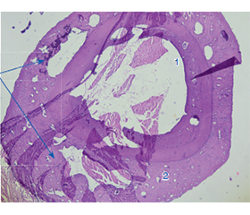
Актуальність. Дефекти кісток, спричинені хірургічними втручаннями, пораненнями, а також травмами, внаслідок яких відбуваються патологічні переломи, зокрема на тлі остеопорозу й пухлинних уражень, порушують як структуру, так і функцію скелета, серйозно впливаючи на якість життя, фізичне і психічне здоров’я пацієнтів. Таким чином, вивчення можливостей поліпшення репаративного остеогенезу є обґрунтованим завданням із теоретичної, експериментальної та клінічної точок зору. Мета: оцінити гістологічні ознаки й ефективність репаративного остегенезу після застосування модулюючих факторів: лазерного опромінення, кальцитоніну та їх комбінації. Матеріали та методи. В експерименті in vivo досліджували репаративний остеогенез у 32 тварин (щурів Вістар), у яких великогомілкову кістку було пошкоджено стоматологічним буром з використанням кетамінового наркозу. Їх розділили на 4 групи по 8 щурів: група А — контрольна; у групі B місце пошкодження кістки опромінювали інфрачервоним лазером (виробник «Фотоніка Плюс», Україна, модель «Ліка-терапевт М», довжина хвилі 810 нм, щільність потужності 50 мВт/см2) протягом 3 хвилин одразу після травми; у групі C тварини отримували кальцитонін (60 мг/0,1 мл) підшкірно через 2 дні після травми; у групі D щурам вводили кальцитонін та опромінювали місце пошкодження лазером. Результати. Поєднання фотобіомодуляції і кальцитоніну обумовило найбільш завершене формування регенератів, що можна охарактеризувати як повноцінну репаративну активність кісткової тканини після травматичного ушкодження. Великі ділянки регенерації демонстрували збільшену щільність кісткових трабекул, при цьому були наявні ділянки фіброзної тканини з ознаками запальної інфільтрації, що вказує на активний репаративний остеогенез. Про найбільший стимулюючий вплив у групі D, у якій був застосований комбінований вплив лазера й кальцитоніну, свідчать отримані показники щільності кісткової тканини. Висновки. Інфрачервоне лазерне опромінення й кальцитонін ефективно стимулювали репаративний остеогенез порівняно з контролем. Найбільший стимулюючий вплив спостерігали в групі D, у якій був застосований комбінований вплив лазера й кальцитоніну.
Background. Bone defects caused by surgical interventions, injuries, as well as traumas, resulting in pathological fractures, particularly against the background of osteoporosis and tumor lesions, disrupt both the structure and function of the skeleton, seriously affecting the quality of life, physical and mental health of patients. Thus, studying the possibilities of improving the processes of reparative osteogenesis is a justified task from a theoretical, experimental and clinical point of view. The purpose was to investigate the histological signs and effectiveness of reparative osteogenesis after the use of modulating factors: laser irradiation, calcitonin and their combination. Materials and methods. In an in vivo experiment, reparative osteogenesis was studied in 32 animals (Wistar rats) whose tibia was injured with a dental drill using ketamine anesthesia. They were divided into 4 groups of 8 rats each: group А — control; in group B, the site of bone damage was irradiated with an infrared laser (manufacturer Fotonika Plus, Ukraine, model Lika-terapevt M, wavelength 810 nm, power density 50 mW/cm2) for 3 minutes immediately after the injury; in group C, the animals were injected with calcitonin (60 mg/0.1 ml subcutaneously, 2 days after the injury); in group D, rats were administered calcitonin and the injury site was irradiated with a laser. Results. The combination of photobiomodulation and calcitonin led to the most complete formation of regenerates, which can be characterized as a full-fledged reparative activity of bone tissue after traumatic injury. Large areas of regeneration demonstrated increased density of bone trabeculae, while there were areas of fibrous tissue with signs of inflammatory infiltration, indicating active reparative osteogenesis. The greatest stimulating effect in group D, in which the combined effect of laser and calcitonin was applied, is evidenced by the obtained indicators of specific bone density. Conclusions. Infrared laser irradiation and calcitonin effectively stimulated reparative osteogenesis compared to the control. But the greatest stimulating effect was recorded in group D, in which the combined effect of laser and calcitonin was applied.
травми кісток; остеопороз; остеолітичні метастази в кістки; патологічні переломи; остеобласти; репаративний остеогенез; фотобіомодуляція; метаболізм кальцію; кальцитонін
bone injuries; osteoporosis; osteolytic bone metastases; pathological fractures; osteoblasts; reparative osteogenesis; photobiomodulation; calcium metabolism; calcitonin
Для ознакомления с полным содержанием статьи необходимо оформить подписку на журнал.
- Jannesar AM, Bigham-Sadegh A, Oryan A, Maffulli N. Enhancing bone healing through osteogenic medium components: biologi–cal roles, mechanisms, and clinical applications. Br Med Bull. 2026 Mar 21;158(1):ldag012. doi: 10.1093/bmb/ldag012.
- Drobotun ОV, Коnovalenko SV, Теrnovyy МК. The use of a training 3D-model in the treatment of a patient with a pathological fracture of the proximal part of the femur (case from practice). Orthopaedics, traumatology and prosthetics. 2024;1:53-58. doi: 10.15674/0030-59872024153-58.
- Kottmann V, Nienhaus M, Drees P, Gercek E, Ritz U. From bone homeostasis to skeletal metastasis and osteosarcoma: Insights into osteoclast and osteoblast roles in bone remodelling and cancer. Biochim Biophys Acta Rev Cancer. 2026 Apr;1881(2):189551. doi: 10.1016/j.bbcan.2026.189551.
- Zervou Z, Mabrouk R, van Velsen EFS, Bevers MSAM, Ali–zadeh E, van den Bergh JP, Zillikens MC. Relationship between bone mineral density and strength derived from 3D-shaper and HR-pQCT in patients with X-linked osteoporosis related to PLS3. Arch Osteoporos. 2026 Jan 21;21(1):27. doi: 10.1007/s11657-026-01656-2.
- Hao Y, Yang N, Sun M, Yang S, Chen X. The role of calcium channels in osteoporosis and their therapeutic potential. Front Endocrinol. 2024;15:1450328. doi: 10.3389/fendo.2024.1450328.
- Konovalenko S, Protsenko V, Tykhonov V, Solonitsyn Y, Hrebennikov Y, Konovalenko V, Ternovyi M. Study of the modulating factors combined action on reparative osteogenesis. Trauma. 2025;26(4):240-248. doi: 10.22141/1608-1706.4.26.2025.1028.
- Todosiuk T, Chemerovskiy V, Rublenko А, Ulianchych N, Kolomiiets V, Firstov S. Reparative osteogenesis markers during bone defects substitution with germanium-doped ceramics under experimental osteoporosis. Ukr Biochem J. 2026;98(1):98-107. doi: 10.15407/ubj98.01.098.
- Bonura A, Brunelli N, Marcosano M, Iaccarino G, Fofi L, Vernieri F, Altamura C. Calcitonin Gene-Related Peptide Systemic Effects: Embracing the Complexity of Its Biological Roles — A Narrative Review. International Journal of Molecular Sciences. 2023;24(18):13979.
- Keller J, et al. Calcitonin controls bone formation by inhibiting the release of sphingosine 1-phosphate from osteoclasts. Nat Commun. 2014 Oct 21;5:5215. doi: 10.1038/ncomms6215.
- Weiss RE, Singer FR, Gorn AH, Hofer DP, Nimni ME. Calcitonin stimulates bone formation when administered prior to initiation of osteogenesis. J Clin Invest. 1981 Sep;68(3):815-8. doi: 10.1172/jci110319.
- Roy A, Thulasiraman S, Panneerselvam E, Thulasi Doss G, Selvaraj MN, Ganesh SK, et al. Evaluation of the efficacy of salmon calcitonin nasal spray on bone healing following open reduction and internal fixation of mandibular fractures — a randomized controlled trial. J Craniomaxillofac Surg. 2021 Dec;49(12):1151-1157. doi: 10.1016/j.jcms.2021.08.002.
- Lyritis G, Boscainos PJ. Calcitonin effects on cartilage and fracture healing. J Musculoskelet Neuronal Interact. 2001 Dec;2(2):137-42.
- De Freitas LC, Kawamoto EL, Souza AMA, Kawakami PY, Gonçalves AS, Azevedo LH. Use of Phototherapy and Er-YAG Laser in the Management of Mandible Osteoradionecrosis: A Case Report. J Lasers Med Sci. 2023;14:e58.
- El Mobadder M, Grzech-Lesniak Z, El Mobadder W, Rifai M, Ghandour M, Nammour S. Management of Medication-Related Osteonecrosis of the Jaw with Photobiomodulation and Minimal Surgical Intervention. Dent J. 2023;11:127.
- Chen AC, Arany PR, Huang YY, Tomkinson EM, Sharma SK, Kharkwal GB, et al. Low-level laser therapy activates NF-kB via ge–neration of reactive oxygen species in mouse embryonic fibroblasts. PLoS One. 2011;6:e22453.
- Rola P, Włodarczak S, Lesiak M, Doroszko A, Włodarczak A. Changes in Cell Biology under the Influence of Low-Level Laser The–rapy. Photonics. 2022;9:502.
- Wan Z, Zhang P, Lv L, Zhou Y. NIR light-assisted photothe–rapies for bone-related diseases and bone tissue regeneration: A syste–matic review. Theranostics. 2020 Sep 26;10(25):11837-11861.
- Fu J, Liu X, Tan L, Cui Z, Zheng Y, Liang Y, et al. Photoelectric-responsive extracellular matrix for bone engineering. ACS Nano. 2019;13:13581-94. doi: 10.1021/acsnano.9b08115.
- Sanchez-Casanova S, Martin-Saavedra FM, Escudero-–Duch C, Falguera Uceda MI, Prieto M, Arruebo M, et al. Local delivery of bone morphogenetic protein-2 from near infrared-responsive hydrogels for bone tissue regeneration. Biomaterials. 2020;241:119909.
- Berni M, Brancato AM, Torriani C, Bina V, Annunziata S, Cornella E, et al. The Role of Low-Level Laser Therapy in Bone Hea–ling: Systematic Review. International Journal of Molecular Sciences. 2023;24(8):7094.
- Luca RE, Todea CD, Duma VF, Bradu A, Podoleanu AG. Quantitative assessment of rat bone regeneration using complex master-slave optical coherence tomography. Quant Imaging Med Surg. 2019 May;9(5):782-798. doi: 10.21037/qims.2019.05.03.
- Ozawa Y, Shimizu N, Kariya G, Abiko Y. Low-energy laser irradiation stimulates bone nodule formation at early stages of cell culture in rat calvarial cells. Bone. 1998 Apr;22(4):347-54.
- Shymon VM, Kubash VI, Stoika VV. Influence of different laser modes with thermoelectric effect on compact bone tissue structure and regeneration of bone in white laboratory rats. Problems of Clinical Pediatrics. 2017;3–4(37–38):72-78 (in Ukrainian).
- Polo TOB, Fonseca-Santos JM, Momesso GAC, da Silva WPP, Barbosa S, Santos AMS, et al. Single intraoperative infrared laser optimized bone repair in rat femoral osteotomies with experimentally induced osteoporosis. Lasers Med Sci. 2023 Mar 20;38(1):87. doi: 10.1007/s10103-023-03746-w.
- Shrikar R. Desai, et al. Effect of irradiation of 810nm laser on bone for 10 sec: A rabbit histological study. IOSR Journal of Dental and Medical Sciences. 2019;18(3):14-20. doi: 10.9790/0853-1803121420.
- Pires-Oliveira DA, Oliveira RF, Amadei SU, Pacheco-–Soares C, Rocha RF. Laser 904 nm action on bone repair in rats with osteoporosis. Osteoporos Int. 2010 Dec;21(12):2109-14.
- Mikhail FF, El-Din M, Ibrahim T, Zekry K, Nemat A, Nasry S. Effect of Laser Therapy on the Osseointegration of Immediately Loaded Dental Implants in Patients under Vitamin C, Omega-3 and Calcium Therapy. Open Access Maced J Med Sci. 2018 Aug 15;6(8):1468-1474. doi: 10.3889/oamjms.2018.291.
- Nesioonpour S, Mokmeli S, Vojdani S, Mohtadi A, Akhondzadeh R, et al. The Effect of Low-Level Laser on Postoperative Pain After Tibial Fracture Surgery: A Double-Blind Controlled Randomized Clinical Trial. Anesth Pain Med. 2014;4(3):e17350. doi: 10.5812/aapm.17350.
- Momeni M, Safari M, Sarrafzadeh A, Momeni E. Effect of Low-Level Laser Therapy on the Osseointegration of Immediately Loaded Implants with a Connective Tissue Graft: A Randomized Cli–nical Trial. J Lasers Med Sci. 2024 Jun 30;15:e19. doi: 10.34172/jlms.2024.19.
- Moscatel MBM, Pagani BT, Trazzi BF de M, Reis CHB, Ribeiro CA, Buchaim DV, Buchaim RL. Effects of Photobiomodulation in Association with Biomaterials on the Process of Guided Bone Rege–neration: An Integrative Review. Ceramics. 2025;8(3):94.
- Fitzgerald C, Burley C, Wright K, McLeod K, Asadi S, Parmenter B. Exercise improves quality of life, mental health and pain in people living with osteoporosis: a systematic review and meta-analysis. Osteoporos Int. 2026 Feb 20. doi: 10.1007/s00198-026-07876-z.
- Yang DH, Yang MY. The Role of Macrophage in the Pathogenesis of Osteoporosis. Int J Mol Sci. 2019 Apr 28;20(9):2093. doi: 10.3390/ijms20092093.
- Parithimarkalaignan S, Padmanabhan TV. Osseointegration: an update. J Indian Prosthodont Soc. 2013 Mar;13(1):2-6. doi: 10.1007/s13191-013-0252-z.
- Buhrmann C, Busch F, Shayan P, Shakibaei M. Sirtuin-1 (SIRT1) is required for promoting chondrogenic differentiation of me–senchymal stem cells. J Biol Chem. 2014;289:22048-62.
- Dvir-Ginzberg M, Steinmeyer J. Towards elucidating the role of SirT1 in osteoarthritis. Front Biosci. 2013;18:343-55.
- Liu CF, Lefebvre V. The transcription factors SOX9 and SOX5/SOX6 cooperate genome-wide through super-enhancers to drive chondrogenesis. Nucleic Acids Res. 2015;43:8183-203.
- Di Giacomo V, Cataldi A, Sancilio S. Biological Factors, Me–tals, and Biomaterials Regulating Osteogenesis through Autophagy. Int J Mol Sci. 2020 Apr 17;21(8):2789. doi: 10.3390/ijms21082789.
- Bigi A, Boanini E. Functionalized biomimetic calcium phosphates for bone tissue repair. J Appl Biomater Funct Mater. 2017;15:e313-e325. doi: 10.5301/jabfm.5000367.
- Oliveira WF, Arruda IRS, Silva GMM, Machado G, Coelho L, Correia MTS. Functionalization of titanium dioxide nanotubes with biomolecules for biomedical applications. Mater Sci Eng C Mater Biol Appl. 2017;81:597-606. doi: 10.1016/j.msec.2017.08.017.
- Husain A, Jeffries MA. Epigenetics and bone remodeling. Curr Osteoporos Rep. 2017;15:450-458. doi: 10.1007/s11914-017-0391-y.
- Torres HM, Arnold KM, Oviedo M, Westendorf JJ, Wea–ver SR. Inflammatory Processes Affecting Bone Health and Repair. Curr Osteoporos Rep. 2023 Dec;21(6):842-853. doi: 10.1007/s11914-023-00824-4.
- Kara C, Selamet H, Gökmenoğlu C, Kara N. Low level laser therapy induces increased viability and proliferation in isolated cancer cells. Cell Prolif. 2018 Apr;51(2):e12417. doi: 10.1111/cpr.12417.
- Kim H, Kim Y, Kim TH, Heo SY, Jung WK, Kang HW. Sti–mulatory effects of wavelength-dependent photobiomodulation on proli–feration and angiogenesis of colorectal cancer. J Photochem Photo–biol B. 2022 Sep;234:112527. doi: 10.1016/j.jphoto–biol.2022.112527.
- Rhee YH, Moon JH, Choi SH, Ahn JC. Low-Level Laser Therapy Promoted Aggressive Proliferation and Angiogenesis Through Decreasing of Transforming Growth Factor-β1 and Increasing of Akt/Hypoxia Inducible Factor-1α in Anaplastic Thyroid Cancer. Photomed Laser Surg. 2016 Jun;34(6):229-35. doi: 10.1089/pho.2015.3968.
- Taradaj J, Shay B, Dymarek R, Sopel M, Walewicz K, Beeckman D, et al. Effect of laser therapy on expression of angio- and fibrogenic factors, and cytokine concentrations during the healing process of human pressure ulcers. Int J Med Sci. 2018 Jul 13;15(11):1105-1112. doi: 10.7150/ijms.25651.
- Hazrati P, Azadi A, Fekrazad S, Wang HL, Fekrazad R. The effect of photobiomodulation therapy on fracture healing: a systematic review and meta-analysis of animal studies. Lasers Med Sci. 2025 Feb 28;40(1):121. doi: 10.1007/s10103-025-04376-0.
- Sasso Stuani MB, Sasso Stuani A, Leite Pedroso G, da Silva Mira PC, Gollino S, et al. The effect of low-level laser therapy after rapid maxillary expansion: Micro-CT analysis. Lasers Med Sci. 2025 May 27;40(1):245. doi: 10.1007/s10103-025-04497-6.