Резюме
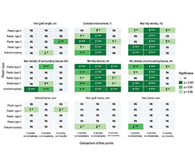
Актуальність. Значні за обсягом ушкодження кінцівок після мінно-вибухової травми потребують не лише вдалої реконструктивної пластики, але й об’єктивного інструментального контролю перебігу післяопераційної тканинної перебудови. Одним із таких засобів, що дозволяє виконати це, є комп’ютерна томографія (КТ), яка надає інформацію про стан клаптя, навколишніх тканин і судинного компонента реконструйованого сегмента. Мета: вивчити особливості КТ-показників післяопераційного клаптя в пацієнтів із масивними дефектами кінцівок після мінно-вибухових уражень протягом 6 місяців спостереження при застосуванні різних типів реконструктивної пластики. Матеріали та методи. У досліджені взяли участь 110 осіб із масивною мінно-вибуховою травмою кінцівки, яким виконували 5 видів реконструктивної пластики. У післяопераційному періоді одразу після операції, через 3 та 6 місяців пацієнтам проводили КТ-дослідження на томографі Aquilion Prime SP TSX-303B/5C з аналізом таких показників, як щільність клаптя й навколишніх тканин, просвіт артерії і вени, контрастне накопичення, довжина та просвіт венграфта. Результати. Аналіз змішаних моделей показав, що час спостереження вірогідно впливав практично на всі досліджувані показники, окрім просвіту артерії. Тип пластики був значущим для показників щільності клаптя, максимальної щільності навколишніх тканин, просвіту вени, контрастного накопичення й просвіту венграфта. Водночас взаємодія типу пластики з часом не була статистично значущою для жодного показника. При аналізі часових змін показників встановлено, що незалежно від виду пластики через 6 місяців мінімальна щільність клаптя зростала на 9,470 одиниці Гаунсфілда (HU), максимальна щільність клаптя — на 7,961 HU, мінімальна щільність навколишніх тканин — на 8,391 HU, а контрастне накопичення — на 11,537 %. Просвіт артерії істотно не змінювався, тоді як просвіт вени й венграфта зменшувався. Найсильніший зв’язок з типом пластики встановлено для контрастного накопичення: = 0,455 одразу після операції, = 0,405 через 3 місяці та = 0,339 через 6 місяців. Усі кореляції досліджуваних показників із функціональністю сегмента були слабкими. Найбільше за модулем значення мало контрастне накопичення через 3 місяці спостереження (r = –0,211). Висновки. Зміни в досліджуваних КТ-показниках відображають переважно морфологічну й судинно-тканинну перебудову після реконструктивної пластики, найбільш інформативними серед них є щільність тканин і контрастне накопичення.
Background. Extensive limb injuries after mine-blast trauma require not only successful reconstructive plastic surgery but also objective instrumental monitoring of postoperative tissue remodeling. One such tool that allows this to be performed is computed tomography (CT), which provides information on the condition of the flap, surrounding tissues, and the vascular component of the reconstructed segment. The purpose was to investigate the features of CT indicators of the postoperative flap in patients with massive limb defects after mine-blast injuries during 6 months of follow-up when using different types of reconstructive plastic surgery. Materials and methods. The study included 110 patients with massive mine-blast trauma of the limb. They underwent 5 different types of reconstructive plastic surgery. In the postoperative period, immediately after surgery, after 3 and 6 months, the patients underwent CT examination using an Aquilion Prime SP TSX-303B/5C scanner, with analysis of indicators, such as density of the flap and surrounding tissues, lumen of the artery and vein, contrast enhancement, length and lumen of the vein graft. Results. Mixed model analysis showed that the observation time significantly affected practically all studied indicators, except for the arterial lumen. The type of plastic surgery was significant for the indicators of flap density, maximum density of surrounding tissues, venous lumen, contrast enhancement, and vein graft lumen. At the same time, the interaction between the type of plastic surgery and time was not statistically significant for any indicator. When analyzing temporal changes in the indicators, it was found that, regardless of the type of plastic surgery, after 6 months the minimum flap density increased by 9.470 Hounsfield units (HU), the maximum flap density by 7.961 HU, the minimum density of surrounding tissues by 8.391 HU, and contrast enhancement by 11.537 %. The arterial lumen did not change significantly, whereas the lumen of the vein and vein graft decreased. The strongest association with the type of plastic surgery was established for contrast enhancement: = 0.455 immediately after surgery, = 0.405 after 3 months, and = 0.339 after 6 months. All correlations of the studied indicators with segment functionality were weak. The highest absolute value was observed for contrast enhancement at the 3-month follow-up period (r = –0.211). Conclusions. Changes in the studied CT indicators mainly reflect morphological and vascular-tissue remodeling after reconstructive plastic surgery; among them, tissue density and contrast enhancement are the most informative ones.
Список литературы
1. Bendavid E, Boerma T, Akseer N, Langer A, Malembaka EB, et al. The effects of armed conflict on the health of women and children. The Lancet. 2021 Feb 6;397(10273):522-32. doi: 10.1016/S0140-6736(21)00131-8.
2. Chevalley K, Zimmerman J, Mittendorf A, Sennersten F, Dalman A, et al. Civilian pattern of injuries in armed conflicts — a systematic review. Scandinavian Journal of Trauma, Resuscitation and Emergency Medicine. 2024 Dec 4;32(1):125. doi: 10.1186/s13049-024-01299-7.
3. Roy CM, Garbern SC, Relan P, Bills CB, Schultz ML, et al. Acute Facility Management of Blast Injuries In Low- and Middle-Income Countries: A Systematic Review and Meta-Analysis. Prehospital and Disaster Medicine. 2025 Jun 30:1-3. doi: 10.1017/S1049023X25101222.
4. El-Taji O, Ali A, Alser O, Ghali A, Jomaa A, et al. Patterns of war related trauma in Gaza during armed conflict: survey study of international healthcare workers. BMJ. 2025 Sep 25;390. doi: 10.1136/bmj-2025-087524.
5. Trutyak I, Malickii V, Samotowka M, Trunkvalter V, Trutyak R, Ivaschenko V. Problematic issues of limb amputation in wounded with combat trauma. Proceeding of the Shevchenko Scientific Society. Medical Sciences. 2023 Dec 22;72(2). doi: 10.25040/ntsh2023.02.08.
6. Neeman U, Hashavia E, Soffer D, Timor I, Zeltser D, et al. Blast injuries: the experience of a level 1 trauma center. Injury. 2025 Jan 1;56(1):111839. doi: 10.1016/j.injury.2024.111839.
7. Farhat T, Nahouli H, Hajjar M, Abdul-Sater Z, Kobeissi E, et al. Characteristics of injuries during the 2006 Lebanon conflict: a three-center retrospective study of survivors, 16 years after the conflict. Frontiers in public health. 2024 May 28;12:1382514. doi: 10.3389/fpubh.2024.1382514.
8. Yuste V, Rodero MD, Bernal A, Gomez-Escolar L, Sevilla JJ, Garin M. The role of plastic surgery in the management of war injuries from the Russo-Ukrainian War. European Journal of Plastic Surgery. 2024 Sep 6;47(1):91. doi: 10.1007/s00238-024-02236-8.
9. Ahmed AK, Bakhiet MY, Mohamed HH. Plastic surgery interventions for war-related injuries: a review of 300 cases from Sudan, a cohort study. BMC surgery. 2025 Oct 3;25(1):442. doi: 10.1186/s12893-025-03181-4.
10. Khomenko IP, Lurin IA, Gumeniuk KV, McKnight G, Makarov VV, et al. Telemedicine support aids lower limb reconstruction after combat injury in Ukraine. Journal of Surgical Case Reports. 2023 Jul;2023(7):rjad403. doi: 10.1093/jscr/rjad403.
11. Dreizin D, Smith EB, Champ K, Morrison JJ. Roles of trauma CT and CTA in salvaging the threatened or mangled extremity. Radiographics. 2022 Mar;42(2):E50-67. doi: 10.1148/rg.210092.
12. Tamburrini S, Lassandro G, Tiralongo F, Iacobellis F, Ronza FM, et al. CTA imaging of peripheral arterial injuries. Diagnostics. 2024 Jun 26;14(13):1356. doi: 10.3390/diagnostics14131356.
13. Tirrell AR, Kim KG, Rashid W, Attinger CE, Fan KL, Evans KK. Patient-reported outcome measures follo–wing traumatic lower extremity amputation: a systematic review and meta-analysis. Plastic and Reconstructive Surgery — Global Open. 2021 Nov 1;9(11):e3920. doi: 10.1097/GOX.0000000000003920.
14. Schober TL, Abrahamsen C. Patient perspectives on major lower limb amputation — a qualitative systematic review. International Journal of Orthopaedic and Trauma Nursing. 2022 Jul 1;46:100958. doi: 10.1016/j.ijotn.2022.100958.
15. Ghai S, Hitzig SL, Eberlin L, Melo J, Mayo AL, et al. Reporting of rehabilitation outcomes in the traumatic lower limb amputation literature: a systematic review. Archives of Physical Medicine and Rehabilitation. 2024 Jun 1;105(6):1158-70. doi: 10.1016/j.apmr.2023.08.028.
16. Paes FM, Munera F. Computer tomography angiography of peripheral vascular injuries. Radiologic Clinics. 2023 Jan 1;61(1):141-50. doi: 10.1016/j.rcl.2022.08.006.
17. Joseph TI, Ratnakanthan PJ, Paul E, Clements W. Utility of computed tomography angiography in traumatic lower limb injury: Review of clinical impact in level 1 trauma centre. Injury. 2021 Oct 1;52(10):3064-7. doi: 10.1016/j.injury.2021.02.047.
18. Warwick H, Cherches M, Shaw C, Toogood P. Comparison of computed tomography angiography and physical exam in the evaluation of arterial injury in extremity trauma. Injury. 2021 Jul 1;52(7):1727-31. doi: 10.1016/j.injury.2021.04.057.
19. Du Plessis J, Kalu A, McKee H, Mohammed A, Maroun R, et al. Computed tomography angiography utilization in lower extremity trauma: insights from a Canadian Level I Trauma Centre. Canadian Association of Radiologists Journal. 2026 Feb;77(1):187-94. doi: 10.1177/08465371251342064.
20. Stefanou N, Arnaoutoglou C, Papageorgiou F, Matsagkas M, Varitimidis SE, Dailiana ZH. Update in combined musculoskeletal and vascular injuries of the extremities. World Journal of Orthopedics. 2022 May 18;13(5):411. doi: 10.5312/wjo.v13.i5.411.
21. Zeiderman MR, Pu LL. Contemporary approach to soft-tissue reconstruction of the lower extremity after trauma. Burns & Trauma. 2021;9:tkab024. doi: 10.1093/burnst/tkab024.
22. Othman S, Azoury SC, Klifto KM, Levin LS, Kovach SJ. Soft-tissue management and reconstruction of lower-extremity trauma. Plastic and Aesthetic Research. 2022 Apr 9;9:29. doi: 10.20517/2347-9264.2021.135.
23. Evans BG, Colen DL. The evolution of lower extre–mity reconstruction. Plastic and Aesthetic Research. 2022 May 6;9(5):34. doi: 10.20517/2347-9264.2021.134.
24. Jenny LY, Tolley PD, Kneib C, Miller EA, Crowe CS. Current concepts in microsurgical soft tissue reconstruction of lower extremity trauma in a single-vessel extremity. Plastic and Aesthetic Research. 2022 May 17;9(5):37. doi: 10.20517/2347-9264.2021.124.
25. Li B, Tian X, Fei H, Li G, Wu X. Management of suspected peripheral vascular injuries in orthopedic trauma. Orthopaedic Surgery. 2024 Jul;16(7):1548-54. doi: 10.1111/os.14079.
26. Gyri E, Fast A, Resch A, Rath T, Radtke C. Reconstruction of traumatic and non-traumatic lower extremity defects with local or free flaps. European Surgery. 2022 Feb;54(1):44-9. doi: 10.1007/s10353-021-00704-0.
27. Mert S, Heidekrueger PI, Fuchs B, Nuernberger T, Haas-Ltzenberger EM, et al. Microvascular Muscle vs. Fascio-Cutaneous Free Flaps for Reconstruction of Plantar Load-Bearing Foot Defects — An International Survey. Journal of Clinical Medicine. 2024 Feb 24;13(5):1287. doi: 10.3390/jcm13051287.
28. List EB, Hahn BA, Qiu SS, de Jong T, Rakhorst HA, et al. Free fasciocutaneous versus muscle flaps in lower extremity reconstruction: Implications for functionality and quality of life. Journal of Reconstructive Microsurgery. 2025 Oct;41(08):641-8. doi: 10.1055/a-2483-5388.
29. Koster IT, Borgdorff MP, Jamaludin FS, de Jong T, Botman M, Driessen C. Strategies following free flap failure in lower extremity trauma: a systematic review. JPRAS open. 2023 Jun 1;36:94-104. doi: 10.1016/j.jpra.2023.03.002.
30. Dzgn S, zdemir MT, Manti N, lgen NK, Akkurt MO. Optimizing outcomes in mangled lower extremity reconstruction: insights from a retrospective study of 93 patients and their functional scores. Journal of Clinical Medicine. 2025 Feb 21;14(5):1436. doi: 10.3390/jcm14051436.