
Журнал «Травма» Том 27, №4, 2026
Вернуться к номеру
Біомеханічне обґрунтування місця проведення пластини при хірургічній корекції лійкоподібної деформації грудної клітки
Авторы: Камінська М.О. (1), Дігтяр В.А. (1), Яресько О.В. (2)
(1) - Дніпровський державний медичний університет, м. Дніпро, Україна
(2) - ДУ «Інститут патології хребта та суглобів ім. проф. М.І. Ситенка НАМН України», м. Харків, Україна
Рубрики: Травматология и ортопедия
Разделы: Клинические исследования
Версия для печати
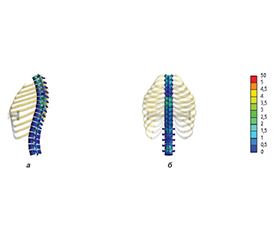
Актуальність. Мінімально інвазивна торакопластика за методом D. Nuss є стандартом лікування лійкоподібної деформації грудної клітки, проте взаємодія імплантату з каркасом часто викликає ускладнення (зміщення пластини, хронічний біль). Більшість сучасних досліджень напружено-деформованого стану фокусується лише на комплексі «грудина — ребра», не враховуючи хребетний стовп та мобільність реберно-хребцевих суглобів. Мета: провести порівняльний аналіз особливостей розподілу напружено-деформованого стану в біомеханічній системі «грудина — ребра — хребет» при хірургічній корекції лійкоподібної деформації грудної клітки залежно від анатомічного рівня проведення пластини. Матеріали та методи. Створено математичну скінченно-елементну модель грудної клітки з урахуванням хребетного стовпа та фізіологічної рухливості реберно-хребцевих з’єднань. Проведено порівняння двох варіантів торакопластики: традиційного проведення короткої пластини через IV–V міжребер’я та використання пластини власної конструкції, встановленої через VI–VII міжребер’я. Моделювання виконували в середовищах SolidWorks та ANSYS. Оцінювання напруженого стану проводили за критерієм еквівалентних напружень за Мізесом. Результати. При традиційному проведенні пластини через IV–V міжребер’я максимальні напруження в опорних ребрах досягали 22,4 МПа, а в структурах хребта — 9,9 МПа. При використанні пластини власної конструкції на рівні VI–VII міжребер’я максимальні напруження в ребрах знижувалися до 14,0 МПа, а в хребцях — до 2,4 МПа. Встановлено більш рівномірний розподіл навантаження в системі «грудина — ребра — хребет» та зменшення перевантаження реберно-хребцевих суглобів. Помірне підвищення напружень у зоні контакту «пластина — грудина» не мало критичного впливу на кісткові структури. Висновки. Проведення коригувальної пластини через VI–VII міжребер’я забезпечує більш сприятливі біомеханічні показники порівняно з традиційною методикою. Використання пластини власної конструкції дозволяє знизити напруження в ребрах та структурах хребта, поліпшити розподіл навантаження й потенційно зменшити ризик післяопераційних ускладнень при хірургічній корекції лійкоподібної деформації грудної клітки.
Background. Minimally invasive thoracoplasty using the D. Nuss technique remains the standard treatment for pectus excavatum. However, interaction between the implant and the thoracic framework is frequently associated with complications, including bar displacement and chronic pain syndrome. Most contemporary studies investigating the stress-strain state focus primarily on the “sternum-ribs” complex without considering the spinal column and the mobility of the costovertebral joints. Objective: to perform a comparative analysis of stress-strain distribution patterns in the biomechanical “sternum-ribs-spine” system during surgical correction of pectus excavatum depending on the anatomical level of bar placement. Materials and methods. A finite element mathematical model of the chest wall was developed, incorporating the spinal column and physiological mobility of the costovertebral articulations. Two thoracoplasty techniques were compared: conventional placement of a short bar through the IV–V intercostal spaces and placement of a custom-designed bar through the VI–VII intercostal spaces. Modeling was performed using SolidWorks and ANSYS software. Stress distribution was evaluated according to the von Mises equivalent stress criterion. Results. With conventional bar placement through the IV–V intercostal spaces, maximum stresses in the supporting ribs reached 22.4 MPa, while stresses in the spinal structures reached 9.9 MPa. In contrast, placement of the custom-designed bar at the VI–VII intercostal level reduced maximum stresses to 14.0 MPa in the ribs and 2.4 MPa in the vertebrae. A more uniform load distribution within the “sternum-ribs-spine” system and reduced overload of the costovertebral joints were observed. Moderate stress elevation in the “bar-sternum” contact zone did not produce a critical effect on osseous structures. Conclusions. Placement of the corrective bar through the VI–VII intercostal spaces provides more favorable biomechanical characteristics compared with the conventional technique. The use of a custom-designed bar reduces stress levels in the ribs and spinal structures, improves load distribution, and may potentially decrease the risk of postoperative complications following surgical correction of pectus excavatum.
лійкоподібна деформація грудної клітки; торакопластика; метод скінченних елементів; напружено-деформований стан; хребет; пластина
pectus excavatum; thoracoplasty; finite element method; stress-strain state; spine; bar