
Журнал «Травма» Том 27, №4, 2026
Вернуться к номеру
Комп’ютерне моделювання напружено-деформованого стану системи фіксації перелому проксимального відділу стегнової кістки
Авторы: Козін О.А. (1), Нестеренко М.М. (2), Павленко С.М. (1)
(1) - Полтавський державний медичний університет, м. Полтава, Україна
(2) - Національний університет «Полтавська політехніка імені Юрія Кондратюка», м. Полтава, Україна
Рубрики: Травматология и ортопедия
Разделы: Клинические исследования
Версия для печати
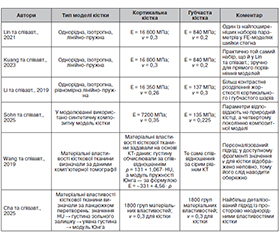
Актуальність. Дослідження біомеханічних властивостей проксимального відділу стегнової кістки є важливим етапом у розробці ефективних методик хірургічного лікування переломів даної ділянки скелета. У більшості робіт з комп’ютерного моделювання гвинтового остеосинтезу шийково-діафізарний кут або приймається як усереднена геометрична константа, або взагалі не розглядається як самостійний фактор, хоча саме він визначає нахил шийки, довжину плеча навантаження, напрям передачі сил через зону перелому. Мета: провести комп’ютерне моделювання напружено-деформованого стану системи фіксації перелому проксимального відділу стегнової кістки з урахуванням різних шийково-діафізарних кутів та конструктивних схем остеосинтезу. Матеріали та методи. Аналіз сучасних наукових джерел був спрямований на визначення основних вимог до комп’ютерної моделі проксимального відділу стегнової кістки, зокрема щодо індивідуальних геометричних параметрів кістки та її щільності, варіантів виконання віртуальної остеотомії і розташування фіксаторів, а також напрямків і величин прикладених навантажень. Модель перелому формували шляхом віртуальної остеотомії шийки стегнової кістки в межах внутрішньосуглобового ушкодження з несприятливою до зсуву орієнтацією площини перелому типу Pauwels II–III. Після завершення геометричного моделювання та складання вузла виконували підготовку розрахункової моделі в SolidWorks Simulation. Для цього для кожної деталі визначали матеріальні характеристики, а між елементами задавали контактні взаємодії. Результати. Встановлені основні недоліки та переваги поширених комп’ютерних моделей проксимального відділу стегнової кістки за умови її ситуаційного ушкодження. При порівнянні роботи канюльованих гвинтів за різних шийково-діафізарних кутів спочатку методично виправданим було використання стандартизованих матеріальних властивостей кортикальної та губчастої кістки. На наступному етапі модель можна було ускладнити, перейшовши до CT-based material mapping, якщо було потрібно оцінити ще й вплив остеопенії або індивідуальної неоднорідності кісткової тканини. Для моделювання системи фіксації перелому шийки стегна канюльованими гвинтами з урахуванням різних шийково-діафізарних кутів перевагу надали використанню дворівневої схеми навантаження. На першому етапі доцільно застосувати 2100 Н як базове навантаження одноопорного стояння, що дозволяє коректно порівнювати результати з літературними даними. На другому етапі для поглибленого аналізу доцільно вводити або фази ходи з різними кратностями маси тіла, або більш складну м’язово-суглобову схему навантаження. Висновки. Сучасне планування хірургічного лікування переломів проксимального відділу стегнової кістки повинно базуватися на даних комп’ютерного дослідження з подальшим вибором індивідуальної тактики оперативного втручання та ефективної фіксації.
Background. The study on the biomechanical properties of the proximal femur is an important stage in developing effective methods of surgical treatment for fractures of this part of the skeleton. Most works on computer modeling of screw osteosynthesis consider the caput-collum-diaphyseal (CCD) angle as an averaged geometric constant or not consider it as an independent factor at all, although it determines the inclination of the neck, the length of the load arm, and the direction of force transmission through the fracture zone. The purpose was to perform the computer modeling of the stress-strain state of the proximal femoral fracture fixation system taking into account different CCD angles and structural schemes of osteosynthesis. Materials and methods. Analysis of data from modern scientific sources was aimed at identifying the main requirements to the computer model of the proximal femur, namely individual geometric parameters of the bone and its density, variability of virtual osteotomy and location of fixators, as well as directions and values of loads. The fracture model was formed by virtual osteotomy of the femoral neck within the intra-articular injury with an unfavorable fracture plane orientation of Pauwels type II–III fracture predisposing to displacement. After completing geometric modeling and assembly of the unit, the computational model was prepared in SolidWorks Simulation. To do this, material characteristics were assigned to each part, and contact interactions between the elements were defined. Results. The main disadvantages and advantages of common computer models of the proximal femur under the condition of its situational damage were established. When comparing the performance of cannulated screws at different CCD angles, it was methodologically justified to use standardized material properties of cortical and cancellous bone at first. At the next stage, the model could be complicated by moving to CT-based material mapping, if it was also necessary to assess the influence of osteopenia or individual bone heterogeneity. To model the femoral neck fracture fixation system with cannulated screws, taking into account different CCD angles, preference was given to a two-level loading scheme. At the first stage, it is advisable to apply 2100 Н as the basic load of single-support standing, which will allow for correct comparison of the results with the literature data. At the second stage, for in-depth analysis, it is advisable to introduce either gait phases with different multiplicity of body weight, or a more complex muscle-joint loading scheme. Conclusions. Modern planning of surgical treatment for proximal femoral fractures should be based on computerized imaging data with subsequent selection of individual surgical tactics and selection of effective fixation.
комп’ютерне моделювання; проксимальний відділ стегнової кістки; шийково-діафізарний кут; переломи; остеосинтез
computer modeling; proximal femur; caput-collum-diaphyseal angle; fractures; osteosynthesis