
Інформація призначена тільки для фахівців сфери охорони здоров'я, осіб,
які мають вищу або середню спеціальну медичну освіту.

Підтвердіть, що Ви є фахівцем у сфері охорони здоров'я.

Журнал «Травма» Том 27, №4, 2026

Вернуться к номеру

Архітектоніка губчастої речовини п’яткової кістки: пілотне дослідження
Авторы: V.H. Hryn (1), O.S. Maksymenko (1), Ya.O. Brovarnyk (1), V.S. Drabovskiy (1), S.V. Davydenko (2), A.V. Piliuhin (1), Ya.A. Tarasenko (1), R.L. Ustenko (1), M.V. Hryn (1), H.N. Hoduadze (1), Ye.E. Chip (1), S.V. Kononenko (1), F. Paulsen (3)
(1) - Poltava State Medical University, Poltava, Ukraine
(2) - I.I. Schmalhausen Institute of Zoology of National Academy of Sciences of Ukraine, Kyiv, Ukraine
(3) - Friedrich-Alexander-Universitt Erlangen-Nrnberg, Erlangen, Germany

Рубрики: Травматология и ортопедия

Разделы: Клинические исследования

Версия для печати


Резюме

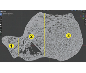
Актуальність. П’яткова кістка є ключовим структурним компонентом стопи, який відіграє ключову роль у забезпеченні опори тіла та передачі навантаження. Її губчаста речовина формує складну тривимірну трабекулярну архітектоніку, що забезпечує ефективний розподіл механічного навантаження. Особливий інтерес становить нейтральна зона (трикутник Ворда) — ділянка зі зниженою щільністю трабекул, функціональне значення якої залишається недостатньо з’ясованим. Сучасні методи тривимірної візуалізації відкривають нові можливості для детального аналізу структурної організації губчастої речовини п’яткової кістки та її морфометричних характеристик. Мета: вивчити структурну організацію і морфометричні особливості губчастої речовини п’яткової кістки. Матеріали та методи. У дослідженні використано анатомічні препарати п’яткових кісток дорослих людей (n = 5), отриманих на кафедрі анатомії людини Полтавського державного медичного університету. Зразки не мали патологічних ознак або травматичних пошкоджень. Результати. У губчастій речовині п’яткової кістки виявлено дві основні трабекулярні системи — компресійну і тензійну, які формують біомеханічно ефективну конструкцію для оптимального розподілу механічного навантаження. Між ними визначається центральна нейтральна зона (трикутник Ворда), що характеризується зниженою щільністю трабекул та помірною варіабельністю геометричних параметрів. При цьому основні морфометричні характеристики найбільш виражених трабекул залишаються відносно стабільними. Тривимірна реконструкція, виконана за допомогою сканера Artec 3D Artec Micro, підтвердила адаптивну орієнтацію трабекул відповідно до функціонального навантаження: у ділянці п’яткового горба переважають масивні короткі трабекули, у центральній частині спостерігається більш розріджене розташування трабекул, а передній відділ характеризується пластинчасто-лінійною архітектонікою. Висновки. Трабекулярна архітектоніка п’яткової кістки представлена складною системою компресійних і тензійних трабекул, орієнтованих відповідно до напрямків дії механічних сил. Нейтральна зона (трикутник Ворда) є тривимірною морфологічною структурою, що відіграє ключову роль у перерозподілі навантаження.

Background. The calcaneus is a key structural component of the foot that plays a crucial role in weight bearing and load transmission. Its trabecular bone forms a complex three-dimensional architecture that ensures efficient distribution of mechanical stresses. Of particular interest is the neutral zone (Ward’s triangle), an area of reduced trabecular density whose functional significance remains incompletely understood. Modern three-dimensional imaging techniques provide new opportunities for detailed analysis of the structural organization and morphometric characteristics of the trabecular bone of the calcaneus. Therefore, the aim is to investigate structural organization and morphometric features of the trabecular architecture of the calcaneus. Materials and methods. The study used anatomical specimens of calcanei from adult humans (n = 5) obtained from the Department of Human Anatomy of Poltava State Medical University. The bones showed no signs of pathological changes or traumatic damage. Results. Two principal trabecular systems — compressive and tensile — were identified in the trabecular bone of the calcaneus, forming a biomechanically efficient framework for optimal load distribution. Between these systems, a central neutral zone (Ward’s triangle) was observed as an area of reduced trabecular density with moderate variability in its geometric parameters. At the same time, the main morphometric characteristics of the most prominent trabeculae remained relatively stable among the examined specimens. Three-dimensional reconstruction performed using the Artec 3D Artec Micro scanner confirmed the adaptive orientation of trabeculae in accordance with functional loading patterns: dense, short trabeculae predominated in the calcaneal tuberosity, a more sparse trabecular arrangement was observed in the central region, and the anterior part exhibited a plate-like linear architecture. Conclusions. The trabecular architectonics of the calcaneus is represented by a complex system of compression and tensile trabeculae, oriented according to the directions of mechanical forces. The neutral zone (Ward’s zone) is a three-dimensional morphological structure that plays a key role in load redistribution.


Ключевые слова

кістка; стопа; п’яткова кістка; трикутник Ворда; закон Вольфа; трабекулярна архітектоніка; губчаста речовина кістки; контрфорси

bone; foot; calcaneus; Ward’s triangle; Wolff’s law; trabecular architectonics; cancellous bone; buttresses


Для ознакомления с полным содержанием статьи необходимо оформить подписку на журнал.


Список литературы

1. Gupton M, zdemir M, Terreberry RR. Anatomy, Bony Pelvis and Lower Limb: Calcaneus. In: StatPearls. Treasure Island, FL: StatPearls Publishing LLC; 2025.
2. Hall RL, Shereff MJ. Anatomy of the calcaneus. Clinical orthopaedics and related research. 1993;290:27-35.
3. Hryn V, Ryabushko M, Bilash V, Svintsytska N, Ustenko R, Piliuhin A, et al. Structural characteristics of the calcaneus and their clinical relevance in orthopaedics and rehabilitation. Bulletin of problems in biology and medicine. 2025;3(178):19-27. doi: 10.29254/2077-4214-2025-3-178-19-27.
4. Hryn V, Tikhonova O, Tarasenko Y, Bilash V, Severyn Y, Strizhenok V, et al. Bone support of the foot: anatomy and variability of the calcaneus. Morphologia. 2025;19(2):82-7. doi: 10.26641/1997-9665.2025.2.82-87.
5. Von Meyer GH. The classic: The architecture of the trabecular bone (tenth contribution on the mechanics of the human skeletal framework). Clinical orthopaedics and related research. 2011;469(11):3079-84. doi: 10.1007/s11999-011-2042-4.
6. Von Meyer GH. Die Architectur der Spongiosa, Achiv fur Anatomie. Physiologie und Wissenschaftliche Medizin. 1867;34:615-25.
7. Wolff J. The classic: on the inner architecture of bones and its importance for bone growth. 1870. Clinical orthopaedics and related research. 2010;468(4):1056-65. doi: 10.1007/s11999-010-1239-2.
8. Chen JH, Liu C, You L, Simmons CA. Boning up on Wolff’s Law: mechanical regulation of the cells that make and maintain bone. Journal of biomechanics. 2010;43(1):108-18. doi: 10.1016/j.jbiomech.2009.09.016.
9. Ruff C, Holt B, Trinkaus E. Who’s afraid of the big bad Wolff? “Wolff’s law” and bone functional adaptation. American journal of physical anthropology. 2006;129(4):484-98. doi: 10.1002/ajpa.20371.
10. Wang ZJ, Huang XL, Chu YC, Zou YW. Applied ana–tomy of the calcaneocuboid articular surface for internal fixation of calcaneal fractures. Injury. 2013;44(11):1428-30. doi: 10.1016/j.injury.2012.08.023.
11. Yu Q, Li Z, Li J, Yu Q, Zhang L, Liu D, et al. Calcaneal fracture maps and their determinants. Journal of orthopaedic surgery and research. 2022;17(1):39. doi: 10.1186/s13018-022-02930-y.
12. Cardadeiro G, Baptista F, Zymbal V, Rodrigues LA, Sardinha LB. Ward’s area location, physical activity, and body composition in 8- and 9-year-old boys and girls. Journal of bone and mineral research. 2010;25(11):2304-12. doi: 10.1002/jbmr.229.
13. Koh D, Tan B, Mehta K, Loh J, Chong LR, Kon Kam King C. Morphometric Analysis of the Calcaneus in a Southeast Asian Population. Cureus. 2024;16(4):e58899. doi: 10.7759/cureus.58899.
14. Amuti T, Muuthuri N, Nichome L, Ouko I, Misiani M, Olabu B, et al. Morphometric Dimensions of the Calcaneus. The journal of foot and ankle surgery. 2020;59(5):949-52. doi: 10.1053/j.jfas.2019.09.040.
15. Anbumani T, Sridharan R, Thamarai Selvi A. An anato–mical study of morphology and morphometric analysis of calcaneum and its talar articular surfaces. International Journal of Anatomy and Research. 2017;5:4223-9. doi: 10.16965/ijar.2017.291.
16. Bajraliu M, Walley KC, Kwon JY. Rationale for the hypodense calcaneal region of Ward’s neutral triangle. The Orthopaedic Journal at Harvard Medical School. 2016;17:63-7.
17. Saers JPP, Ryan TM, Stock JT. Baby steps towards lin–king calcaneal trabecular bone ontogeny and the development of bipedal human gait. Journal of anatomy. 2020;236(3):474-92. doi: 10.1111/joa.13120.
18. Kivell TL. A review of trabecular bone functional adaptation: what have we learned from trabecular analyses in extant hominoids and what can we apply to fossils? Journal of anatomy. 2016;228(4):569-94. doi: 10.1111/joa.12446.
19. Gefen A, Seliktar R. Comparison of the trabecular architecture and the isostatic stress flow in the human calcaneus. Medical engineering & physics. 2004;26(2):119-29. doi: 10.1016/j.medengphy.2003.10.003.
20. Sabry FF, Ebraheim NA, Mehalik JN, Rezcallah AT. Internal architecture of the calcaneus: implications for calcaneus fractures. Foot & ankle international. 2000;21(2):114-8. doi: 10.1177/107110070002100204.
21. Sposeto RB, Matas-Joannas G, Godoy-Santos AL. Current Concepts in Intra-articular Calcaneus Fractures. Revista brasileira de ortopedia. 2025;60(3):s00451809886. doi: 10.1055/s-0045-1809886.
22. Ward F. On the internal architecture of the human femur. London Med Gaz. 1838;23:28-92.
23. Charnley J. Compression arthrodesis: Including central dislocation as a principle in hip surgery. Edinburgh: Livingstone E & S; 1953. 264 p.
24. Pauwels F. Biomechanics of the locomotor apparatus: contributions on the functional anatomy of the locomotor apparatus. Springer Verlag Berlin Heidelberg; 1980.
25. Kummer FJ. The neutral zone in bone structure. Clinical orthopaedics and related research. 1972;88:36-41.
26. Cowin SC. Bone Mechanics Handbook. CRC Press; 2001.
27. Oftadeh R, Perez-Viloria M, Villa-Camacho JC, Vaziri A, Nazarian A. Biomechanics and mechanobiology of trabecular bone: a review. Journal of biomechanical engineering. 2015;137(1):0108021-01080215. doi: 10.1115/1.4029176.
28. Xie S, Wallace RJ, Callanan A, Pankaj P. From Tension to Compression: Asymmetric Mechanical Behaviour of Trabe–cular Bone’s Organic Phase. Annals of biomedical engineering. 2018;46(6):801-9. doi: 10.1007/s10439-018-2009-7.
29. Jia P, Yang Y, Fang Z, Tang X. The contribution of trabeculae and the failure initiation in the proximal femur: a finite element analysis. Journal of orthopaedic surgery and research. 2025;20(1):1022. doi: 10.1186/s13018-025-06447-y.
30. Zhang J, Li H, Zhou Y, Chen S, Rong Q. An Analysis of Trabecular Bone Structure Based on Principal Stress Trajectory. Bioengineering. 2023;10(10):1224.
31. Chavarri-Prado D, Brizuela-Velasco A, lvarez-Are-nal , Dieguez-Pereira M, Prez-Pevida E, Viteri-Agustn I, et al. The Bone Buttress Theory: The Effect of the Mechanical Loading of Bone on the Osseointegration of Dental Implants. Biology. 2021;10(1):12. doi: 10.3390/biology10010012.

Вернуться к номеру