Резюме
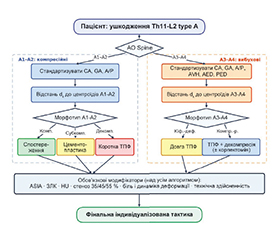
Актуальність. Вибір тактики лікування при ушкодженнях типу A грудопоперекового переходу (ГПП, Th11-L2) залишається дискусійним. Класифікація AO Spine описує морфологію та слугує міжнародною мовою спілкування, проте її недостатньо для індивідуального вибору втручання, особливо у складних реконструктивних ситуаціях. Це обґрунтовує виділення внутрішньотипових кількісних морфотипів і побудову індивідуалізованого алгоритму. Мета: виконати морфофункціональну стратифікацію ушкоджень типу A ГПП: виділити КТ-морфометричні морфотипи, що відображають співвідношення локальної морфології перелому й деформаційної відповіді сегмента, і побудувати алгоритм, у якому морфотип формалізує базовий тактичний сценарій, а кількісні порогові значення — його модифікацію. Матеріали та методи. За даними КТ-морфометрії (CA, GA, A/P ratio, AVH ratio, AED, PED, HU) методами k-means і Gaussian mixture model виділено морфотипи; HU застосовано як модифікатор якості кістки. Кластеризацію оцінювали за silhouette, BIC і клінічною інтерпретованістю, відмінності — за критеріями Краскела — Уолліса/Манна — Уїтні та , дискримінаційну здатність — ROC-аналізом, зв’язок із результатом — відношенням шансів. Алгоритм побудовано за принципом «тип AO Spine морфотип базова тактика кількісні модифікатори». Результати. Усередині A1-A2 виділено три морфотипи — компенсований, субкомпенсований і декомпенсований (градієнт втрати передньої опори); усередині A3-A4 — два: кіфотико-деформаційний і компресійно-ретропульсійний. Морфотип тісно пов’язаний із реалізованою тактикою: при A1-A2 — перехід від спостереження до цементопластики та короткої транспедикулярної фіксації (ТПФ); при A3-A4 — вибір між довгою ТПФ і декомпресією з фіксацією. Порогові значення (CA > 14°/GA > 25° для довжини конструкції; рівень-залежний орієнтир стенозу 35/45/55 % для відкритої vs непрямої декомпресії; HU < 150 за аугментацію) уточнювали рішення. Кіфотико-деформаційний морфотип A3-A4 в усіх 40 спостереженнях лікували довгою ТПФ, компресійно-ретропульсійний — варіативно. Висновки. Морфотип доцільно використовувати як кількісне клініко-тактичне уточнення підтипу AO Spine: AO Spine задає морфологічну вісь, морфотип — рівень ухвалення базового рішення, а ASIA, задній лігаментозний комплекс і якість кістки залишаються обов’язковими модифікаторами. Алгоритм є дериваційною моделлю та потребує проспективної валідації.
Background. The choice of treatment strategy for type A injuries of the thoracolumbar junction (T11-L2) remains controversial. The AO Spine classification describes injury morphology and serves as an international language for communication; however, it is insufficient for individualized selection of a specific intervention, especially in complex reconstructive scenarios. This supports the identification of intratype quantitative morphotypes and the development of an individualized treatment algorithm. The purpose was to perform morphofunctional stratification of type A thoracolumbar junction injuries by identifying CT morphometric morphotypes that reflect the relationship between local fracture morphology and the deformation response of the spinal segment, as well as to develop an algorithm in which the morphotype formalizes the baseline tactical scenario, while quantitative threshold values modify it. Materials and methods. Morphotypes were identified using CT morphometric parameters (CA, GA, A/P ratio, AVH ratio, AED, PED, and HU) with k-means clustering and Gaussian mixture model; HU was used as a modifier of bone quality. Clustering was assessed using silhouette, BIC, and clinical interpretability. Differences were evaluated using the Kruskal-Wallis/Mann-Whitney tests and test; discriminatory ability was assessed by ROC analysis, and the association with outcome was evaluated using odds ratios. The algorithm was constructed according to the principle “AO Spine type morphotype baseline treatment strategy quantitative modifiers”. Results. Within A1-A2 injuries, three morphotypes were identified: compensated, subcompensated, and decompensated, reflecting a gradient of anterior support loss. Within A3-A4 injuries, two morphotypes were identified: kyphotic-deformational and compression-retropulsion. The morphotype was closely associated with the implemented treatment strategy: in A1-A2 injuries, there was a transition from observation to cementoplasty and short-segment transpedicular fixation (TPF); in A3-A4 injuries, the choice was between long-segment TPF and decompression with fixation. Threshold values, including CA > 14°/GA > 25° for construct length, level-dependent canal stenosis thresholds of 35/45/55 % for open versus indirect decompression, and HU < 150 as an argument for augmentation, refined the baseline decision. The kyphotic-deformational A3-A4 morphotype was treated with long-segment TPF in all 40 cases, whereas treatment of the compression-retropulsion morphotype was variable. Conclusions. The morphotype may be used as a quantitative clinical and tactical refinement of the AO Spine subtype: AO Spine defines the morphological axis, the morphotype provides the level for baseline decision-making, while ASIA grade, posterior ligamentous complex, and bone quality remain mandatory modifiers. The proposed algorithm is a derivation model and requires prospective validation.
Список литературы
1. Ambulgekar RK, Ghag NS. Epidemiological study of thoracolumbar spine fracture patients reported to tertiary care center of Central India. International Journal of Research in Orthopaedics. 2023;9(2):261-266. doi: 10.18203/issn.2455-4510.IntJResOrthop20230344.
2. Wendt K, Nau C, Jug M, Pape HC, Kdolsky R, Thomas S, et al. ESTES recommendation on thoracolumbar spine fractures : January 2023. Eur J Trauma Emerg Surg. 2024;50(4):1261-1275. doi: 10.1007/s00068-023-02247-3, PMID: 37052627.
3. Denis F. The three column spine and its significance in the classification of acute thoracolumbar spinal injuries. Spine (Phila Pa 1976). 1983;8(8):817-831. doi: 10.1097/00007632-198311000-00003, PMID: 6670016.
4. Magerl F, Aebi M, Gertzbein SD, Harms J, Nazarian S. A comprehensive classification of thoracic and lumbar injuries. Eur Spine J. 1994;3(4):184-201. doi: 10.1007/bf02221591, PMID: 7866834.
5. Vaccaro AR, Oner C, Kepler CK, Dvorak M, Schnake K, Bellabarba C, et al. AOSpine thoracolumbar spine injury classification system: fracture description, neurological status, and key modifiers. Spine (Phila Pa 1976). 2013;38(23):2028-2037. doi: 10.1097/BRS.0b013e3182a8a381, PMID: 23970107.
6. Vaccaro AR, Lehman RA, Jr., Hurlbert RJ, Anderson PA, Harris M, Hedlund R, et al. A new classification of thoracolumbar injuries: the importance of injury morphology, the integrity of the posterior ligamentous complex, and neurologic status. Spine (Phila Pa 1976). 2005;30(20):2325-2333. doi: 10.1097/01.brs.0000182986.43345.cb, PMID: 16227897.
7. McCormack T, Karaikovic E, Gaines RW. The load sha–ring classification of spine fractures. Spine (Phila Pa 1976). 1994;19(15):1741-1744. doi: 10.1097/00007632-199408000-00014, PMID: 7973969.
8. Rajasekaran S, Vaccaro AR, Kanna RM, Schroeder GD, Oner FC, Vialle L, et al. The value of CT and MRI in the classification and surgical decision-making among spine surgeons in thoracolumbar spinal injuries. Eur Spine J. 2017;26(5):1463-1469. doi: 10.1007/s00586-016-4623-0, PMID: 27250728.
9. Falco L, Ohannesian VA, Monteiro PQ, Silva RAS, Menezes IR, Suruagy Motta RFO, et al. Accuracy of CT Scan for Detecting Posterior Ligamentous Complex Injury in Traumatic Thoracolumbar Fractures: A Systematic Review and Meta-Analysis. Global Spine J. 2025;15(7):3531-3543. doi: 10.1177/21925682251343525, PMID: 40380790.
10. Dunsmuir RA, Price MJ, Rankine JJ. "Dirty Fat Pad" Sign: A novel computed tomography (CT) indicator of injury to the posterior ligamentous complex in acute fractures of the thoracolumbar spine. Injury. 2026;57(7):113110. doi: 10.1016/j.injury.2026.113110, PMID: 41927418.
11. Schnake KJ, Blattert TR, Hahn P, Franck A, Hartmann F, Ullrich B, et al. Classification of Osteoporotic Thoracolumbar Spine Fractures: Recommendations of the Spine Section of the German Society for Orthopaedics and Trauma (DGOU). Global Spine J. 2018;8(2 Suppl):46S-49S. doi: 10.1177/2192568217717972, PMID: 30210960.
12. Komadina R, Bloemers FW, Jug M, Wendt KW, Nau C, Pape HC. Fractures of the thoracolumbar spine in osteoporosis. Eur J Trauma Emerg Surg. 2024;50(5):1977-1984. doi: 10.1007/s00068-024-02625-5, PMID: 39254697.
13. Buyukayten V, Ridderikhof ML, Twisk JWR, Groot OQ, Doornberg JN, Bloemers FW. The AO Spine TL Injury Classification System Drives Clinical Decision Making in Acute Thoracolumbar Fractures: A Retrospective Evaluation of Clinical Implementation. Global Spine J. 2026:21925682261432541. doi: 10.1177/21925682261432541, PMID: 41797480.
14. Dandurand C, ner CF, Hazenbiller O, Bransford RJ, Schnake K, Vaccaro AR, et al. Understanding Decision Ma–king as It Influences Treatment in Thoracolumbar Burst Fractures Without Neurological Deficit: Conceptual Framework and Methodology. Global Spine J. 2024;14(1_suppl):8s-16s. doi: 10.1177/21925682231210183, PMID: 38324598.
15. Aly MM, Abdelaziz M, Alfaisal FA, Alrumian RA, Espinoza XAS, Gutirrez-Gonzlez R, et al. Multicenter External Validation of the Accuracy of Computed Tomography Criteria for Detecting Thoracolumbar Posterior Ligamentous Complex Injury. Neurosurgery. 2025;96(6):1236-1248. doi: 10.1227/neu.0000000000003263, PMID: 39636120.
16. Joaquim AF, Patel AA. Thoracolumbar spine trauma: Evaluation and surgical decision-making. J Craniovertebr Junction Spine. 2013;4(1):3-9. doi: 10.4103/0974-8237.121616, PMID: 24381449.
17. Kepler CK, Vaccaro AR, Schroeder GD, Koerner JD, Vialle LR, Aarabi B, et al. The Thoracolumbar AOSpine Injury Score. Global Spine J. 2016;6(4):329-334. doi: 10.1055/s-0035-1563610, PMID: 27190734.
18. Aly MM, Al-Shoaibi AM, Abduraba Ali S, Al Fattani A, Eldawoody H. How Often Would MRI Change the Thoracolumbar Fracture Classification or Decision-Making Compared to CT Alone? Global Spine J. 2024;14(1):11-24. doi: 10.1177/21925682221089579, PMID: 35382642.
19. Altay M, Ozkurt B, Aktekin CN, Ozturk AM, Dogan O, Tabak AY. Treatment of unstable thoracolumbar junction burst fractures with short- or long-segment posterior fixation in ma–gerl type a fractures. Eur Spine J. 2007;16(8):1145-1155. doi: 10.1007/s00586-007-0310-5, PMID: 17252216.
20. Aono H, Ishii K, Tobimatsu H, Nagamoto Y, Takenaka S, Furuya M, et al. Temporary short-segment pedicle screw fixation for thoracolumbar burst fractures: comparative study with or without vertebroplasty. Spine J. 2017;17(8):1113-1119. doi: 10.1016/j.spinee.2017.03.022, PMID: 28373079.
21. McLain RF. The biomechanics of long versus short fixation for thoracolumbar spine fractures. Spine (Phila Pa 1976). 2006;31(11 Suppl):S70-79; discussion S104. doi: 10.1097/01.brs.0000218221.47230.dd, PMID: 16685240.
22. Yu D, Zhang Y, Li X, Wang W, Li Z, Xiao J. Efficacy and safety of posterior short-segment versus long-segment pedicle screws fixation for thoracolumbar burst fractures: A systematic review and meta-analysis. Medicine (Baltimore). 2025;104(23):e42699. doi: 10.1097/MD.0000000000042699, PMID: 40489858.
23. Canseco JA, Reinhold M, Dalton J, Dandurand C, Oner CF, Dvorak M, et al. Acute thoracolumbar burst fractures (AO types A3/A4) with and without concomitant posterior ligamentous complex injury: treatment outcomes in surgically and nonsurgically managed patients. A multi-center prospective study. Eur Spine J. 2026. doi: 10.1007/s00586-025-09680-5, PMID: 41854908.
24. Wood KB, Buttermann GR, Phukan R, Harrod CC, Mehbod A, Shannon B, et al. Operative compared with nonoperative treatment of a thoracolumbar burst fracture without neurological deficit: a prospective randomized study with follow-up at sixteen to twenty-two years. J Bone Joint Surg Am. 2015;97(1):3-9. doi: 10.2106/JBJS.N.00226, PMID: 25568388.
25. Scherer J, Joaquim A, Vaccaro A, Kanna R, El-Sharkawi M, Takahata M, et al. AO Spine-DGOU Osteoporotic Fracture Classification System: Internal Validation by the AO Spine Knowledge Forum Trauma. Global Spine J. 2025;15(4):2152-2157. doi: 10.1177/21925682241288187, PMID: 39327898.
26. Nekhlopochyn OS, Verbov VV, Cheshuk IV, Vorodi MV. Assessment of risk factors for the vertebral body kyphotic deformity progression in patients with type A1 injuries of the thoracolumbar junction. Ukrainian Neurosurgical Journal. 2023;29(3):26-33. doi: 10.25305/unj.278927.
27. Nekhlopochyn O, Verbov V, Cheshuk I, Vorodi M. Ana–lysis of the effectiveness of indirect spinal canal decompression in the treatment of burst fractures at the thoracolumbar junction. Orthopaedics Traumatology and Prosthetics. 2024(3):13-21. doi: 10.15674/0030-59872024313-21.
28. Nekhlopochyn OS, Verbov VV, Cheshuk IV, Vorodi MV, Karpinsky MY, Yaresko OV. Short-segment stabilization techniques for burst fractures of the thoracolumbar junction: a finite element study under lateral flexion. Ukrainian Neurosurgical Journal. 2025;31(4):44-54. doi: 10.25305/unj.331033.
29. Xu C, Bai X, Ruan D, Zhang C. Comparative finite ele–ment analysis of posterior short segment fixation constructs with or without intermediate screws in the fractured vertebrae for the treatment of type a thoracolumbar fracture. Comput Methods Biomech Biomed Engin. 2024;27(11):1398-1409. doi: 10.1080/10255842.2023.2243360, PMID: 37553841.
30. Pidd KT, Sadauskas D, Tomatis V, Knight EJ. Which is the Superior Thoracolumbar Injury Classification Tool? TLICS Versus AOSpine 2013: A Systematic Review. Global Spine J. 2025;15(4):2536-2546. doi: 10.1177/21925682241311303, PMID: 39722528.