
Архив офтальмологии Украины Том 14, №2, 2026
Вернуться к номеру
Експресія miR-126 та її зв’язок зі стадіями діабетичної ретинопатії у пацієнтів із цукровим діабетом 2-го типу
Авторы: Кір’ян Є.П.
Національний медичний університет імені О.О. Богомольця, м. Київ, Україна
Рубрики: Офтальмология
Разделы: Клинические исследования
Версия для печати
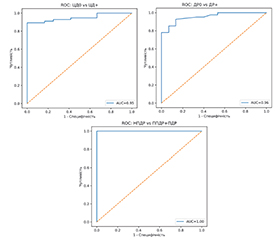
Актуальність. МікроРНК є важливими епігенетичними регуляторами експресії генів, які беруть участь у контролі ангіогенезу, запалення, апоптозу та ремоделювання тканин. Накопичені дані свідчать про їхню суттєву роль у розвитку цукрового діабету (ЦД) та його мікросудинних ускладнень, зокрема діабетичної ретинопатії (ДР). Особливий інтерес становить miR-126 — одна з основних ендотеліальних мікроРНК, що регулює ангіогенез і підтримання цілісності судинної стінки через VEGF-залежні сигнальні шляхи. Мета: встановити діагностичне значення експресії miR-126 та її зв’язок зі стадіями ДР у пацієнтів із ЦД 2-го типу. Матеріали та методи. Обстежено 68 пацієнтів: осіб без ЦД (контроль), хворих на ЦД 2-го типу без ДР, а також пацієнтів із непроліферативною (НПДР), препроліферативною (ППДР) та проліферативною (ПДР) ДР. Експресію miR-126 визначали методом кількісної ПЛР у реальному часі. Статистичний аналіз включав критерій Крускала — Уолліса, post-hoc аналіз Данна, кореляційний аналіз Спірмана, логістичну регресію та ROC-аналіз. Результати. Встановлено послідовне зниження експресії miR-126 відповідно до прогресування ДР. Порівняно з контрольною групою її експресія була знижена при НПДР у 1,7 раза, при ППДР — у 6,4 раза та при ПДР — у 10,6 раза (р < 0,05). Найбільш виражене зниження показника спостерігалося при переході від НПДР до ППДР/ПДР. Рівень експресії miR-126 не залежав від статі та віку пацієнтів, але мав сильний зворотний зв’язок із тривалістю ЦД (ρ = ‒0,83; p < 0,0001). Зниження експресії miR-126 було незалежним предиктором ДР (OR = 0,056; 95% ДІ 0,007–0,470; p = 0,0079). ROC-аналіз показав високу діагностичну ефективність маркера для виявлення ЦД 2-го типу (AUC = 0,95), ДР (AUC = 0,96) та тяжких стадій ДР (AUC = 1,00). Встановлено дуже сильний прямий зв’язок між експресією miR-126 та miR-146a-5p (ρ = 0,912; p < 0,001). Висновки. Експресія miR-126 прогресивно знижується відповідно до тяжкості ДР та характеризується високою діагностичною цінністю щодо виявлення ДР і її тяжких стадій. Тісний зв’язок між miR-126 та miR-146a-5p свідчить про взаємопов’язане залучення механізмів ендотеліальної дисфункції та хронічного запалення у патогенезі прогресування ДР.
Background. MicroRNAs are important epigenetic regulators of gene expression involved in the control of angiogenesis, inflammation, apoptosis, and tissue remodeling. Accumulated data indicate their essential role in the development of diabetes mellitus (DM) and its microvascular complications, in particular diabetic retinopathy (DR). Of particular interest is miR-126, one of the main endothelial miRNAs that regulates angiogenesis and maintenance of vascular wall integrity through VEGF-dependent signaling pathways. Objective: to establish the diagnostic significance of miR-126 expression and its relationship with DR stages in patients with type 2 DM. Materials and methods. Sixty-eight patients were examined: individuals without DM (control), patients with type 2 diabetes without DR, as well as those with non-proliferative (NPDR), pre-proliferative (PPDR), and proliferative diabetic retinopathy (PDR). miR-126 expression was determined by real-time quantitative polymerase chain reaction. Statistical analysis included Kruskal-Wallis test, Dunn’s post-hoc analysis, Spearman correlation analysis, logistic regression and ROC analysis. Results. A consistent decrease in miR-126 expression was found with the progression of DM. Compared to the control group, its expression was reduced in NPDR by 1.7 times, in PPDR by 6.4 times and in PDR by 10.6 times (p < 0.05). The most pronounced decrease was observed when switching from NPDR to PPDR/PDR. The level of miR-126 expression did not depend on the sex and age of the patients, but had a strong inverse relationship with DM duration (ρ = –0.83; p < 0.0001). Decreased miR-126 expression was an independent predictor of DR (OR = 0.056; 95% CI 0.007–0.470; p = 0.0079). ROC analysis showed high diagnostic efficiency of the marker for the detection of type 2 diabetes (AUC = 0.95), DR (AUC = 0.96) and severe stages of DR (AUC = 1.00). A very strong direct correlation was found between the expression of miR-126 and miR-146a-5p (ρ = 0.912; p < 0.001). Conclusions. The expression of miR-126 progressively decreases according to DR severity and is characterized by high diagnostic value int terms of detecting DR and its severe stages. The close relationship between miR-126 and miR-146a-5p suggests the interconnected involvement of endothelial dysfunction and chronic inflammation mechanisms in DR progression.
цукровий діабет 2-го типу; діабетична ретинопатія; miR-126; miR-146a-5p; ендотеліальна дисфункція; ангіогенез; біомаркери
type 2 diabetes; diabetic retinopathy; miR-126; miR-146a-5p; endothelial dysfunction; angiogenesis; biomarkers
Для ознакомления с полным содержанием статьи необходимо оформить подписку на журнал.
- Teo ZL, Tham YC, Yu M, Chee ML, Rim TH, Cheung N, et al. Global prevalence of diabetic retinopathy and projection of burden through 2045: systematic review and meta-analysis. Ophthalmology. 2021;128(11):1580-1591. doi: 10.1016/j.ophtha.2021.04.027.
- Flaxman SR, Bourne RRA, Resnikoff S, et al. Global causes of blindness and distance vision impairment 1990-2020: a systematic review and meta-analysis. Lancet Glob Health. 2017;5(12):e1221-e1234. doi: 10.1016/S2214-109X(17)30393-5.
- Kaur A, Kumar R, Sharma A. Diabetic Retinopathy Leading to Blindness- A Review. Curr Diabetes Rev. 2024;20(9):e240124225997. doi: 10.2174/0115733998274599231109034741.
- Pei X, Huang D, Li Z. Genetic insights and emerging therapeutics in diabetic retinopathy: from molecular pathways to persona–lized medicine. Front Genet. 2024 Aug 22;15:1416924. doi: 10.3389/fgene.2024.1416924.
- Bhatwadekar AD, Shughoury A, Belamkar A, Ciulla TA. Genetics of Diabetic Retinopathy, a Leading Cause of Irreversible Blindness in the Industrialized World. Genes (Basel). 2021 Jul 31;12(8):1200. doi: 10.3390/genes12081200.
- Lu TX, Rothenberg ME. MicroRNA. J Allergy Clin Immunol. 2018 Apr;141(4):1202-1207. doi: 10.1016/j.jaci.2017.08.034.
- Lombardi G, Delvin E. Micro-RNA: A Future Approach to Personalized Diagnosis of Bone Diseases. Calcif Tissue Int. 2023 Feb;112(2):271-287. doi: 10.1007/s00223-022-00959-z.
- Kaur P, Kotru S, Singh S, Munshi A. Role of miRNAs in diabetic neuropathy: mechanisms and possible interventions. Mol Neurobiol. 2022 Mar;59(3):1836-1849. doi: 10.1007/s12035-021-02662-w.
- Ismail A, El-Mahdy HA, Eldeib MG, Doghish AS. miRNAs as cornerstones in diabetic microvascular complications. Mol Genet Metab. 2023 Jan;138(1):106978. doi: 10.1016/j.ymgme.2022.106978.
- Theofilis P, Oikonomou E, Vogiatzi G, Sagris M, Antonopoulos AS, Siasos G, et al. The Role of MicroRNA-126 in Atherosclerotic Cardiovascular Diseases. Curr Med Chem. 2023;30(17):1902-1921. doi: 10.2174/0929867329666220830100530.
- Lampsas S, Agapitou C, Chatzirallis A, Papavasileiou G, Poulakis D, Pegka S, et al. MicroRNA (miRNA) in the Pathogenesis of Diabetic Retinopathy: A Narrative Review. Genes (Basel). 2025 Sep 9;16(9):1060. doi: 10.3390/genes16091060.
- Smit-McBride Z, Morse LS. MicroRNA and diabetic retinopathy—biomarkers and novel therapeutics. Ann Transl Med 2021;9(15):1280. doi: 10.21037/atm-20-5189.
- Ghaneh T, Zeinali F, Babini H, Astaraki S, Hassan-Zadeh V. An increase in the expression of circulating miR30d-5p and miR126-3p is associated with intermediate hyperglycaemia in Iranian population. Arch Physiol Biochem. 2023 Apr;129(2):489-496. doi: 10.1080/13813455.2020.1839105.
- Nemecz M, Stefan DS, Comarița IK, Constantin A, Tanko G, Guja C, Georgescu A. Microvesicle-associated and circulating microRNAs in diabetic dyslipidemia: miR-218, miR-132, miR-143, and miR-21, miR-122, miR-155 have biomarker potential. Cardiovasc Diabetol. 2023 Sep 25;22(1):260. doi: 10.1186/s12933-023-01988-0.
- Wei L, Sun X, Fan C, Li R, Zhou S, Yu H. The pathophy–siological mechanisms underlying diabetic retinopathy. Front Cell Dev Biol. 2022;10:963615. doi: 10.3389/fcell.2022.963615.
- Kumari N, Karmakar A, Ganesan SK. Targeting epigenetic modifications as a potential therapeutic option for diabetic retinopathy. J Cell Physiol. 2020 Mar;235(3):1933-1947. doi: 10.1002/jcp.29180.
- Wang W, Lo A. Diabetic retinopathy: Pathophysiology and treatments. Int J Mol Sci. 2018;19(6):E1816. doi: 10.3390/ijms19061816.
- Brownlee J. One-vs-Rest and One-vs-One for Multi-Class Classification [Internet]. Machine Learning Mastery; 2021 [cited 2025 May 28]. Available from: https://machinelearningmastery.com/one-vs-rest-and-one-vs-one-for-multi-class-classification.
- Robin X, Turck N, Hainard A, Tiberti N, Lisacek F, Sanchez J-C, Müller M. pROC: an open-source package for R and S+ to analyze and compare ROC curves. BMC Bioinformatics. 2011;12(77):1-8. doi: 10.1186/1471-2105-12-77.
- Kiryan EP. MicroRNA-146a-5p expression at different stages of diabetic retinopathy in patients with type 2 diabetes. Medical Science of Ukraine. 2025;3(21):85-93. [Ukrainian]. doi: 10.32345/2664-4738.3.2025.09.
- Keng-Lim BW, Chen-Ng KY, Yee-Lee J, Hsein-Chen C, Ling-Chang W, Yian-Koh R, et al. miRNAs as potential biomarkers for early detection of prediabetes among obesity: a systematic review. Biosci Rep. 2025 Dec 16;45(12):BSR20253283. doi: 10.1042/BSR20253283.
- Taheri M, Eghtedarian R, Ghafouri-Fard S, Omrani MD. Non-coding RNAs and type 2 diabetes mellitus. Arch Physiol Biochem. 2023 Apr;129(2):526-535. doi: 10.1080/13813455.2020.1843498.
- Wang P, Wen Y. The inflammatory-miRNA axis in diabetic microvascular complications: a meta-analysis highlighting synergistic effects in concurrent retinopathy and nephropathy. J Diabetes Metab Disord. 2025 Dec 11;24(2):290. doi: 10.1007/s40200-025-01809-z.
- Lou Z, Li Q, Wang C, Li Y. The effects of microRNA-126 reduced inflammation and apoptosis of diabetic nephropathy through PI3K/AKT signalling pathway by VEGF. Arch Physiol Biochem. 2022 Oct;128(5):1265-1274. doi: 10.1080/13813455.2020.1767146.
- Zhao W, Liu Y, Li C, Yin Z, Liu Y, An M. Mechanisms of MALAT1 regulating proliferative diabetic retinopathy via targeting miR-126-5p. Am J Transl Res. 2023 May 15;15(5):3279-3289. PMID: 37303661; PMCID: PMC10251023.
- Wang Y, Yan H. MicroRNA-126 contributes to Niaspan treatment induced vascular restoration after diabetic retinopathy. Sci Rep. 2016 May 26;6:26909. doi: 10.1038/srep26909.
- Santovito D, Toto L, De Nardis V, Marcantonio P, D’Aloisio R, Mastropasqua A, et al. Plasma microRNA signature associated with retinopathy in patients with type 2 diabetes. Sci Rep. 2021 Feb 18;11(1):4136. doi: 10.1038/s41598-021-83047-w.
- Monfared YK, Mirzaii-Dizgah MR, Khodabandehloo E, Sarookhani MR, Hashemipour S, Mirzaii-Dizgah I. Salivary microRNA-126 and 135a: a potentially non-invasive diagnostic biomarkers of type- 2 diabetes. J Diabetes Metab Disord. 2021 Oct 28;20(2):1631-1638. doi: 10.1007/s40200-021-00914-z.
- Margaritis K, Margioula-Siarkou G, Giza S, Kotanidou EP, Tsinopoulou VR, Christoforidis A, Galli-Tsinopoulou A. Micro-RNA Implications in Type-1 Diabetes Mellitus: A Review of Literature. Int J Mol Sci. 2021 Nov 10;22(22):12165. doi: 10.3390/ijms222212165.
- Qin LL, An MX, Liu YL, Xu HC, Lu ZQ. MicroRNA-126: a promising novel biomarker in peripheral blood for diabetic retino–pathy. Int J Ophthalmol. 2017 Apr 18;10(4):530-534. doi: 10.18240/ijo.2017.04.05.