
Архив офтальмологии Украины Том 14, №2, 2026
Вернуться к номеру
Особливості вітреоретинальних втручань при ідіопатичних епіретинальних мембранах
Авторы: Панченко Ю.О. (1), Гуліда А.О. (1), Лисенко Н.Р. (2), Вербицький І.В. (1), Могілевський С.Ю. (1)
(1) - Національний університет охорони здоров’я України імені П.Л. Шупика, м. Київ, Україна
(2) - Офтальмологічний центр ТОВ «Новий зір м. Кривий Ріг», м. Кривий Ріг, Україна
Рубрики: Офтальмология
Разделы: Клинические исследования
Версия для печати
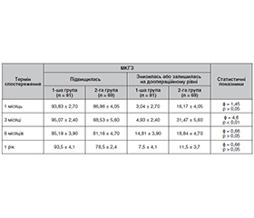
Актуальність. Ідіопатичні епіретинальні мембрани (ЕRМ) належать до найбільш поширених захворювань вітреомакулярного інтерфейсу та є однією з провідних причин зниження центрального зору у пацієнтів старших вікових груп. За даними сучасних досліджень, поширеність ЕRМ становить від 7 до 39 %. З огляду на старіння населення кількість пацієнтів із симптоматичними ЕRМ постійно зростає, що визначає високу медико-соціальну значущість проблеми та необхідність удосконалення сучасних підходів до їх лікування. Мета: дослідити ефективність сучасних комбінових та двохетапних методів хірургічного лікування епіретинальних мембран у поєднанні з віковою катарактою. Матеріали та методи. Під спостереженням у дослідженні перебували 150 пацієнтів (150 очей) з ERM та початковою віковою катарактою, які були розподілені у 2 групи. Вік досліджуваних пацієнтів становив 61,0 ± 2,3 року. У 1-й групі (81 око) виконали закриту субтотальну вітректомію 25Ga (ЗСВ) з пілінгом ERM. Факоемульсифікацію катаракти (ФЕК) проводили через 3 місяці. У 2-й групі (69 очей) — ЗСВ з пілінгом ERM/внутрішньої пограничної мембрани (ILM) з ФЕК та імплантацією інтраокулярної лінзи. Усім пацієнтам виконували оптичну когерентну томографію макулярної ділянки, за результатами якої визначали центральну товщину сітківки, оцінювали морфологічні зміни макулярної ділянки, наявність епімакулярної мембрани. Обстеження проводили до оперативного лікування, а також через 1, 3, 6 та 12 місяців після операції. Результати. Комбіноване виконання ФЕК під час вітреоретинального втручання супроводжується більшою частотою розвитку макулярного набряку у ранньому післяопераційному періоді. Водночас зазначені зміни були тимчасовими. Уже через три місяці їх частота суттєво зменшувалася, а через один рік клінічно значущий макулярний набряк був відсутній у всіх пацієнтів незалежно від виду оперативного лікування. Це свідчить про сприятливий довгостроковий прогноз після обох варіантів втручання. Висновки. Проведені нами дослідження показали, що ЗСВ з видаленням виключно ERM та ЗСВ з подвійним пілінгом ERM/ILM без хірургії та з хірургією катаракти є ефективними методами лікування пацієнтів з ERM, що сприяють поліпшенню зорових функцій при терміні спостереження 1 рік. ФЕК суттєво не поліпшувала гостроту зору, а, навпаки, була причиною резистентного макулярного набряку у терміни спостереження 1 та 3 місяці. Пілінг ILM не поліпшує функціональні результати після операції EРM, але запобігає виникненню рецидивів ERM.
Background. Idiopathic epiretinal membranes (ERM) are among the most common disorders of the vitreomacular interface and are one of the leading causes of reduced central vision in older patients. According to recent studies, the prevalence of ERM ranges from 7 to 39 %. With the aging of the population, the number of patients with symptomatic ERMs continues to increase, highlighting the high medical and social significance of this condition and the need to improve current treatments. Objective: to investigate the efficacy of contemporary combined and two-stage surgical approaches for the treatment of epiretinal membranes associated with age-related cataract. Materials and methods. The study included 150 patients (150 eyes) with ERM and initial age-related cataract, who were divided into two study groups. The mean age of the participants was 61.0 ± 2.3 years. In group 1 (81 eyes), 25-gauge closed subtotal vitrectomy (CSV) with ERM peeling was performed, followed by phacoemulsification 3 months later. In group 2 (69 eyes), CSV with combined ERM/internal limiting membrane (ILM) peeling was performed simultaneously with phacoemulsification and intraocular lens implantation. All patients underwent optical coherence tomography of the macular region, which was used to determine central retinal thickness, assess morphological changes in the macula, and detect the presence of an epiretinal membrane. Examinations were performed before surgery and at 1, 3, 6, and 12 months postoperatively. Results. Phacoemulsification in combination with vitreoretinal surgery is associated with a higher incidence of macular edema in the early postoperative period. However, these changes were transient; their incidence decreased significantly after 3 months, and at one year, clinically significant macular edema was absent in all patients, regardless of the type of surgical intervention. This indicates a favorable long-term prognosis for both surgical approaches. Conclusions. Our studies demonstrated that CSV with ERM peeling only and CSV with combined ERM/ILM peeling, performed either without or with simultaneous cataract surgery, are effective methods for the treatment of ERM and result in improved visual function over a 1-year follow-up period. Phacoemulsification did not significantly improve visual acuity and, conversely, was associated with a higher incidence of persistent macular edema at 1 and 3 months postoperatively. ILM peeling did not improve functional outcomes after ERM surgery but reduced the risk of ERM recurrence.
ідіопатична епіретинальна мембрана; внутрішня погранична мембрана; макула; вітреоретинальна хірургія; факоемульсифікація; макулярний набряк; оптична когерентна томографія; максимально коригована гострота зору
idiopathic epiretinal membrane; internal limiting membrane; macula; vitreoretinal surgery; phacoemulsification; macular edema; optical coherence tomography; best-corrected visual acuity
Для ознакомления с полным содержанием статьи необходимо оформить подписку на журнал.
- Bailey ST, Vemulakonda GA, Kim SJ, Kovach JL, Lim JI, Ying GS, et al.; American Academy of Ophthalmology Retina/Vitreous Committee. Idiopathic Epiretinal Membrane and Vitreomacular Traction Preferred Practice Pattern®. Ophthalmology. 2025;132(4):P197-P233. doi: 10.1016/j.ophtha.2024.12.019.
- Путієнко О.О., Парій І.О. Аналіз результатів вітреоретинальної хірургії ідіопатичних епіретинальних мембран із застосуванням газової тампонади. Клінічна офтальмологія. 2025;13(1):6-12. doi: 10.22141/2309-8147.13.1.2025.399.
- Holzwarth J, Krohne TU, Lommatzsch A, Priglinger SG, Hattenbach LO. Epiretinal membrane: diagnostics, indications and surgical treatment. Ophthalmologie. 2024;121(6):443-451. doi: 10.1007/s00347-024-02055-z.
- Quiroz-Reyes MA, Quiroz-Gonzalez EA, Quiroz-Gonzalez MA, Lima-Gomez V. Surgical management, use and efficacy of adjuvant dyes in idiopathic epiretinal membranes: a systemic review with network meta-analysis. Int J Retina Vitreous. 2023 Dec 6;9(1):77. doi: 10.1186/s40942-023-00515-3. PMID: 38057831; PMCID: PMC10702105.
- Chi YC, Chu WL, Chen KJ, Cheng KC. Morphological Change in Optical Coherence Tomography and Functional Outcomes in Epiretinal Membrane Peeling with or without SF6 Tamponade. Diagnostics. 2024;14(14):1483. doi: 10.3390/diagnostics14141483.
- Matsuoka N, Hasegawa Y, Takahashi H, et al. Effects of Internal Limiting Membrane Peeling Combined with Removal of Idiopathic Epiretinal Membrane: A Systematic Review of Literature and Meta-Analysis. Retina. 2017;37(10):1813-1819. doi: 10.1097/IAE.0000000000001537.
- Mihalache A, Huang RS, Ahmed H, et al. Pars Plana Vitrectomy with or without Internal Limiting Membrane Peel for Epiretinal Membrane: A Systematic Review and Meta-Analysis. Ophthalmologica. 2024;247(1):30-43. doi: 10.1159/000534851.
- Christodoulou E, Batsos G, Christou EE, et al. Internal limiting membrane peeling during vitrectomy for idiopathic epiretinal membrane: a meta-analysis. Acta Ophthalmologica. 2021. doi: 10.1111/aos.0265.
- Kim BH, Yoon CK, Bae K, Lee EK, Lee CH, Kim DI, et al. Influence of internal limiting membrane peeling during idiopathic epiretinal membrane removal: a randomized controlled trial. Scientific Reports. 2025;15:17499. doi: 10.1038/s41598-025-01987-z.
- Парій І.О., Путієнко О.О., Денисюк Л.І. Вивчення механізму дії перфторпропану (C₃F₈) на макулярну зону сітківки у вітреоретинальній хірургії ідіопатичних епіретинальних мембран. Клінічна офтальмологія. 2026;14(1):39-44. doi: 10.22141/2309-8147.14.1.2026.434.
- Sere AM, Muntean GA, Cristea AP, Nicoară SD. OCTA Biomarkers Underlying Structure-Function Correlations in Idiopathic Epiretinal Membrane: A Systematic Review. Diagnostics. 2025;15(20):2596. doi: 10.3390/diagnostics15202596.
- Li H, Zhang C, Li H, et al. Effects of disorganization of retinal inner layers for idiopathic epiretinal membrane surgery: the surgical status and prognosis. BMC Ophthalmology. 2023;23:108. doi: 10.1186/s12886-023-02856-x.
- Mahmoudzadeh R, Israilevich R, Salabati M, Hsu J, Garg SJ, Regillo CD, et al. Pars Plana Vitrectomy for Idiopathic Epiretinal Membrane: OCT Biomarkers of Visual Outcomes in 322 Eyes. Ophthalmol Retina. 2022 Apr;6(4):308-317. doi: 10.1016/j.oret.2021.10.008. Epub 2021 Oct 27. PMID: 34718218.
- Gelormini F, Ricardi F, Parisi G, Vallino V, Ghezzo B, Cucciarelli C, et al. Visual Performance and Predictive OCT Biomarkers in Epiretinal Membrane Assessment: Beyond Distance Visual Acuity. Investigative Ophthalmology & Visual Science. 2025;66(1):31. doi: 10.1167/iovs.66.1.31.
- Luan R, Wang M, Gong Y, et al. Optical coherence tomography biomarkers as outcome predictors to guide dexamethasone implant use in patients with iERM: a randomized controlled trial. BMC Ophthalmol. 2024;24(1):193. doi: 10.1186/s12886-024-03429-2.
- Lin HL, Tseng PC, Hsu MH, Peng SJ. Using a deep learning model to predict postoperative visual outcomes of idiopathic epiretinal membrane surgery. Am J Ophthalmol. 2025;272:67-78. doi: 10.1016/j.ajo.2025.01.003.
- Ferro Desideri L, Hinrichsen L, Eldridge N, Chou HD, Chang YC, Subhi Y, et al. Artificial intelligence analysis of OCT biomarkers to predict visual outcomes following vitrectomy for epire–tinal membrane. Int J Retina Vitreous. 2025 Oct 14;11(1):106. doi: 10.1186/s40942-025-00735-9. PMID: 41088490; PMCID: PMC12522735.
- Vounotrypidis E, Meyer J, Vogt D, et al. The impact of epire–tinal membrane stage and postoperative treatment on visual and anatomical outcomes following vitrectomy in eyes with preexisting macular edema. Int J Retina Vitreous. 2025;11(1):74. doi: 10.1186/s40942-025-00697-y.
- Post M, Cicinelli MV, Zanzottera EC, et al. Prevalence and risk factors of ellipsoid zone damage after pars plana vitrectomy for idiopathic epiretinal membrane. Retina. 2022;42(2):256-264. doi: 10.1097/IAE.0000000000003321.
- Fouad YA, Soliman MK, Elhusseiny A, et al. Visual outcomes and complications of combined versus sequential pars plana vitrectomy and phacoemulsification for epiretinal membrane. Eye (Lond). 2024;38(9):1707-1713. doi: 10.1038/s41433-024-03004-5.
- Combined versus Sequential Pars Plana Vitrectomy and Phacoemulsification for Macular Hole and Epiretinal Membrane: A Systematic Review and Meta-Analysis. Ophthalmology Retina. 2023;7(9):880-883. doi: 10.1016/j.oret.2023.03.017.
- Kunavisarut P, Supawongwattana M, Patikulsila D, Choovuthayakorn J, Watanachai N, Chaikitmongkol V, et al. Idiopathic Epiretinal Membranes: Visual Outcomes and Prognostic Factors. Turk J Ophthalmol. 2022 Apr 28;52(2):109-118. doi: 10.4274/tjo.galenos.2021.09258. PMID: 35481732; PMCID: PMC9069092.
- Розанова З.А., Довгань І.П., Левицька Г.В., Уманець М.М. Пролонгована газова тампонада як метод лікування персистуючих ідіопатичних розривів макули. Ukrainian Journal of Ophthalmology. 2025;6(527):24-28. https://doi.org/10.31288/oftalmolzh202562428.
- Govetto A, Lalane RA III, Sarraf D, Figueroa MS, Hubschman JP. Insights Into Epiretinal Membranes: Presence of Ectopic Inner Foveal Layers and a New Optical Coherence Tomography Staging Scheme. Am J Ophthalmol. 2017;175:99-113. doi: 10.1016/j.ajo.2016.12.006.
- Englmaier VA, Storp JJ, Dierse S, Eter N, Al-Nawaiseh S. Idiopathic Epiretinal Membranes: Pathophysiology, Classifications and OCT Biomarkers. Klin Monbl Augenheilkd. 2024;241(5):666-674. doi: 10.1055/a-2043-4662.
- Eude Y, Poinas A, Volteau C, Lebreton O, Bonissent A, Fossum P, et al. Spontaneous Peeling of the Internal Limiting Membrane during Epiretinal Membrane Surgery: Lessons from the PEELING Clinical Trial. Ophthalmol Retina. 2025 Oct;9(10):934-942. doi: 10.1016/j.oret.2025.04.015. Epub 2025 Apr 22. PMID: 40274190.
- Shettigar MP, Dave VP, Chou HD, et al. Vitreous substitutes and tamponades: A review of types, applications and future directions. Indian J Ophthalmol. 2024;72(8):1102-1111. doi: 10.4103/IJO.IJO_2417_23.
- Chung H-S, Lee D, Park J-H. Clinical Outcomes of Combined Phacoemulsification, Extended Depth-of-Focus Intraocular Lens Implantation, and Epiretinal Membrane Peeling Surgery. Journal of Clinical Medicine. 2025;14(7):2423. https://doi.org/10.3390/jcm14072423.
- Fung AT, Galvin J, Tran T. Epiretinal membrane: A review. Clin Exp Ophthalmol. 2021;49(3):289-308. doi: 10.1111/ceo.13914.
- Reichel FF, Labbe E, Gelisken F, Seitz IP, Hagazy S, et al. Persistence and recurrence after removal of idiopathic epire–tinal membrane. Eye. 2025;39:314-319. doi: 10.1038/s41433-024-03429-y.