
Архив офтальмологии Украины Том 14, №2, 2026
Вернуться к номеру
Результати вітреоретинальної хірургії наскрізних ідіопатичних розривів макули з діаметром основи більше ніж 1000 мкр із застосуванням газово-повітряної суміші перфторпропану, що слабо розширюється
Авторы: Путієнко О.О. (1), Руднєв М.О. (1), Варивончик Д.В. (2), Денисюк Л.І. (1), Лисенко М.Г. (1)
(1) - Національний університет охорони здоров’я України імені П.Л. Шупика, м. Київ, Україна
(2) - ДНТ «Інститут оцінювання функціонування» (Український державний науково-дослідний інститут
медико-соціальних проблем інвалідності), м. Дніпро, Україна
Рубрики: Офтальмология
Разделы: Клинические исследования
Версия для печати
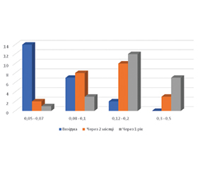
Актуальність. Стандартні методи вітреоретинальних втручань наскрізних макулярних розривів (МР) великого розміру дозволяють здійснити повне закриття розриву тільки у 77–87 % випадків, тому є актуальним пошук нових підходів до лікування. Мета: оцінити ефективність вітреоретинальної хірургії наскрізних ідіопатичних МР з діаметром основи більше ніж 1000 мкр із застосуванням газово-повітряної суміші перфторпропану, що слабо розширюється. Матеріали та методи. Під спостереженням перебували 22 хворі (24 ока) із наскрізними ідіопатичними МР з діаметром основи більше ніж 1000 мкр. Мінімальний діаметр МР був у діапазоні 650–960 мкр, максимальний — 1105–1585 мкр. Вітреоретинальне втручання виконували калібром 25G, за стандартною методикою із видаленням внутрішньої пограничної мембрани (ВПМ) діаметром 2,0 диска зорового нерва, із пасивним масажем країв розриву протягом 5 хвилин і тампонадою вітреальної порожнини 20% газово-повітряною сумішшю перфторпропану. Термін спостереження становив 2 місяці та 1 рік. Результати. Через 2 місяці повне закриття розривів було на 21 оці (91,7 %), через рік після повторних втручань повне закриття МР було досягнуто на 22 очах (95,9 %). У термін 2 місяці мінімальна товщина сітківки у фовеа була в середньому 80,4 (SD 8,32) мкр, товщина центрального сектора макули становила в середньому 178,7 (SD 10,4) мкр, товщина всієї ділянки в задньому полюсі ока, що вимірювали, — 260,1 (SD 23,1) мкр, об’єм ділянки, яку вимірювали, був у середньому 7,30 (SD 0,11) мм3. Через рік після операції ці показники значуще збільшилися на 13,9; 14,9; 11,2 мкр та 0,11 мм3 відповідно. Відновлення зовнішньої пограничної мембрани через 2 місяці було на 10 очах (47,6 %), а через 1 рік вже на 18 (81,8 %), різниця статистично значуща (χ2 = 5,53, р = 0,018). Відновлення еліпсоїдної зони фоторецепторів мали 8 очей (38,1 %) через 2 місяці та 17 очей (77,3 %) через 1 рік (χ2 = 6,78, р = 0,009). Гострота зору значуще покращилася як через 2 місяці (χ2 = 9,58, р = 0,002), так і через рік після втручання (χ2 = 21,8, р = 0,0001). Висновки. Застосування 20% газово-повітряної суміші перфторпропану у вітреоретинальній хірургії наскрізних ідіопатичних МР із діаметром основи більше ніж 1000 мкр є високоефективним — 95,9 % повного закриття розриву через рік після операції. Отримані через 2 місяці результати відновлення рельєфу сітківки, зовнішньої пограничної мембрани, еліпсоїдної зони фоторецепторів і гостроти зору є проміжними, оскільки значуще покращуються через рік після втручання, що свідчить про велику тривалість відновлення макулярної зони у випадках хірургії МР великого розміру.
Background. The use of standard vitreoretinal surgical techniques for large full-thickness macular holes (MHs) results in their complete closure in only 77–87 % of cases; therefore, the search for new treatment approaches remains a priority. Objective: to evaluate the efficacy of vitreoretinal surgery for full-thickness idiopathic MHs with a base diameter above 1,000 µm using a gas-air mixture of low-expansion perfluoropropane. Materials and methods. The study included 22 patients (24 eyes) with full-thickness idiopathic MHs with a base diameter greater than 1,000 µm. The minimum MHs diameter ranged from 650 to 960 µm, and the maximum from 1,105 to 1,585 µm. Vitreoretinal surgery was performed using a 25G, following a standard technique involving the removal of the internal limiting membrane with a diameter of 2.0 optic disc, passive massage of the tear edges for 5 minutes and tamponade of the vitreous cavity with a 20% perfluoropropane gas-air mixture. The follow-up period was 2 months and 1 year. Results. After 2 months, complete closure of the holes was achieved in 21 eyes (91.7 %); one year after repeat procedures, complete closure of the MHs was achieved in 22 eyes (95.9 %). In 2-month follow up, the minimum retinal thickness in the fovea averaged 80.4 (SD 8.32) µm; the thickness of the central macular sector averaged 178.7 (SD 10.4) µm; the thickness of the entire measured area in the posterior pole of the eye was 260.1 (SD 23.1) µm; and the volume of the measured area averaged 7.30 (SD 0.11) mm³. One year after surgery, these parameters had increased significantly by 13.9, 14.9, 11.2 µm and 0.11 mm³, respectively. Recovery of the external limiting membrane was observed in 10 eyes (47.6 %) after 2 months, and in 18 eyes (81.8 %) after 1 year; the difference was statistically significant (χ² = 5.53, p = 0.018). Recovery of the ellipsoid zone of photoreceptors was noted in 8 eyes (38.1 %) after 2 months and in 17 eyes (77.3 %) after 1 year (χ² = 6.78, p = 0.009). Visual acuity improved significantly both 2 months (χ² = 9.58, p = 0.002) and 1 year after the procedure (χ² = 21.8, p = 0.0001). Conclusions. The use of a 20% perfluoropropane gas-air mixture in vitreoretinal surgery for full-thickness idiopathic MHs with a base diameter above 1,000 µm is highly effective, achieving a 95.9 % rate of complete hole closure one year after surgery. The results obtained after 2 months regarding the restoration of retinal anatomy, the external limiting membrane, the ellipsoid zone and visual acuity are interim, as they improve significantly one year after the procedure, which indicates that recovery of the macular area takes a long time in cases of surgery for large-sized MHs.
наскрізні ідіопатичні макулярні розриви; діаметр основи більше 1000 мкр; 20% газово-повітряна суміш перфторпропану; зовнішня погранична мембрана; еліпсоїдна зона; гострота зору
full-thickness idiopathic macular holes; base diameter greater than 1,000 µm; 20% perfluoropropane gas-air mixture; external limiting membrane; ellipsoid zone; visual acuity
Для ознакомления с полным содержанием статьи необходимо оформить подписку на журнал.
- Steel DH, Donachie PHJ, Aylward GW, Laidlaw DA, Williamson TH, Yorston D, BEAVRS Macular hole outcome group. Factors affecting anatomicaland visual outcome after macular hole surgery: findings from a largeprospective UK cohort. Eye (Basingstoke). 2021;35:316-25. https://doi.org/10.1038/s41433-020-0844-x.
- Frisina R, Gius I, Tozzi L, Midena E. Refractory full thickness macular hole: current surgical management. Eye (Basingstoke). 2021. https://doi.org/10.1038/s41433-020-01330-y.
- Maguire MJ, Steel DH, Yorston D, et al. Outcome of revision procedures for failed primary macular hole surgery. Retina. 2020. https://doi.org/10.1097/iae.0000000000003072.
- Geng X-Y, Wu H-Q, Jiang J-H, et al. Area and volume ratios for prediction of visual outcome in idiopathic macular hole. Int J Ophthalmol. 2017. https://doi.org/10.18240/ijo.2017.08.12.
- Kaźmierczak K, Stafiej J, Stachura J, Żuchowski P, Malukiewicz G. Long-term anatomic and functional outcomes after macular hole surgery. J Ophthalmol 2018;2018:3082194.
- Ch’ng SW, Patton N, Ahmed M, Ivanova T, Baumann C, Charles S, et al. The Manchester large macular hole study: Is it time to reclassify large macular holes? Am J Ophthalmol 2018;195:36-42.
- Frisina R, Gius I, Tozzi L, Midena E. Refractory full thickness macular hole: current surgical management. Eye. 2022;36:1344-1354.
- Michalewska Z, Michalewski J, RA, Nawrocki J. Inverted internal limiting membrane flap technique for large macular holes. Ophthalmology. 2010;117:2018-25. https://doi.org/10.1016/j.ophtha.2010.02.011.
- Michalewska Z, Michalewski J, Dulczewska-Cichecka K, Adelman RA, Nawrocki J. Temporal inverted internal limiting membrane flap technique versus classic inverted internal limiting membrane flap technique. Retina. 2015;35:1844-50. https://doi.org/10.1097/IAE. 00000 00000 000555.
- Mahalingam P. Surgical outcomes of inverted internal limiting membrane flap technique for large macular hole. Indian J Ophthalmol. 2013;61:601-603.
- Chen Z, Zhao C, Ye JJ, Wang XQ, Sui RF. Inverted internal limiting membrane flap technique for repair of large macular holes: a shortterm follow-up of anatomical and functional outcomes. Chin Med J. 2016;129:511-7. https://doi.org/10.4103/0366-6999.176988.
- Meyer CH, Szurman P, Haritoglou C, et al. Application of subretinal fluid to close refractory full thickness macular holes: treatment strategies and primary outcome: APOSTEL study. Graefe Arch Clin Exp Ophthalmol. 2020;258:2151-61. https://doi.org/10.1007/s00417-020-04735-3.
- Primavera V, Centoducati T, Agea L, Zucchiatti I, Querques L, Querques G. Pilot evaluation of a new surgical technique for persistent or recurrent large macular holes. Ophthalmic Surg Lasers Imaging Retina. 2018;49:266-8. https://doi.org/10.3928/23258160-20180329-09.
- Wong R, Howard C, Orobona GD. Retina expansion technique for macular hole apposition report 2. Retina. 2018;38:660-3. https://doi.org/10.1097/IAE.0000000000001705.
- Osman Abdelzaher Mohammed et al. New Surgical Technique for Management of Recurrent Macular Hole. Middle East Afr J Ophthalmol. 2017;24(1):61-63. DOI: 10.4103/meajo.MEAJO_211_15
- Charles S, Randolph JC, Neekhra A, Salisbury CD, Littlejohn N, Calzada JI. Arcuate retinotomy for the repair of large ma–cular holes. Ophthalmic Surg Lasers Imaging Retina. 2013;44(1):69-72.
- Rizzo S, Caporossi T, Tartaro R, et al. A human amniotic membrane plug to promote retinal breaks repair and recurrent macular hole closure. Retina. 2019;39:S95-103. https://doi.org/10.1097/IAE.00000 00000 002320.
- Dervenis N, Dervenis P, Sandinha T, Murphy DC, Steel DH. Intraocular Tamponade Choice with Vitrectomy and Internal Limiting Membrane Peeling for Idiopathic Macular Hole: A Systematic Review and Meta-analysis. Ophthalmol Retina. 2022;6(6):457-468. doi: 10.1016/j.oret.2022.01.023.
- Venkatesh R, Mohan A, Sinha S, Aseem A, Yadav NK. Newer indices for predicting macular hole closure in idiopathic ma–cular holes: A retrospective, comparative study. Indian J Ophthalmol. 2019;67:1857-62.
- Liu L, Enkh-Amgalan I, Wang N-K, et al. Results of macular hole surgery Evaluation Based on the International Vitreomacular Traction Study Classification. Retina. 2018;38:900-6. https://doi.org/10.1097/IAE.0000000000001647.
- Flavio A. Rezende, Bruna G. Ferreira, Emmanouil Rampakakis, et al. Surgical classification for large macular hole: based on different surgical techniques results: the CLOSE study group. International Journal of Retina and Vitreous. 2023;9:4. https://doi.org/10.1186/s40942-022-00439-4.
- Landa G, Gentile RC, Garcia PMT, Muldoon TO and Rosen RB. External limiting membrane and visual outcome in ma–cular hole repair: spectral domain OCT analysis. Eye. 2012;26,61-69.
- Shimozono M, Oishi A, Hata M, Kurimoto Y. Restoration of the photoreceptor outer segment and visual outcomes after macular hole closure: Spectral-domain optical coherence tomography analysis. Graefes Arch Clin Exp Ophthalmol. 2011;249:1469-76.
- Vishal MY, Naresh Babu, Piyush Kohli, Anand Rajendran, Kim Ramasamy. Retrospective study of changes in ocular coherence tomography characteristics after failed macular hole surgery and outcomes of fluid gas exchange for persistent macular hole. Indian Journal of Ophthalmology. 2018;66(8):1130-1135.
- Lee MW, Kim TY, Song YY, Baek SK, Lee YH. Changes in each retinal layer and ellipsoid zone recovery after full thickness macular hole surgery. Sci Rep. 2021;11:11351. doi: 10.1038/s41598‑021‑90955‑4.
- Bacherini D, Savastano MC, Dragotto F, et al. Morpho-functional evaluation of full-thickness macular holes by the integration of optical coherence tomography angiography and microperimetry. J Clin Med. 2020;9:229. https://doi.org/10.3390/jcm9010229.
- Ito Y, Terasaki H, Takahashi A, Yamakoshi T, Kondo M, Nakamura M. Dissociated optic nerve fiber layer appearance after internal limiting membrane peeling for idiopath ic macular holes. Ophthalmology. 2005;112:1415-20.
- Mitamura Y, Ohtsuka K. Relationship of dissociated optic nerve fiber layer appearance to internal limiting membrane peeling. Ophthalmology. 2005;112:1766-70.
- Figen Batıoğlu, Emin Özmert. Evaluation of Ganglion Cell-Inner Plexiform Layer Thickness after Vitreoretinal Surgery with Internal Limiting Membrane Peeling in Cases with Idiopathic Macular Hole. Turk J Ophthalmol. 2017;47:138-143. DOI: 10.4274/tjo.34545.
- Xin Ye, Jiahao Xu, Shucheng He, Jun Wang, Jinglei Yang, Jiwei Tao, Yiqi Chen and Lijun Shen. Quantitative evaluation of dissociated optic nerve fibre layer (DONFL) following idiopathic macular hole surgery. Eye. 2023;37:1451-1457. doi.org/10.1038/s41433-022-02150.
- Spaid RF. Dissociated optic nerve fiber layer appearance after internal limiting membrane removal is inner retinal dimpling. Retina. 2012;32:1719-1726.