
Архив офтальмологии Украины Том 14, №2, 2026
Вернуться к номеру
Сироваткові L- та E-селектини як незалежні предиктори несприятливого розвитку та перебігу діабетичної ретинопатії при цукровому діабеті 2-го типу
Авторы: A.V. Serdiuk (1), S.Yu. Mogilevskyy (2), V.M. Serdiuk (1), M.S. Babenko (3), S.V. Ziablitsev (3)
(1) - Dnipro State Medical University, Dnipro, Ukraine
(2) - Shupyk National Healthcare University of Ukraine, Kyiv, Ukraine
(3) - Bogomolets National Medical University, Kyiv, Ukraine
Рубрики: Офтальмология
Разделы: Клинические исследования
Версия для печати
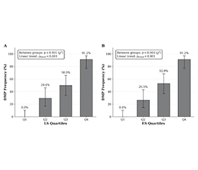
Актуальність. Активація ендотелію, взаємодія лейкоцитів та ендотелію є центральними подіями за діабетичної ретинопатії (ДР) і можуть лежати в основі діабетичної макулопатії (ДМП). Сироваткові маркери стратифікації макулярного ризику вивчені недостатньо. Мета: оцінити сироваткові рівні L-селектину (LS) та E-селектину (ES) як незалежні предиктори несприятливого розвитку й перебігу діабетичної ретинопатії при цукровому діабеті 2-го типу. Матеріали та методи. Базовий перехресний аналіз включав 136 пацієнтів із цукровим діабетом 2-го типу та ДР. ДМП оцінювали як наявну/відсутню (+/–); кінцевими точками оптичної когерентної томографії були центральні товщина (ЦТС) та об’єм сітківки (ЦОС). LS та ES вимірювали за допомогою імуноферментного методу. Лінійна й логістична регресія (на +10 нг/мл) були скориговані за віком, тривалістю діабету, систолічним артеріальним тиском та стадією ДР; дискримінацію порогів оцінювали в ROC-аналізі. Результати. ДМП відмічено в 42,6 % пацієнтів (58/136). LS та ES у групі DMP+ були вищими, ніж у групі DMP– (p < 0,001). Після коригування обидва біомаркери незалежно асоціювалися з вищими ЦТС та ЦОС (p < 0,001) та прогнозованою ДМП (LS: відношення шансів 4,14; ES: відношення шансів 5,92 на кожні 10 нг/мл, p < 0,001). Дискримінація була відмінною (показник AUC становив 0,900 для LS і 0,902 для ES); оптимальними пороговими значеннями були LS ≥ 49,0 нг/мл та ES ≥ 55,9 нг/мл. Для суворо визначеного діабетичного макулярного набряку асоціації послаблювалися після коригування на стадію ДР. Висновки. LS та ES є сильними незалежними предикторами ДМП та гірших структурних результатів оптичної когерентної томографії при ДР і можуть підтримувати прагматичну стратифікацію ризику.
Background. Endothelial activation and leukocyte-endothelium interactions are central to diabetic retinopathy (DR) and may underlie diabetic maculopathy (DMP). Serum markers for macular risk stratification remain limited. Aim of the study was to evaluate serum L- (LS) and E-selectins (ES) as independent predictors of the adverse development and course of diabetic retinopathy in type 2 diabetes mellitus. Materials and methods. Baseline cross-sectional analysis included 136 patients with type 2 diabetes and DR. DMP was assessed as present/absent (+/–); optical coherence tomography endpoints were central retinal thickness (CRT) and volume (CRV). LS and ES were measured by enzyme-linked immunosorbent assay. Linear and logistic regression (per +10 ng/mL) were adjusted for age, diabetes duration, systolic blood pressure, and DR stage; ROC analysis assessed discrimination. Results. DMP was present in 42.6 % of patients (58/136). LS and ES were higher in DMP+ than in DMP– (p < 0.001). After adjustment, both biomarkers were independently associated with higher CRT and CRV (p < 0.001) and predicted DMP (LS: odds ratio 4.14; ES: odds ratio 5.92 per +10 ng/mL; p < 0.001). Discrimination was excellent (AUC 0.900 for LS; 0.902 for ES); optimal cutoffs were LS ≥ 49.0 ng/mL and ES ≥ 55.9 ng/mL. For strictly defined diabetic macular edema, associations attenuated after adjustment for DR stage. Conclusions. LS and ES are strong independent predictors of DMP and worse optical coherence tomography structural outcomes in DR and may support pragmatic risk stratification.
цукровий діабет 2-го типу; діабетична ретинопатія; макулярний набряк; томографія; селектини; L-селектин; E-селектин; біомаркери
type 2 diabetes; diabetic retinopathy; macular edema; tomography; selectins; L-selectin; E-selectin; biomarkers
Для ознакомления с полным содержанием статьи необходимо оформить подписку на журнал.
- Lim JI, Kim SJ, Bailey ST, Kovach JL, Vemulakonda GA, Ying GS, et al.; American Academy of Ophthalmology Preferred Practice Pattern Retina/Vitreous Committee. Diabetic Retinopathy Preferred Practice Pattern®. Ophthalmology. 2025 Apr;132(4):P75-P162. doi: 10.1016/j.ophtha.2024.12.020.
- Amoaku WM, Ghanchi F, Bailey C, Banerjee S, Banerjee S, Downey L, et al. Diabetic retinopathy and diabetic macular oedema pathways and management: UK Consensus Working Group. Eye (Lond). 2020 Jun;34(Suppl 1):1-51. doi: 10.1038/s41433-020-0961-6.
- McAllister M, Ko S, Bawa K, Mearns E, Tabano D, Martinez A, et al. Clinical practice guidelines for the treatment and management of diabetic macular oedema: a systematic review. Eye (Lond). 2025 Dec;39(17):3121-3128. doi: 10.1038/s41433-025-04043-2.
- Szeto SK, Lai TY, Vujosevic S, Sun JK, Sadda SR, Tan G, et al. Optical coherence tomography in the management of diabetic macular oedema. Prog Retin Eye Res. 2024 Jan;98:101220. doi: 10.1016/j.preteyeres.2023.101220.
- Schmidt-Erfurth U, Garcia-Arumi J, Bandello F, Berg K, Chakravarthy U, Gerendas BS, et al. Guidelines for the Management of Diabetic Macular Edema by the European Society of Retina Specialists (EURETINA). Ophthalmologica. 2017;237(4):185-222. doi: 10.1159/000458539.
- Ferrara M, Loda A, Coco G, Grassi P, Cestaro S, Rezzola S, et al. Diabetic Retinopathy: Soluble and Imaging Ocular Biomarkers. J Clin Med. 2023 Jan 24;12(3):912. doi: 10.3390/jcm12030912.
- Wang X, Liu W, Zheng X, Yang MM. New insights of potential biomarkers in diabetic retinopathy: integrated multi-omic analyses. Front Endocrinol (Lausanne). 2025 Jul 1;16:1595207. doi: 10.3389/fendo.2025.1595207.
- Ren J, Zhang S, Pan Y, Jin M, Li J, et al. Diabetic retinopathy: Involved cells, biomarkers, and treatments. Front Pharmacol. 2022 Aug 9;13:953691. doi: 10.3389/fphar.2022.953691.
- Ivetic A, Hoskins Green HL, Hart SJ. L-selectin: A Major Regulator of Leukocyte Adhesion, Migration and Signaling. Front Immunol. 2019 May 14;10:1068. doi: 10.3389/fimmu.2019.01068.
- Kovoor E, Chauhan SK, Hajrasouliha A. Role of inflammatory cells in pathophysiology and management of diabetic retinopathy. Surv Ophthalmol. 2022 Nov-Dec;67(6):1563-1573. doi: 10.1016/j.survophthal.2022.07.008.
- Wang W, Lo ACY. Diabetic Retinopathy: Pathophysiology and Treatments. Int J Mol Sci. 2018 Jun 20;19(6):1816. doi: 10.3390/ijms19061816.
- Srejovic JV, Muric MD, Jakovljevic VL, Srejovic IM, Sreckovic SB, Petrovic NT, et al. Molecular and Cellular Mechanisms Involved in the Pathophysiology of Retinal Vascular Disease — Interplay Between Inflammation and Oxidative Stress. Int J Mol Sci. 2024 Nov 4;25(21):11850. doi: 10.3390/ijms252111850.
- Zhang J, Huang S, Zhu Z, Gatt A, Liu J. E-selectin in vascular pathophysiology. Front Immunol. 2024 Jul 19;15:1401399. doi: 10.3389/fimmu.2024.1401399.
- Rahman I, Collado Sánchez A, Davies J, Rzeniewicz K, Abukscem S, Joachim J, et al. L-selectin regulates human neutrophil transendothelial migration. J Cell Sci. 2021 Feb 8;134(3):jcs250340. doi: 10.1242/jcs.250340.
- Tang Q, Buonfiglio F, Böhm EW, Zhang L, Pfeiffer N, Korb CA, et al. Diabetic Retinopathy: New Treatment Approaches Targeting Redox and Immune Mechanisms. Antioxidants (Basel). 2024 May 12;13(5):594. doi: 10.3390/antiox13050594.
- Siddiqui K, George TP, Mujammami M, Isnani A, Alfadda AA. The association of cell adhesion molecules and selectins (VCAM-1, ICAM-1, E-selectin, L-selectin, and P-selectin) with microvascular complications in patients with type 2 diabetes: A follow-up study. Front Endocrinol (Lausanne). 2023 Feb 9;14:1072288. doi: 10.3389/fendo.2023.1072288.
- Blum A, Pastukh N, Socea D, Jabaly H. Levels of adhesion molecules in peripheral blood correlat with stages of diabetic retino–pathy and may serve as bio markers for microvascular complications. Cytokine. 2018 Jun;106:76-79. doi: 10.1016/j.cyto.2017.10.014.
- Kasza M, Meleg J, Vardai J, Nagy B Jr, Szalai E, Damjanovich J, et al. Plasma E-selectin levels can play a role in the development of diabetic retinopathy. Graefes Arch Clin Exp Ophthalmol. 2017 Jan;255(1):25-30. doi: 10.1007/s00417-016-3411-1.
- Ekelund C, Dereke J, Nilsson C, Landin-Olsson M. Are soluble E-selectin, ICAM-1, and VCAM-1 potential predictors for the development of diabetic retinopathy in young adults, 15-34 years of age? A Swedish prospective cohort study. PLoS One. 2024 Jun 6;19(6):e0304173. doi: 10.1371/journal.pone.0304173.
- World Health Organization. Prevention of blindness from diabetes mellitus: report of a WHO consultation in Geneva, Switzerland, 9-11 November 2005. Geneva: World Health Organization; 2006.
- Harding S, Greenwood R, Aldington S, Gibson J, Owens D, Taylor R, et al.; Diabetic Retinopathy Grading and Disease Management Working Party. Grading and disease management in national screening for diabetic retinopathy in England and Wales. Diabet Med. 2003 Dec;20(12):965-71. doi: 10.1111/j.1464-5491.2003.01077.x.
- Wilkinson CP, Ferris FL 3rd, Klein RE, Lee PP, Agardh CD, Davis M, et al.; Global Diabetic Retinopathy Project Group. Proposed international clinical diabetic retinopathy and diabetic macular edema disease severity scales. Ophthalmology. 2003 Sep;110(9):1677-82. doi: 10.1016/S0161-6420(03)00475-5.
- Kanda Y. Investigation of the freely available easy-to-use software EZR for medical statistics. Bone Marrow Transplant. 2013 Mar;48(3):452-8. doi: 10.1038/bmt.2012.244.
- Joussen AM, Poulaki V, Le ML, Koizumi K, Esser C, Janicki H, et al. A central role for inflammation in the pathogenesis of diabetic retinopathy. FASEB J. 2004 Sep;18(12):1450-2. doi: 10.1096/fj.03-1476fje.
- Kusuhara S, Fukushima Y, Ogura S, Inoue N, Uemura A. Pathophysiology of Diabetic Retinopathy: The Old and the New. Diabetes Metab J. 2018 Oct;42(5):364-376. doi: 10.4093/dmj.2018.0182.
- Yue T, Shi Y, Luo S, Weng J, Wu Y, Zheng X. The role of inflammation in immune system of diabetic retinopathy: Molecular mecha–nisms, pathogenetic role and therapeutic implications. Front Immunol. 2022 Dec 13;13:1055087. doi: 10.3389/fimmu.2022.1055087.
- Forrester JV, Kuffova L, Delibegovic M. The Role of Inflammation in Diabetic Retinopathy. Front Immunol. 2020 Nov 6;11:583687. doi: 10.3389/fimmu.2020.583687.
- Karadayi K, Top C, Gülecek O. The relationship between soluble L-selectin and the development of diabetic retinopathy. Ocul Immunol Inflamm. 2003 Jun;11(2):123-9. doi: 10.1076/ocii.11.2.123.15920.
- Teo ZL, Tham YC, Yu M, Chee ML, Rim TH, Cheung N, et al. Global Prevalence of Diabetic Retinopathy and Projection of Burden through 2045: Systematic Review and Meta-analysis. Ophthalmology. 2021 Nov;128(11):1580-1591. doi: 10.1016/j.ophtha.2021.04.027.
- Im JHB, Jin YP, Chow R, Yan P. Prevalence of diabetic ma–cular edema based on optical coherence tomography in people with diabetes: A systematic review and meta-analysis. Surv Ophthalmol. 2022 Jul-Aug;67(4):1244-1251. doi: 10.1016/j.survophthal.2022.01.009.
- Hui VWK, Szeto SKH, Tang F, Yang D, Chen H, Lai TYY, et al. Optical Coherence Tomography Classification Systems for Diabetic Macular Edema and Their Associations With Visual Outcome and Treatment Responses — An Updated Review. Asia Pac J Ophthalmol (Phila). 2022 Jun 1;11(3):247-257. doi: 10.1097/APO.0000000000000468.
- Tryggestad JB, Shah RD, Braffett BH, Bacha F, Gidding SS, Gubitosi-Klug RA, et al.; TODAY Study Group. Circulating adhesion molecules and associations with HbA1c, hypertension, nephro–pathy, and retinopathy in the Treatment Options for type 2 Diabetes in Adolescent and Youth study. Pediatr Diabetes. 2020 Sep;21(6):923-931. doi: 10.1111/pedi.13062.
- Xu Y, Hou H, Zhao L. The role of VCAM-1 in diabetic retinopathy: A systematic review and meta-analysis. J Diabetes Complications. 2023 Jan;37(1):108380. doi: 10.1016/j.jdiacomp.2022.108380.
- Stratton IM, Kohner EM, Aldington SJ, Turner RC, Holman RR, Manley SE, et al. UKPDS 50: risk factors for incidence and progression of retinopathy in type II diabetes over 6 years from diagnosis. Diabetologia. 2001 Feb;44(2):156-63. doi: 10.1007/s001250051594.
- Li Z, Yuan Y, Qi Q, Wang Q, Feng L. Relationship between dyslipidemia and diabetic retinopathy in patients with type 2 diabetes mellitus: a systematic review and meta-analysis. Syst Rev. 2023 Aug 24;12(1):148. doi: 10.1186/s13643-023-02321-2.
- ACCORD Study Group; ACCORD Eye Study Group; Chew EY, Ambrosius WT, Davis MD, Danis RP, Gangaputra S, Greven CM, Hubbard L, et al. Effects of medical therapies on retinopathy progression in type 2 diabetes. N Engl J Med. 2010 Jul 15;363(3):233-44. doi: 10.1056/NEJMoa1001288.
- Agouti I, Masson E, Loundou A, Jean E, Arnaud L, Abdili E, et al. Plasma levels of E-selectin are associated with retinopathy in sickle cell disease. Eur J Haematol. 2023 Mar;110(3):271-279. doi: 10.1111/ejh.13902.
- Bianco L, Arrigo A, Aragona E, Antropoli A, Berni A, Saladino A, et al. Neuroinflammation and neurodegeneration in diabetic retinopathy. Front Aging Neurosci. 2022 Aug 16;14:937999. doi: 10.3389/fnagi.2022.937999.
- Mimura T, Noma H. Oxidative Stress in Diabetic Retinopathy: A Comprehensive Review of Mechanisms, Biomarkers, and Therapeutic Perspectives. Antioxidants (Basel). 2025 Oct 4;14(10):1204. doi: 10.3390/antiox14101204.