
Архив офтальмологии Украины Том 14, №2, 2026
Вернуться к номеру
Особливості використання оптичної когерентної томографії для діагностики положення інтраокулярної лінзи
Авторы: Скрипник Р.Л., Павленко Р.О., Жабоєдова Н.В., Задорожна А.І.
Національний медичний університет імені О.О. Богомольця, м. Київ, Україна
Рубрики: Офтальмология
Разделы: Клинические исследования
Версия для печати
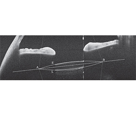
Актуальність. Катаракта залишається частою причиною зниження зору. Сучасна катарактальна хірургія спрямована на отримання високого стабільного рефракційного результату та якості зору. Імплантація інтраокулярних лінз (ІОЛ) потребує точної центрації та їх мінімального нахилу для уникання коматичних аберацій, астигматизму та дисфотопсій. Оптична когерентна томографія (ОКТ) переднього сегмента є високоточним неінвазивним методом оцінки положення ІОЛ. Це обстеження важливе при рефракційних невідповідностях, субʼєктивних скаргах на якість зору, у випадках ризику неспроможності зв’язкового апарату кришталика (псевдоексфоліативний синдром, висока міопія). Мета даного дослідження — підвищити ефективність лікування катаракти шляхом аналізу стабільності положення інтраокулярної лінзи в капсульному мішку за допомогою оптичної когерентної томографії переднього відрізка. Матеріали та методи. Проведено дослідження, що містило дані 31 пацієнта (19 жінок, 12 чоловіків, середній вік 63,0 ± 8,8 року) після неускладненої факоемульсифікації з імплантацією ІОЛ. Проведено офтальмологічне обстеження, що включало візометрію, авторефрактометрію, тонометрію, ОКТ переднього сегмента. На отриманих зображеннях у перпендикулярних осях шляхом апроксимації поверхонь ІОЛ дугами еліпса визначено площину лінзи та її децентрацію відносно зіниці. Загальний кут нахилу ІОЛ розраховано за формулою додавання векторів у двох меридіанах. Результати. Кут нахилу ІОЛ становив в середньому 3,38° (від 1,01 до 5,67°, стандартне відхилення 1,21°). Медіана децентрації відносно рогівкової топографічної осі становила 0,24 мм (95% ДІ 0,17‒0,29 мм). Не виявлено статистично значимої кореляції між значеннями децентрації та нахилу ІОЛ. Максимальна коригована гострота зору (МКГЗ) становила в середньому 0,15 ± 0,07 logMAR. Значення МКГЗ статистично не відрізнялися між двома моделями ІОЛ (p > 0,05) і не залежали від параметрів їхнього просторового позиціонування. Висновки. Досліджені моделі ІОЛ продемонстрували високу стабільність положення в капсульному мішку. Відсутність впливу помірного нахилу (до 5°) та децентрації (до 0,4 мм) на гостроту зору свідчить про значну оптичну толерантність обох монофокальних моделей до незначних анатомічних відхилень. Включення цього виду обстеження в післяопераційні візити пацієнтів може вчасно показати потенційний ризик клінічно значущого зміщення ІОЛ.
Background. Cataract remains a common cause of vision loss. Modern cataract surgery aims to deliver stable, high-quality refractive and visual outcomes. Intraocular lens (IOL) implantation requires precise centration and minimal tilt to avoid comatic aberrations, astigmatism, and dysphotopsias. Anterior segment optical coherence tomography (OCT) is a highly accurate non-invasive method for assessing IOL position. This examination is important in cases of refractive discrepancies, subjective complaints regarding visual quality, and conditions associated with a risk of zonular weakness (pseudoexfoliation syndrome, high myopia). The aim: to improve cataract treatment outcomes by analyzing the stability of IOL position in the capsular bag using anterior segment OCT. Materials and methods. This study included data from 31 patients (19 women, 12 men, mean age 63.0 ± 8.8 years) after uncomplicated phacoemulsification with IOL implantation. Comprehensive ophthalmic examination was conducted, including best corrected visual acuity (BCVA), autorefractometry, tonometry, anterior segment OCT. On the images obtained, in perpendicular axes, the lens plane and its decentration relative to the pupil were determined by approximating IOL surfaces with elliptical arcs. We calculated the total IOL tilt using the formula of adding vectors in two meridians. Results. The IOL tilt was on average 3.38° (from 1.01 to 5.67°, standard deviation 1.21°). The median decentration relative to the corneal topographic axis was 0.24 mm (95% CI 0.17–0.29 mm). No statistically significant correlation was found between the decentration and IOL tilt. BCVA was on average 0.15 ± 0.07 logMAR, its values did not differ statistically between two IOLs models (p > 0.05) and didn’t depend on the parameters of their positioning. Conclusions. The evaluated IOL models demonstrated high stability of the position in the capsular bag. No effect of mild tilt (< 5°) and decentration (< 0.4 mm) on visual acuity indicates considerable optical tolerance of both monofocal models to minor anatomical deviations. Incorporating this examination into postoperative visits of patients may promote timely identification of a potential risk of clinically significant IOL displacement.
катаракта; оптична когерентна томографія; факоемульсифікація; нахил інтраокулярної лінзи; децентрація інтраокулярної лінзи
cataract; optical coherence tomography; phacoemulsification; intraocular lens tilt; intraocular lens decentration
Для ознакомления с полным содержанием статьи необходимо оформить подписку на журнал.
- Катаракта. Адаптована клінічна настанова, заснована на доказах [Internet]. Державний експертний центр Міністерства охорони здоров’я України; 2015. Available from: https://www.dec.gov.ua/wp-content/uploads/2019/11/2016_49_akn_katarakta.pdf.
- Merino PP. Anterior segment topography and aberrations for clinical applications (Doctoral dissertation, Universidad de Valladolid). https://doi.org/10.35376/10324/16299.
- Miháltz K, Vécsei-Marlovits PV. The impact of visual axis position on the optical quality after implantation of multifocal intraocular lenses with different asphericity values. Graefe’s Archive for Clinical and Experimental Ophthalmology. 2021 Jan 20;259(3):673-83. https://doi.org/10.1007/s00417-020-05052-5.
- Burgmüller M, Mihaltz K, Schütze C, Angermann B, Vécsei-Marlovits V. Assessment of long-term intraocular lens (IOL) decentration and tilt in eyes with pseudoexfoliation syndrome (PES) follo–wing cataract surgery. Graefeʼs Archive for Clinical and Experimental Ophthalmology. 2018 Dec;256(12):2361-7. https://doi.org/10.1007/s00417-018-4132-4.
- Wei D, Wei L, Yanjun H, Zequan X, Lei J, Qiang W. Visual quality analysis using the Chinese Catquest-9SF scale follo–wing different spherical aberration IOL implantation. Frontiers in Public Health. 2022 Nov 3;10:1029002. https://doi.org/10.3389/fpubh.2022.1029002.
- Hu X, Zhang J, Zhang Y, Yu W, Yuan H, Qiu X, et al. Tolerance to intraocular lens decentration and tilt in five presbyopia-correcting intraocular lenses: SS-AS-OCT–based study. Journal of Cataract and Refractive Surgery. 2026 Apr 22;52(5):458. https://doi.org/10.1097/j.jcrs.0000000000001850.
- Rodríguez LM, Tielas LS, Sánchez‐Lozano A, García‐Onrubia L, Velarde‐Rodríguez G, Faria‐Ribeiro MÁ, et al. Clinical impact of intraocular lens tilt and decentration on visual outcome in patients undergoing cataract surgery. Acta Ophthalmologica. 2025 Jan;103. https://doi.org/10.1111/aos.17303.
- Wang SY, Stem MS, Oren G, Shtein R, Lichter PR. Patient-centered and visual quality outcomes of premium cataract surgery: a systematic review. European Journal of Ophthalmology. 2017 Jun 26;27(4):387-401. https://doi.org/10.5301/ejo.5000978.
- Apple DJ, Sims J. Harold Ridley and the invention of the intraocular lens. Survey of ophthalmology. 1996 Jan 1;40(4):279-92. https://doi.org/10.1016/S0039-6257(96)82003-0.
- Son Y, Bang SP, Park CY. Basic optics underlying current intraocular lenses. Journal of Clinical Medicine. 2025 Dec 4;14(23):8608. https://doi.org/10.3390/jcm14238608.
- Vacalebre M, Frison R, Corsaro C, Neri F, Santoro A, Conoci S, et al. Current state of the art and next generation of materials for a customized intraocular lens according to a patient-specific eye power. Polymers. 2023 Mar 22;15(6):1590. https://doi.org/10.3390/polym15061590.
- Pateras E, Karioti M. Contrast sensitivity studies and test — a review. Int J Ophthalmol Clin Res. 2020;7(116):1-7. https://doi.org/10.23937/2378-346X/1410116.
- Lundström M, Pesudovs K. Catquest-9SF patient outcomes questionnaire: nine-item short-form Rasch-scaled revision of the Catquest questionnaire. Journal of Cataract & Refractive Surgery. 2009 Mar 1;35(3):504-13. https://doi.org/10.1016/j.jcrs.2008.11.038.
- Lundstrom M, Behndig A, Kugelberg M, Montan P, Stene–vi U, Pesudovs K. The outcome of cataract surgery measured with the Catquest‐9SF. Acta Оphthalmologica. 2011 Dec;89(8):718-23. https://doi.org/10.1111/j.1755-3768.2009.01801.x.
- Cervantes-Coste G, Tapia A, Corredor-Ortega C, Osorio M, Valdez R, Massaro M, et al. The influence of angle alpha, angle kappa, and optical aberrations on visual outcomes after the implantation of a high-addition trifocal IOL. Journal of Clinical Medicine. 2022 Feb 8;11(3):896. https://doi.org/10.3390/jcm11030896.
- Ferreira TB, Berendschot TT, Ribeiro FJ. Clinical outcomes after cataract surgery with a new transitional toric intraocular lens. Journal of Refractive Surgery. 2016 Jul 1;32(7):452-9. https://doi.org/10.3928/1081597X-20160428-07.
- Siatiri H, Mohammadpour M, Gholami A, Ashrafi E, Siatiri N, Mirshahi R. Optical aberrations, accommodation, and visual acuity with a bioanalogic continuous focus intraocular lens after cataract surgery. Journal of Current Ophthalmology. 2017 Dec 1;29(4):274-81. https://doi.org/10.1016/j.joco.2017.06.007.
- Orts P, Piñero DP, Aguilar S, Tañá P. Efficacy of astigmatic correction after femtosecond laser-guided cataract surgery using intraoperative aberrometry in eyes with low-to-moderate levels of corneal astigmatism. International Ophthalmology. 2020 May;40(5):1181-9. https://doi.org/10.1007/s10792-020-01283-x.
- Li L, Wang K, Yan Y, Song X, Liu Z. Research on calculation of the IOL tilt and decentration based on surface fitting. Computational and Mathematical Methods in Medicine. 2013;2013(1):572530. https://doi.org/10.1155/2013/572530. PMID: 23935701; PMCID: PMC3725812.
- Ang RE, Doroy ZA, Yao JA, Cruz EM. Correlation of angle kappa and angle alpha on visual outcomes in eyes implanted with three types of multifocal intraocular lenses. Scientific Reports. 2024 Nov 5;14(1):26732. https://doi.org/10.1038/s41598-024-77699-7.
- Wanten JC, Bauer NJ, Boonstra A, Berendschot TT, Nuijts RM. Association of intraocular lens tilt and decentration with visual acuity using SS-OCT-based analysis. Graefeʼs Archive for Clinical and Experimental Ophthalmology. 2025 Dec;263(12):3423-32. http://doi.org/10.1007/s00417-025-06965-9.
- Langenbucher A, Szentmáry N, Cayless A, Wendelstein J, Hoffmann P. Prediction of IOL decentration, tilt and axial position using anterior segment OCT data. Graefeʼs Archive for Clinical and Experimental Ophthalmology. 2024 Mar;262(3):835-46. https://doi.org/10.1007/s00417-023-06208-9.
- Chen XY, Wang YC, Zhao TY, Wang ZZ, Wang W. Tilt and decentration with various intraocular lenses: a narrative review. World Journal оf Clinical Cases. 2022 Apr 26;10(12):3639. https://doi.org/10.12998/wjcc.v10.i12.3639.
- Lin L, Liang Y, Jiang G, Gan Q, Yang T, Liao P, Liang H. Global, regional, and national burden of cataract: A comprehensive analysis and projections from 1990 to 2021. PLoS One. 2025 Jun 23;20(6):e0326263. doi: 10.1371/journal.pone.0326263. PMID: 40549768; PMCID: PMC12185006.
- Hashemi H, Fayaz F, Hashemi A, Khabazkhoob M. Global prevalence of cataract surgery. Curr Opin Ophthalmol. 2025 Jan 1;36(1):10-17. doi: 10.1097/ICU.0000000000001092. Epub 2024 Sep 25. PMID: 39638415.
- Leffler CT, Spalton D, Schwartz SG, Grzybowski A, Maloney RK. Sir Harold Ridley (1906-2001) and His Cure for Aphakia: New Historical Insights Into the Invention of the Intraocular Lens. Am J Ophthalmol. 2025 May;273:167-175. doi: 10.1016/j.ajo.2025.02.020. Epub 2025 Feb 19. PMID: 39983943.
- Bradley A, Xu R, Wang H, Jaskulski M, Hong X, Brink N, Van Noy S. The Impact of IOL Abbe Number on Polychromatic Image Quality of Pseudophakic Eyes. Clin Ophthalmol. 2020 Aug 11;14:2271-2281. doi: 10.2147/OPTH.S233099. PMID: 32848357; PMCID: PMC7429240.
- Narang R, Agarwal A. Refractive cataract surgery. Curr Opin Ophthalmol. 2024 Jan 1;35(1):23-27. doi: 10.1097/ICU.0000000000001005. Epub 2023 Nov 14. PMID: 37962881.
- Lee NS, Ong K. Factors contributing to long-term refractive error after cataract surgery. Int Ophthalmol. 2023 Jul;43(7):2335-2340. doi: 10.1007/s10792-022-02630-w. Epub 2023 Jan 2. PMID: 36592262.
- Gupta V, Pal H, Sawhney S, Aggarwal A, Vanathi M, Luthra G. Optimization of biometry for best refractive outcome in ca–taract surgery. Indian J Ophthalmol. 2024 Jan 1;72(1):29-43. doi: 10.4103/IJO.IJO_1219_23. Epub 2023 Dec 22. PMID: 38131567; PMCID: PMC10841781.
- Wu KY, Khammar R, Sheikh H, Marchand M. Innovative Polymeric Biomaterials for Intraocular Lenses in Cataract Surgery. J Funct Biomater. 2024 Dec 23;15(12):391. doi: 10.3390/jfb15120391. PMID: 39728191; PMCID: PMC11678174.
- Musat AAM, Tataru CP, Musat GC, Nimigean VR, Preda MA, Musat O. High-Order Aberrations in Cataract Surgery: Current Status and Future Perspectives: A Scoping Review. Medicina (Kaunas). 2026 Mar 10;62(3):512. doi: 10.3390/medicina62030512. PMID: 41901592; PMCID: PMC13027784.
- Fernández-Vigo JI, Serrano González-Peramato MT, Nunila Gómez-de-Liaño C, Sánchez-Guillén I, Fernández-Vigo JÁ, Macarro-Merino A. Glistening on intraocular lenses: A review. Arch Soc Esp Oftalmol (Engl Ed). 2023 Sep;98(9):493-506. doi: 10.1016/j.oftale.2023.06.016. Epub 2023 Jun 25. PMID: 37369324.
- Cho JY, Won YK, Park J, Nam JH, Hong JY, Min S, et al. Visual Outcomes and Optical Quality of Accommodative, Multifocal, Extended Depth-of-Focus, and Monofocal Intraocular Lenses in Presbyopia-Correcting Cataract Surgery: A Systematic Review and Bayesian Network Meta-analysis. JAMA Ophthalmol. 2022 Nov 1;140(11):1045-1053. doi: 10.1001/jamaophthalmol.2022.3667. PMID: 36136323; PMCID: PMC9501783.
- Bai G, Li X, Zhang S, Wang Q, Liu G. Analysis of visual quality after multifocal intraocular lens implantation in post-LASIK cataract patients. Heliyon. 2023 Apr 22;9(5):e15720. doi: 10.1016/j.heliyon.2023.e15720. PMID: 37139295; PMCID: PMC10149396.
- Pedrotti E, Bonacci E, Kilian R, Pagnacco C, Anastasi M, Ventura M, et al. Quality of vision and outcomes after bilateral implantation of pseudo-non diffracting beam IOL. Front Med (Lausanne). 2023 Mar 6;10:1085280. doi: 10.3389/fmed.2023.1085280. PMID: 36950509; PMCID: PMC10025297.
- Oli A, Yadav A, Babu JG, Balakrishnan D. Scheimpflug ima–ging for evaluation of intraocular lens position in modified flanged scleral fixated intraocular lens. Ther Adv Ophthalmol. 2023 Jan 12;15:25158414221147208. doi: 10.1177/25158414221147208. PMID: 36654884; PMCID: PMC9841840.
- Van Vught L, Dekker CE, Stoel BC, Luyten GPM, Beenak–ker JM. Evaluation of intraocular lens position and retinal shape in negative dysphotopsia using high-resolution magnetic resonance ima–ging. J Cataract Refract Surg. 2021 Aug 1;47(8):1032-1038. doi: 10.1097/j.jcrs.0000000000000576. PMID: 33577270.
- João MD, Costa JV, Monteiro T, Franqueira N, Faria Correia F, Vaz F. Intraocular Lens Position and Anterior Chamber Parameters Evaluation After Nd:YAG Laser Posterior Capsulotomy for Posterior Capsular Opacification Using Anterior Segment Swept-Source Optical Coherence Tomography. Clin Ophthalmol. 2022 Jan 18;16:153-159. doi: 10.2147/OPTH.S349649. PMID: 35082482; PMCID: PMC8785224.
- Na KS, An D, Kim HS, Kim EC. Relation between change of effective lens position and toric IOL rotation after toric IOL implantation. Can J Ophthalmol. 2025 Feb;60(1):8-14. doi: 10.1016/j.jcjo.2024.05.011. Epub 2024 Jun 15. PMID: 38889881.
- Kang C, Lee MJ, Chomsky A, Oetting TA, Greenberg PB. Risk factors for complications in resident-performed cataract surgery: A systematic review. Surv Ophthalmol. 2024 Jul-Aug;69(4):638-645. doi: 10.1016/j.survophthal.2024.04.002. Epub 2024 Apr 20. PMID: 38648911.
- Martinez DL, Ahmed IIK, Schlenker MB. Risk communication in cataract surgery. BMJ Open Ophthalmol. 2024 May 9;9(1):e001613. doi: 10.1136/bmjophth-2023-001613. PMID: 38729627; PMCID: PMC11086490.
- Yong GY, Mohamed-Noor J, Salowi MA, Adnan TH, Zaha–ri M. Risk factors affecting cataract surgery outcome: The Malaysian cataract surgery registry. PLoS One. 2022 Sep 21;17(9):e0274939. doi: 10.1371/journal.pone.0274939. PMID: 36129906; PMCID: PMC9491522.