
Архив офтальмологии Украины Том 14, №2, 2026
Вернуться к номеру
Більше ніж амбліопія: рутинна периметрія як ключ до правильного менеджменту пацієнта при прихованій демієлінізації
Авторы: Вітовська О.П. (1, 2), Саргош В.В. (1, 2)
(1) - Німецька очна клініка (Deutsche Augenklinik), м. Київ, Україна
(2) - Національний медичний університет імені О.О. Богомольця, м. Київ, Україна
Рубрики: Офтальмология
Разделы: Справочник специалиста
Версия для печати
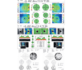
Актуальність. Затримка в діагностиці розсіяного склерозу залишається значною проблемою, попри доступність нейровізуалізації та оновлення діагностичних критеріїв. Субклінічний або атиповий дебют захворювання без гострого оптичного невриту може призводити до помилкового трактування зорових порушень як функціональної патології, зокрема амбліопії. Мета: продемонструвати на прикладі клінічного випадку необхідність статичної периметрії у диференціальній діагностиці між рефракційною амбліопією та ранніми проявами органічного ураження зорових шляхів при розсіяному склерозі за відсутності структурних змін сітківки та зорового нерва на оптичній когерентній томографії. Матеріали та методи. Описаний клінічний випадок пацієнтки П., 35 років, яка звернулась з метою проведення ексимерлазерної корекції зору, але у зв’язку з невідповідністю даних стандартного огляду та суб’єктивних скарг до діагностичного алгоритму було залучено додаткові функціональні та структурні методи дослідження (комп’ютерна статична периметрія, оптична когерентна томографія, електроретинографія та магнітно-резонансна томографія головного мозку з контрастуванням). Результати. Наведений клінічний випадок спонукає до індивідуалізованого підходу до обстеження пацієнтів із нетиповими скаргами або невідповідністю між клінічною картиною та результатами стандартного офтальмологічного огляду. Висновки. Комплексне оцінювання результатів стандартного офтальмологічного обстеження, анамнезу, функціональних (статична периметрія) і структурних (оптична когерентна томографія) методів дослідження у пацієнтів із нетиповим симптомокомплексом є ключовим для своєчасної діагностики латентного органічного ураження та запобігання помилковим оперативним втручанням.
Background. Delayed diagnosis of multiple sclerosis remains a significant challenge despite the accessibility of neuroimaging and updated diagnostic criteria. Subclinical or atypical disease onset without acute optic neuritis can lead to the misinterpretation of visual impairments as functional conditions, particularly amblyopia. Objective: using a clinical case report, to demonstrate the necessity of automated static perimetry in the differential diagnosis between refractive amblyopia and early organic visual pathway impairment in multiple sclerosis, even in the absence of structural retinal and optic nerve changes on optical coherence tomography. Materials and methods. We report a clinical case of a 35-year-old female patient P. who presented for excimer laser vision correction. But, due to the discrepancy between routine ophthalmic examination findings and subjective complaints, additional functional and structural diagnostic modalities — including automated static perimetry, optical coherence tomography, electroretinography, and contrast-enhanced brain magnetic resonance imaging — were added to the diagnostic algorithm. Results. This clinical case highlights the need for an individualized diagnostic approach in patients presenting with atypical symptoms or a discrepancy between clinical picture and routine ophthalmic examination findings. Conclusion. Comprehensive evaluation of routine ophthalmic examination data, medical history, functional (static perimetry) and structural (optical coherence tomography) diagnostic modalities in patients with an atypical symptom complex is crucial for the timely diagnosis of latent organic lesions and the prevention of inappropriate surgical interventions.
розсіяний склероз; комп’ютерна статична периметрія; оптична когерентна томографія; рефракційна хірургія; амбліопія; диференційна діагностика
multiple sclerosis; automated static perimetry; optical coherence tomography; refractive surgery; amblyopia; differential diagnosis
Для ознакомления с полным содержанием статьи необходимо оформить подписку на журнал.
- Walton C, King R, Rechtman L, Kaye W, Leray E, Marrie RA, et al. Rising prevalence of multiple sclerosis worldwide: Insights from the Atlas of MS, third edition. Mult Scler. 2020 Dec;26(14):1816-1821. doi: 10.1177/1352458520970841. Epub 2020 Nov 11. PMID: 33174475; PMCID: PMC7720355.
- Montague T, Drummond J, Ng K, Parratt J. Advancements in multiple sclerosis. Intern Med J. 2025 Jun;55(6):895-904. doi: 10.1111/imj.70023. Epub 2025 Apr 2. PMID: 40171883; PMCID: PMC12155068.
- Portaccio E, Magyari M, Havrdova EK, Ruet A, Brochet B, Scalfari A, et al. Multiple sclerosis: emerging epidemiological trends and redefining the clinical course. Lancet Reg Health Eur. 2024 Aug 22;44:100977. doi: 10.1016/j.lanepe.2024.100977. PMID: 39444703; PMCID: PMC11496978.
- Marrie RA, Rudick R, Horwitz R, Cutter G, Tyry T, Campagnolo D, Vollmer T. Vascular comorbidity is associated with more rapid disability progression in multiple sclerosis. Neurology. 2010 Mar 30;74(13):1041-7. doi: 10.1212/WNL.0b013e3181d6b125. PMID: 20350978; PMCID: PMC2848107.
- Thompson AJ, Banwell BL, Barkhof F, Carroll WM, Coetzee T, Comi G, et al. Diagnosis of multiple sclerosis: 2017 revisions of the McDonald criteria. Lancet Neurol. 2018 Feb;17(2):162-173. doi: 10.1016/S1474-4422(17)30470-2. Epub 2017 Dec 21. PMID: 29275977.
- Optic Neuritis Study Group. Multiple sclerosis risk after optic neuritis: final optic neuritis treatment trial follow-up. Arch Neurol. 2008 Jun;65(6):727-32. doi: 10.1001/archneur.65.6.727. PMID: 18541792; PMCID: PMC2440583.
- Sørensen TL, Frederiksen JL, Brønnum-Hansen H, Peter–sen HC. Optic neuritis as onset manifestation of multiple sclerosis: a nationwide, long-term survey. Neurology. 1999 Aug 11;53(3):473-8. doi: 10.1212/wnl.53.3.473. PMID: 10449106.
- Toosy AT, Mason DF, Miller DH. Optic neuritis. Lancet Neurol. 2014 Jan;13(1):83-99. doi: 10.1016/S1474-4422(13)70259-X. PMID: 24331795.
- Barin L, Kamm CP, Salmen A, Dressel H, Calabrese P, Pot C, et al.; Swiss Multiple Sclerosis Registry. How do patients enter the healthcare system after the first onset of multiple sclerosis symptoms? The in–fluence of setting and physician specialty on speed of diagnosis. Mult Scler. 2020 Apr;26(4):489-500. doi: 10.1177/1352458518823955. Epub 2019 Jan 18. PMID: 31456464; PMCID: PMC7140343.
- Kaufmann M, Kuhle J, Puhan MA, Kamm CP, Chan A, Salmen A, et al.; Swiss Multiple Sclerosis Registry (SMSR). Factors associated with time from first-symptoms to diagnosis and treatment initiation of Multiple Sclerosis in Switzerland. Mult Scler J Exp Transl Clin. 2018 Dec 6;4(4):2055217318814562. doi: 10.1177/2055217318814562. PMID: 30559972; PMCID: PMC6293378.
- Giovannoni G, Butzkueven H, Dhib-Jalbut S, Hobart J, Kobelt G, Pepper G, et al. Brain health: time matters in multiple sclerosis. Mult Scler Relat Disord. 2016 Sep;9 Suppl 1:S5-S48. doi: 10.1016/j.msard.2016.07.003. Epub 2016 Jul 7. PMID: 27640924.
- Petzold A, Balcer LJ, Calabresi PA, Costello F, Frohman TC, Green AJ, et al.; International Multiple Sclerosis Visual System Consortium. Retinal optical coherence tomography in multiple sclerosis: management and clinical applications. Lancet Neurol. 2017 Sep;16(9):720-733.
- Alcubierre R, Sánchez-Dalmau BF, Muñoz S. Multiple sclerosis in ophthalmology: Beyond optic neuritis. Med Clin. 2020;155:70-6.
- Saidha S, Green AJ, Leocani L, Vidal-Jordana A, Kenney RC, Bsteh G, et al. The use of optical coherence tomography and visual evoked potentials in the 2024 McDonald diagnostic criteria for multiple sclerosis. Lancet Neurol. 2025 Oct;24(10):880-892. doi: 10.1016/S1474-4422(25)00275-3. Erratum in: Lancet Neurol. 2025 Nov;24(11):e13. doi: 10.1016/S1474-4422(25)00349-7. PMID: 40975103.
- Vidović T, Cerovski B, Vidović DH, Cerovski J, Novak-Laus K. Inapparent visual field defects in multiple sclerosis patients. Coll Antropol. 2005;29 Suppl 1:67-73. PMID: 16193681.
- Filippi M, Preziosa P, Banwell BL, Barkhof F, Ciccarelli O, De Stefano N, et al. Assessment of lesions on magnetic resonance imaging in multiple sclerosis: practical guidelines. Brain. 2019 Jul 1;142(7):1858-1875. doi: 10.1093/brain/awz144. PMID: 31209474; PMCID: PMC6598631.
- Gill S, Agarwal M. Multiple Sclerosis Part 1: Essentials and the McDonald Criteria. Magn Reson Imaging Clin N Am. 2024 May;32(2):207-220. doi: 10.1016/j.mric.2023.11.002. Epub 2023 Dec 9. PMID: 38555137.
- Schallhorn JM, Schallhorn SC, Hettinger KA, Venter JA, Pelouskova M, Teenan D, et al. Outcomes and complications of excimer laser surgery in patients with collagen vascular and other immune-mediated inflammatory diseases. J Cataract Refract Surg. 2016 Dec;42(12):1742-1752. doi: 10.1016/j.jcrs.2016.09.018. PMID: 28007105.
- Saad A, Narr J, Frings A, Steinberg J, Katz T, Linke SJ. Surgical outcomes of laser in situ keratomileusis (LASIK) in patients with stable systemic disease. Int Ophthalmol. 2024 Feb 29;44(1):119. doi: 10.1007/s10792-024-02956-7. PMID: 38418603; PMCID: PMC10902021.
- Namdev V, Kaur M, Sharma VK, Mulay A, Raj R, Titiyal JS. Current paradigms in refractive surgery. Med J Armed Forces India. 2024 Sep-Oct;80(5):497-504. doi: 10.1016/j.mjafi.2024.08.003. Epub 2024 Aug 26. PMID: 39309588; PMCID: PMC11411312.
- Williams C. Amblyopia. BMJ Clin Evid. 2009 Sep 16;2009:0709. PMID: 21726480; PMCID: PMC2907781.
- Keltner JL, Johnson CA, Cello KE, Dontchev M, Gal RL, Beck RW; Optic Neuritis Study Group. Visual field profile of optic neuritis: a final follow-up report from the optic neuritis treatment trial from baseline through 15 years. Arch Ophthalmol. 2010 Mar;128(3):330-7. doi: 10.1001/archophthalmol.2010.16. PMID: 20212204; PMCID: PMC4107874.
- Cruz OA, Repka MX, Hercinovic A, Cotter SA, Lambert SR, Hutchinson AK, et al.; American Academy of Ophthalmology Preferred Practice Pattern Pediatric Ophthalmology/Strabismus Panel. Amblyopia Preferred Practice Pattern. Ophthalmology. 2023 Mar;130(3):P136-P178. doi: 10.1016/j.ophtha.2022.11.003. Epub 2022 Dec 14. PMID: 36526450; PMCID: PMC10701408.
- Kadhum A, Tan ETC, Wenner Y, Joosse MV, Loudon SE. Effectiveness of optical treatment in amblyopia and validation of measuring spectacle compliance with the ODM. Ophthalmic Physiol Opt. 2024 Jul;44(5):945-953. doi: 10.1111/opo.13322. Epub 2024 May 17. PMID: 38757545.
- Donaldson L, Margolin E. Visual fields and optical cohe–rence tomography (OCT) in neuro-ophthalmology: Structure-function correlation. J Neurol Sci. 2021 Oct 15;429:118064. doi: 10.1016/j.jns.2021.118064. Epub 2021 Sep 1. PMID: 34488042.
- Filippatou A, Theodorou A, Stefanou MI, Tzanetakos D, Kitsos D, Moschovos C, et al. Optical coherence tomography and angio–graphy in multiple sclerosis: A systematic review and meta-analysis. J Neurol Sci. 2025 Mar 15;470:123422. doi: 10.1016/j.jns.2025.123422. Epub 2025 Feb 11. PMID: 39954575.
- Monsalve B, Ferreras A, Calvo P, Urcola JA, Figus M, Monsal–ve J, et al. Diagnostic ability of Humphrey perimetry, Octopus perimetry, and optical coherence tomography for glaucomatous optic neuropathy. Eye (Lond). 2017 Mar;31(3):443-451. doi: 10.1038/eye.2016.251. Epub 2016 Nov 11. PMID: 27834960; PMCID: PMC5350372.
- Klineova S, Kupersmith M. Promising recovery biomarkers after first event acute demyelinating optic neuritis. Mult Scler Relat Disord. 2020 Oct;45:102400. doi: 10.1016/j.msard.2020.102400. Epub 2020 Jul 15. PMID: 32702643.