
Архів офтальмології та щелепно-лицевої хірургії України Том 3, №1, 2026
Вернуться к номеру
Вплив методики видалення нижнього третього моляра на стан післяекстракційної рани
Авторы: Аветіков Г.Д., Іваницька О.С., Карасюнок А.Є., Гаврильєв В.М., Скікевич М.Г., Локес К.П.
Полтавський державний медичний університет, м. Полтава, Україна
Рубрики: Хирургия, Офтальмология
Разделы: Клинические исследования
Версия для печати
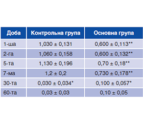
Актуальність. Cучасні тенденції розвитку хірургічної стоматології спрямовані на мінімізацію операційної травми, скорочення тривалості втручання, зниження ризику ушкодження судин і нервів для оптимізації процесів репаративної регенерації післяопераційної рани та профілактики ускладнень при проведенні операції видалення мезіально нахилених нижніх третіх молярів. Мета: проаналізувати вплив використання протоколу, доповненого хірургічною методикою, що базувалася на біомеханічно обґрунтованому положенні інструментів і раціональному формуванні операційного доступу, на клінічні ознаки загоєння лунки видаленого нижнього третього моляра. Матеріали та методи. Проведений аналіз стану лунки видаленого зуба в пацієнтів з мезіально нахиленими нижніми третіми молярами, видалення яких проведено за стандартною та авторською методиками. Результати. На 1-шу добу післяопераційного періоду середній показник стану післяекстракційної рани в пацієнтів 2-ї (основної) клінічної групи був статистично значуще вдвічі меншим, ніж у представників 1-ї (контрольної) групи. На 2-гу добу післяопераційного періоду істотних змін даного показника не відзначали, зберігалася статистично значуща різниця. Середній показник в обох групах збільшувався на 5-ту добу спостереження, проте ці зміни не були статистично значущими. На 7-му добу також зафіксовано незначне збільшення зазначеного показника, що відповідало даним попередніх термінів спостереження. На 30-ту добу зафіксоване істотне вірогідне зменшення цього показника в обох групах з досягненням сталих значень на 60-ту добу. На даному етапі спостереження не було статистично значущої різниці між показниками основної та контрольної групи. Висновки. Застосування протоколу, доповненого хірургічною методикою, що базується на біомеханічно обґрунтованому положенні інструментів і раціональному формуванні операційного доступу при екстракції мезіально нахилених нижніх третіх молярів, приводить до статистично значущого поліпшення стану післяекстракційної рани в ранні терміни репаративної регенерації за відсутності змін на 30-ту добу спостереження.
Background. Current trends in the development of surgical dentistry are aimed at minimizing surgical trauma, reducing the duration of the intervention and the risk of vascular and nerve damage in order to optimize the processes of reparative regeneration of the postoperative wound and prevent complications during the operation to remove mesially inclined lower third molars. Purpose was to analyze the effect of using a protocol with the addition of a surgical technique based on a biomechanically justified position of the instruments and rational formation of surgical access on the clinical signs of healing of the socket of the removed lower third molar. Materials and methods. An analysis of the state of the socket of the extracted tooth was conducted in patients with mesial inclination of lower third molars, whose removal was performed using standard and authors’ methods. Results. On the 1st postoperative day, the difference in the average indicators of the state of the post-extraction wound in patients of the 2nd (main) clinical group was statistically significantly half as small as in representatives of the 1st (control) group. On the 2nd day of the postoperative period, no significant changes in this indicator were noted, and a statistically significant difference remained. The average indicator in both groups increased on the 5th day of observation, but these changes were not statistically significant. On the 7th day, a slight increase in the mentioned parameter was also recorded, which corresponded to the data of the previous observation periods. On the 30th day, a significant and probable decrease in this indicator was recorded in both groups, with stable values reached on the 60th day. At this stage of observation, there was no statistically significant difference between parameters of the main and control groups. Conclusions. The use of the protocol with the addition of a surgical technique based on a biomechanically justified position of the instruments and rational formation of the surgical access during the extraction of mesially inclined lower third molars leads to a statistically significant improvement in the condition of the post-extraction wound in the early stages of reparative regeneration with no changes on the 30th day of observation.
нижній третій моляр; ретенція зуба; видалення зуба; післяопераційні ускладнення
mandibular third molar; tooth impaction; tooth extraction; postoperative complications
Для ознакомления с полным содержанием статьи необходимо оформить подписку на журнал.
- Faustova MO, Ananieva MM, Basarab YO, Dobrobolska OV, Vovk IM, Loban' GA. Bacterial factors of cariogenicity (literature review). Wiadomosci lekarskie. 2018;71(2 pt 2):378-382.
- Sifuentes-Cervantes JS, Carrillo-Morales F, Castro-Núñez J, Cunningham LL, Van Sickels JE. Third molar surgery: Past, present, and the future. Oral Surg Oral Med Oral Pathol Oral Radiol. 2021 Nov;132(5):523-531. doi: 10.1016/j.oooo.2021.03.004.
- Puciło M, Puciło A, Safranow K, Nowicka A. The influence of age, sex, and tooth type on the anatomical relationship between tooth roots and the mandibular canal. Imaging Sci Dent. 2021 Dec;51(4):373-382. doi: 10.5624/isd.20210031.
- Safi Y, Moshfeghi M, Ahsaie MG, Zameni M, Sahafi SA. Relationship between Impacted Mandibular Third Molars and the Mandibular Canal on CBCT Scans. J Long Term Eff Med Implants. 2024;34(3):65-74. doi: 10.1615/JLongTermEffMedImplants.2022042569.
- Yacoub S, Dammak N, Zaalouni S, Hrizi MA, Ben Khelifa M. Prevalence of impacted third molars: a radiographic study among a North African population. Tunis Med. 2024 Jan 5;102(1):32-37. doi: 10.62438/tunismed.v102i1.4461.
- Themkumkwun S, Sawatdeenarunat S, Manosuthi P. Surgical removal of third molars in a young adult: review of indications and surgical techniques. J Korean Assoc Oral Maxillofac Surg. 2023 Aug 31;49(4):184-191. doi: 10.5125/jkaoms.2023.49.4.184.
- Lokes K, Karasiunok A, Smaglyuk L, Voloshyna L, Voronko–va H, Rezvina K. Tooth extraction operation as a component of orthodontical treatment. Ukrainian Dental Almanac. 2024;2:32-35. doi: 10.31718/2409-0255.2.2024.06.
- Mendoza AM, Rodenas YJ, Eidson BK. Improved Wisdom Tooth Diagnosis Can Reduce Dental Disease and Nonbattle Injuries. Mil Med. 2023 Nov 3;188(11-12):e3416-e3422. doi: 10.1093/milmed/usad145.
- Kang FW, Yuan XR, Li GC, Yang YFZ, Zhang XM, Hou GY. Coronectomy in Lower Third Molar Surgery: A Systematic Review and Meta-Analysis. J Oral Maxillofac Surg. 2025 May;83(5):601-615. doi: 10.1016/j.joms.2025.01.014.
- Takadoum S, Douilly G, de Boutray M, Kabani S, Maladiè–re E, Demattei C, Lapeyrie P. Sutureless socket technique after remo–val of third molars: a multicentric, open, randomized controlled trial. BMC Oral Health. 2022 Jun 26;22(1):256. doi: 10.1186/s12903-022-02287-y.
- Faustova MO, Chumak YuV, Lokes KP, Lobanʼ GA, Steblovskyi DV, Voloshyna LІ, Avetikov DS. Decamethoxine redu–ces the adhesive properties of streptococci isolated from patients with dry socket. Kharkiv Dental Journal. 2025;2(4(6)):594-605. doi: 10.26565/3083-5607-2025-6-06.
- Picoli FF, Fontenele RC, Van der Cruyssen F, Ahmadzai I, Trigeminal Nerve Injuries Research Group, Politis C, Silva MAG, Jacobs R. Risk assessment of inferior alveolar nerve injury after wisdom tooth removal using 3D AI-driven models: A within-patient study. J Dent. 2023 Dec;139:104765. doi: 10.1016/j.jdent.2023.104765.
- Kiencało A, Jamka-Kasprzyk M, Panaś M, Wyszyńska-Pawelec G. Analysis of complications after the removal of 339 third molars. Dent Med Probl. 2021 Jan-Mar;58(1):75-80. doi: 10.17219/dmp/127028.
- Alhyari R, Boggon JN, Anderson H, Mclaughlin BA, Khalaf K, Lalli A. Cone-Beam Computed Tomography in Wisdom Tooth Surgery Can Aid Informed Decision-Making and Reduce Presurgical Anxiety. J Oral Maxillofac Res. 2025 Sep 30;16(3):e3. doi: 10.5037/jomr.2025.16303.
- Leung YY, Hung KF, Li DTS, Yeung AWK. Application of Cone Beam Computed Tomography in Risk Assessment of Lower Third Molar Surgery. Diagnostics (Basel). 2023 Mar 1;13(5):919. doi: 10.3390/diagnostics13050919.
- Stefanescu A, Sufaru IG, Chiscop I, Lupu FC, Martu C, Oprisan B, Earar K. Clinical and Molecular Impact of Advanced Platelet-Rich Fibrin on Pain, Swelling, and Distal Periodontal Status of Mandibular Second Molars After Mandibular Third-Molar Extraction. Medicina (Kaunas). 2024 Dec 14;60(12):2062. doi: 10.3390/medicina60122062.
- Khoswanto C, Dewi IK. The role of rhBMP-2 in mandi–bular bone regeneration following tooth extraction through HIF-1α and VEGF-A expression: An Immunohistochemical study. J Oral Biol Craniofac Res. 2025 Mar-Apr;15(2):359-364. doi: 10.1016/j.jobcr.2025.02.001.
- Kong J, Li L, Gao F, Zhang K, Xie J, Cui J. Stromal cell-derived factor-1 regulates expression of vascular endothelial growth factor and osteopontin after tooth extraction. J Periodontol. 2025 Dec;96(12):1391-1399. doi: 10.1002/JPER.24-0785.
- Yin Y, Shuai F, Liu X, Zhao Y, Han X, Zhao H. Biomaterials and therapeutic strategies designed for tooth extraction socket hea–ling. Biomaterials. 2025 May;316:122975. doi: 10.1016/j.biomaterials.2024.122975.
- Diniz JA, Dourado ACAG, Barbirato DDS, da Silveira KG, Vasconcellos RJH, Laureano Filho JR. Effect of simvastatin topical use on alveolar bone after tooth extraction: a scoping review. Clin Oral Investig. 2024 Jan 10;28(1):86. doi: 10.1007/s00784-023-05482-5.
- Park G, Jalkh EB, Boczar D, Bergamo ET, Kim H, Kurgan–sky G, et al. Bone regeneration at extraction sockets filled with leukocyte-platelet-rich fibrin: An experimental pre-clinical study. Med Oral Patol Oral Cir Bucal. 2022 Sep 1;27(5):e468-e475. doi: 10.4317/medoral.25462.