
Архів офтальмології та щелепно-лицевої хірургії України Том 3, №1, 2026
Вернуться к номеру
Комплексна оцінка кісткової тканини альвеолярного відростка за допомогою текстурних біомаркерів: систематичний огляд
Авторы: Буднікова А.В., Шпачинський О.С., Копчак А.В.
Національний медичний університет імені О.О. Богомольця, м. Київ, Україна
Рубрики: Хирургия, Офтальмология
Разделы: Клинические исследования
Версия для печати
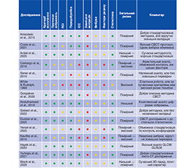
Актуальність. Якість кісткової тканини (КТ) є одним із ключових чинників успішності дентальної імплантації, кісткової пластики та реконструктивних втручань. Традиційна оцінка за даними конусно-променевої комп’ютерної томографії і цифрової рентгенографії не завжди відображає мікроархітектуру трабекулярної кістки. Радіотекстурний аналіз забезпечує кількісну оцінку структурної організації кістки та може доповнювати традиційні методи діагностики. Мета: узагальнити сучасні клінічні докази щодо застосування радіотекстурного аналізу для кількісного аналізу медичних зображень кісткової тканини альвеолярного відростка, визначити діагностичну та прогностичну цінність текстурних параметрів і проаналізувати методологічну якість досліджень. Матеріали та методи. Систематичний огляд виконано відповідно до рекомендацій PRISMA. Пошук проведено у PubMed, Scopus, Web of Science та Google Scholar. До аналізу включено клінічні дослідження, у яких застосовували текстурний аналіз даних конусно-променевої комп’ютерної томографії або цифрової рентгенографії. Методологічну якість оцінювали за JBI Critical Appraisal Checklist. Через значну гетерогенність досліджень метааналіз не проводився. Результати. До якісного синтезу включено 15 клінічних досліджень. Основними параметрами дослідження були entropy, fractal dimension, GLCM (Gray-Level Co-occurrence Matrix) та GLRLM (Gray-Level Run Length Matrix). Ці показники продемонстрували перспективність для оцінки мікроархітектури кістки, ремоделювання, прогнозування первинної стабільності імплантатів і виявлення пародонтальних та остеопоротичних змін. Основними обмеженнями були неоднорідність протоколів, ROI, алгоритмів обробки та відсутність зовнішньої валідації. Висновки. Радіотекстурний аналіз є перспективним допоміжним методом кількісної оцінки альвеолярної кісткової тканини, однак його широке клінічне застосування потребує стандартизації методології та проведення багатоцентрових проспективних досліджень.
Background. Alveolar bone quality is a key determinant of successful dental implant therapy, bone grafting and reconstructive procedures. Conventional assessment using cone beam computed tomography and digital radiography does not always reflect the microarchitecture of trabecular bone. Radiotexture analysis enables quantitative assessment of bone structure beyond conventional diagnostic methods. Objective: to systematize current clinical evidence regarding the use of alveolar bone images, determine the diagnostic and prognostic value of texture features, and to analyze the methodological quality of studies. Materials and methods. A PRISMA-based systematic review was performed using PubMed, Scopus, Web of Science and Google Scholar. The analysis included clinical studies that used texture analysis of cone beam computed tomography or digital radiography data. Methodological quality was assessed with the JBI Critical Appraisal Checklist. Meta-analysis was not performed because of substantial heterogeneity. Results. Fifteen clinical studies were included in the qualitative synthesis. Entropy, fractal dimension, gray-level co-occurrence matrix (GLCM) and gray-level run length matrix (GLRLM) were the main investigated parameters. These indicators showed potential for evaluating bone microarchitecture, remodeling, predicting primary implant stability and identifying periodontal and osteoporotic changes. The main limitations were heterogeneity of protocols, regions of interest, processing algorithms, and lack of external validation. Conclusions. Radiotexture analysis represents a promising adjunctive tool for quantitative alveolar bone assessment; however, its widespread clinical application requires standardization of methodology and multicenter prospective studies.
радіотекстурний аналіз; альвеолярна кістка; КПКТ; текстурний аналіз; дентальна імплантація; GLCM; GLRLM; фрактальна розмірність
radiotexture analysis; alveolar bone; cone beam computed tomography; texture analysis; dental implant therapy; GLCM; GLRLM; fractal dimension
Для ознакомления с полным содержанием статьи необходимо оформить подписку на журнал.
- Kołaciński M, Kozakiewicz M, Materka A. Textural entropy as a potential feature for quantitative assessment of jaw bone healing process. Arch Med Sci. 2015;11(1):78-84.
- Zheng K, Makrogiannis S. Bone texture characterization for osteoporosis diagnosis using digital radiography. In: Proc. 38th IEEE Eng. Med. Biol. Soc. (EMBC) 2016. Р. 1034-1037.
- Colaco LSL, Chhaparwal Y, Patil V, et al. Decoding dental images: a comprehensive review of fractal analysis. BDJ Open. 2025;11:74.
- Borowska M, Antonowicz B, Magnuszewska E, Woźniak Ł, Łukaszuk K, Borys J. Texture components of the radiogra–phic ima–ge assist in the detection of periapical periodontitis. Appl Sci. 2025;15(19):10521.
- Costa ALF, de Souza Carreira B, Fardim KAC, Nussi AD, da Silva Lima VC, Miguel MMV, et al. Texture analysis of cone beam computed tomography images reveals dental implant stability. Int J Oral Maxillofac Surg. 2021;50(12):1609-1616.
- Camargo AJ, Côrtes ARG, Aoki EM, Baladi MG, Arita ES, Watanabe PCA. Analysis of bone quality on panoramic radiograph in osteoporosis research by fractal dimension. Appl Math. 2016;7(4):375-386.
- Wach T, Kozakiewicz M, Michcik A, et al. Three-dimensional analysis of facial skeleton textures in CBCT as an early warning sign of osteoporosis — A pilot study. Diagnostics (Basel). 2026;16(8):1217.
- Sener E, Cinarcik S, Baksi BG. Use of fractal analysis for the discrimination of trabecular changes between individuals with healthy gingiva or moderate periodontitis. J Periodontol. 2015;86(12):1364-1369.
- White SC, Rudolph DJ. Alterations of the trabecular pattern of the jaws in patients with osteoporosis. Oral Surg Oral Med Oral Pathol Oral Radiol Endod. 1999;88(5):628-635.
- Kato CNAO, Barra SG, Tavares NPK, Amaral TMP, Brasileiro CB, Mesquita RA, et al. Use of fractal analysis in dental images: a systematic review. Dentomaxillofac Radiol. 2020;49(2):20180457.
- Haralick RM, Shanmugam K, Dinstein I. Textural features for image classification. IEEE Trans Syst Man Cybern. 1973;SMC-3(6):610-621.
- Galloway MM. Texture analysis using gray level run lengths. Computer Graphics Image Process. 1975;4(2):172-179.
- Kış HC, Güleryüz Gürbulak A. Evaluation of the peri-implant bone trabecular microstructure changes in short implants with fractal analysis. Int J Implant Dent. 2020;6(1):13.
- Gonçalves BC, de Araújo EC, Nussi AD, Bechara N, Sarmento D, Oliveira MS, et al. Texture analysis of cone-beam computed tomography images assists the detection of furcal lesion. J Periodontol. 2020;91(9):1159-1166.
- Keles M, Yuksel IB. Fractal analysis of bone architecture changes following transcrestal sinus lifting with simultaneous implant placement. BMC Oral Health. 2026;26(1):1425.
- Zeytinoğlu M, İlhan B, Dündar N, Boyacioğlu H. Fractal analysis for the assessment of trabecular peri-implant alveolar bone using panoramic radiographs. Clin Oral Investig. 2015;19(2):519-524.
- Sailasri K, Ganji SGMD, Satwalekar P, Nalla S, Basany RB, Krishna SS. Reliability of using texture analysis of periapical radiographs obtained using radiovisiography for assessing bone quality in dental implant planning: a cross-sectional study. Cureus. 2022;14(12):e32860.
- Abdulhameed EA, Al-Rawi NH, Uthman AT, Samsudin AR. Bone texture fractal dimension analysis of ultrasound-treated bone around implant site: a double-blind clinical trial. Int J Dent. 2018;2018:2672659.
- Khaddour AS, Popescu SM, Ionescu M, Sălan AI, Ghiţă RE, Cojocaru MO, et al. Post-extraction bone changes in molars within personalized implant-prosthetic therapy as evaluated with fractal analysis of CBCT. J Pers Med. 2025;15(4):154.
- Torul D, Yilmaz MF, Erzurumlu ZU, Omezli MM. Evaluation of changes in peri-implant bone architecture at different stages of implant treatment with fractal analysis. J Stomatol Oral Maxillofac Surg. 2025;126(6S):102508.
- Özen O, Toy VE, Özen DÇ. Evaluation of trabecular bone structure using fractal analysis in patients with peri-implant health and disease. BMC Oral Health. 2026;26(1):1324.
- Radu A, Ionescu M, Khaddour AS, Munteanu CM, Iacov Crăițoiu MM, Cojocaru MO, et al. Comparative fractal analysis on orthopantomography images of bone remodeling changes at 3 months between natural socket healing and immediate implant placement. Diagnostics (Basel). 2026;16(1):96.
- Rahimi A, Afshari Z. Periodontitis and cardiovascular disease: a literature review. ARYA Atheroscler. 2021;17:1.
- Chapple IL, Hirschfeld J, Cockwell P, et al. Interplay between periodontitis and chronic kidney disease. Nat Rev Nephrol. 2025;21:226-240.
- Araújo VM, Melo IM, Lima V. Relationship between perio–dontitis and rheumatoid arthritis: review of the literature. Mediat Inflamm. 2015;2015:259074.
- Ørstavik D, editor. Essential Endodontology: Prevention and Treatment of Apical Periodontitis. 3rd ed. Hoboken (NJ): Wiley-Blackwell; 2020.
- Papapanou PN, et al. Periodontitis: Consensus report of workgroup 2 of the 2017 World Workshop on the Classification of Periodontal and Peri-Implant Diseases and Conditions. J Clin Periodontol. 2018;45 Suppl 20:S162-S170.
- Kulczyk T, Czajka-Jakubowska A, Przystańska A. A comparison between the implant stability quotient and the fractal dimension of alveolar bone at the implant site. Biomed Res Int. 2018;2018:4357627.
- Urcan SR, Kuran A, Ankaralı DA, Seki U, Coşkunses FM, Tokuc B. Evaluation of the relationship between intake of proton pump inhibitors and peri-implant bone microstructure by using fractal analysis: a retrospective case-control study. Clin Oral Implants Res. 2025;36(12):1586-1594.
- Mu TJ, Lee DW, Park KH, Moon IS. Changes in the fractal dimension of peri-implant trabecular bone after loading: a retrospective study. J Periodontal Implant Sci. 2013;43(5):209-214.
- Şimşek MS, Alan H, Dedeoğlu N. A fractal evaluation study of implant-supported overdentures. Oral Surg Oral Med Oral Pathol Oral Radiol. 2025;140(3):264-270.
- Özgür F, Korkmaz MZ, Bostan SA, Köse TE. Investigation of the effects of two- and three-implant supported overdenture prostheses on peri-implant bone and mandibular trabecular bone using fractal analysis and radiomorphometric methods on panoramic radiographs. BMC Oral Health. 2026;26(1):1257.
- Dundar N, Aslan E, Mutlu O. Fractal dimension, lacunarity, and bone area fraction analysis of peri-implant trabecular bone after prosthodontic loading. Oral Radiol. 2025;41(1):120-130. doi: 10.1007/s11282-024-00784-0.
- Yersel G, Tekce N. Periapical and panoramic radiographs yield non-comparable quantitative texture metrics despite physical ROI standardization: a paired clinical study. J Imaging Inform Med. 2026 Jul 16.
- Kavitha MS, An SY, An CH, Huh KH, Yi WJ, Heo MS, et al. Texture analysis of mandibular cortical bone on digital dental pano–ramic radiographs for the diagnosis of osteoporosis in Korean women. Oral Surg Oral Med Oral Pathol Oral Radiol. 2015;119(3):346-356.
- Veltri M, Ferrari M, Balleri P. Correlation of radiographic fractal analysis with implant insertion torque in a rabbit trabecular bone model. Int J Oral Maxillofac Implants. 2011;26(1):108-114.
- Brennan RM, Genco RJ, Hovey KM, Trevisan M, Wactawski-Wende J. Clinical attachment loss, systemic bone density, and subgingival calculus in postmenopausal women. J Periodontol. 2007;78(11):2104-2111. doi: 10.1902/jop.2007.070155.
- Yurtoglu N, Tozum TF, Uysal S. Evaluation of peri-implant bone changes with fractal analysis. J Clin Med. 2025;14(11):3820.
- Hayek E, Aoun G, Geha H, Nasseh I. Image-based bone density classification using fractal dimensions and histological analysis of implant recipient site. Acta Inform Med. 2020;28(4):272-277.