
Архів офтальмології та щелепно-лицевої хірургії України Том 3, №1, 2026
Вернуться к номеру
Вплив математичного обґрунтування оперативного доступу при остеосинтезі на клінічний перебіг перелому нижньої щелепи
Авторы: Свирида О.С. (1), Свирида Е.О. (2), Стебловський Д.В. (1), Торопов О.А. (1), Яценко П.І. (1), Аветіков Д.С. (1)
(1) - Полтавський державний медичний університет, м. Полтава, Україна
(2) - Медичний лікувально-діагностичний центр «Медіон», м. Полтава, Україна
Рубрики: Хирургия, Офтальмология
Разделы: Клинические исследования
Версия для печати
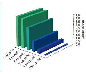
Актуальність. Емпіричне проєктування дизайну розрізу при проведенні інтраорального остеосинтезу досить часто спричиняє ятрогенні пошкодження слизово-окісного клаптя під час його підйому та мобілізації та ініціює локальну ішемію з можливістю подальшого утворення крайового некрозу м’яких тканин. Мета: підвищення ефективності хірургічного лікування та якості функціональної реабілітації пацієнтів із невогнепальними переломами нижньої щелепи шляхом математичного планування оперативного доступу для інтраорального остеосинтезу. Матеріали та методи. Проведений аналіз клінічних показників у пацієнтів, прооперованих за стандартною та авторською методиками. Результати. На першу добу післяопераційного періоду динаміка набряку м’яких тканин свідчить про гостре реактивне запалення в обох клінічних групах, але з мінімальною розбіжністю до мінімуму в першій групі. Це підкреслює наявність початкової ексудації у відповідь на первинну травму та проведення ятрогенних маніпуляцій щодо репозиції кісткових фрагментів. Протягом 3-ї та 5-ї діб спостерігалися відмінності цього показника, з маргінально високими його значеннями у другій клінічній групі, що свідчить про тривале існування інфільтрату, пов’язане з ятрогенним порушенням архітектоніки судинної сітки та розвитком венозного стазу. Висновки. Використання математично обґрунтованого складання плану оперативного втручання з урахуванням біомеханічних параметрів щодо дизайну розрізу з подальшим відшаруванням слизово-окісного клаптя для створення оптимального оперативного доступу, інтраопераційним застосуванням PRF- та PRP-технологій призводить до зниження рівня набряку м’яких тканин та больового синдрому, особливо на 5-ту добу післяопераційного періоду.
Background. Empirical incision design during intraoral osteosynthesis quite often causes iatrogenic damage to the muco-occipital flap during its elevation and mobilization and initiates local ischemia with the risk of further formation of necrosis of soft tissue margins. Purpose was to increase the effectiveness of surgical treatment and the quality of functional rehabilitation of patients with non-gunshot fractures of the mandible by mathematical planning of surgical access for intraoral osteosynthesis. Materials and methods. An analysis of clinical indicators was performed in patients operated on using the standard and authors’ techniques. Results. On the first day of the postoperative period, the dynamics of soft tissue edema indicates acute reactive inflammation in both clinical groups, but with minimal divergence to a minimum in the first group. This fact emphasizes the presence of initial exudation in response to the primary injury and iatrogenic manipulations for reposition of bone fragments. During days 3 and 5, there was a greater discrepancy in this indicator with marginally high values in the second clinical group, which indicates the long-term existence of the infiltrate associated with iatrogenic disruption of the vascular network architectonics and the development of venous stasis. Conclusions. The use of mathematically based planning of the surgical intervention taking into account biomechanical parameters for the design of the incision with subsequent detachment of the muco-occipital flap in order to create optimal surgical access, intraoperative use of PRF and PRP technologies leads to a decrease in the level of soft tissue edema and pain syndrome, especially on day 5 of the postoperative period.
металоостеосинтез; перелом; нижня щелепа; внутрішньоротовий доступ
metal osteosynthesis; fracture; mandible; intraoral access
Для ознакомления с полным содержанием статьи необходимо оформить подписку на журнал.
- Stanford-Moore G, Murr AH. Mandibular Angle Fractures. Facial Plastic Surgery Clinics of North America. 2022;30(1):109-116. https://doi.org/10.1016/j.fsc.2021.08.009.
- Kerdoud O, Aloua R, Kaouani A, Slimani F. Management of mandibular angle fractures through single and two mini-plate fixation systems: Retrospective study of 112 cases. International Journal of Surgery Case Reports. 2021;80:105690. doi: 1016/j.ijscr.2021.105690.
- Padmanaban K, Duraisamy D, Rajiah D, Raghavan P, Annamalai S. Single Versus Two Plate Osteosynthesis for Parasymphysis Fractures of the Mandible: A Prospective Comparative Study. Cureus. 2023 Feb 22;15(2):e35311. doi: 10.7759/cureus.35311.
- Lokes K, Strashko E, Ryabushko M, Baranets I, Yemets A. Early prevention of mandibular contracture caused by maxillofacial phlegmons using physical and rehabilitation medicine. Ukrainian Dental Almanac. 2025;4:51-55. doi: 10.31718/2409-0255.4.2025.06.
- Oksa M, Haapanen A, Kannari L, Furuholm J, Snäll J. Surgical treatment of clinically infected mandibular fractures. Oral Maxillofac Surg. 2024 Jun;28(2):839-847. doi: 10.1007/s10006-024-01213-6.
- Lokes KP, Svyryda OS, Svyryda EO, Steblovskyi DV, Sadyho–va DYu, Yatsenko IV, et al. Ultrasonographic assessment of periodontal tissue microcirculation in patients with mandibular fractures. Kharkiv Dental Journal. 2025;2(4(6)):581-593. doi: 10.26565/3083-5607-2025-6-05.
- Bhagat MJA, Durairaj D, Naganathan V, Nathiya B, Kumar GS, Mariam S. Application of Anatomically Designed 2-Dimensional V Plate in Management of Mandible Fracture: A Pilot Study. J Maxillofac Oral Surg. 2022 Dec;21(4):1363-1368. doi: 10.1007/s12663-021-01558-5.
- Brucoli M, Boffano P, Romeo I, Corio C, Benech A, Ruslin M, et al. Surgical management of unilateral body fractures of the edentulous atrophic mandible. Oral Maxillofac Surg. 2020 Mar;24(1):65-71. doi: 10.1007/s10006-019-00824-8.
- Daqiq O, Roossien CC, Wubs FW, van Minnen B. Bio–mechanical assessment of mandibular fracture fixation using finite ele–ment analysis validated by polymeric mandible mechanical testing. Sci Rep. 2024 May 23;14(1):11795. doi: 10.1038/s41598-024-62011-4.
- Choi TJ, Chung YH, Cho JY, Burm JS. The Use of Microplates for Internal Fixation of Comminuted Mandibular Fractures. Ann Plast Surg. 2019 Jan;82(1):55-61. doi: 10.1097/SAP.0000000000001623.
- Kolisnychenko M, Lokes K. Influence of methods of augmentation of the alveolar process during tooth extraction operation on the stability of dental implants. Ukrainian Dental Almanac. 2024;4:81-85. doi: 10.31718/2409-0255.4.2024.14.
- Purnell CA, Ettinger RE. Mandible Fractures: Consensus and Controversy. Plast Reconstr Surg. 2025 Jul 1;156(1):132e-146e. doi: 10.1097/PRS.0000000000012015.
- Lokes K, Karasiunok A, Smaglyuk L, Voloshyna L, Voronko–va H, Rezvina K. Tooth extraction operation as a component of orthodontical treatment. Ukrainian Dental Almanac. 2024;2:32-35. doi: 10.31718/2409-0255.2.2024.06.
- Oshurko AP, Oliinyk IYu, Kuzniak NB. Anatomical and topographic classification of the mandibular canal with bone atrophy caused by the loss of the masticatory teeth. Romanian Journal of Stomatology. 2022;68(4):153-159. doi: 10.37897/RJS.2022.4.1.
- Sikora M, Baranowska-Bosiacka I, Goschorska M, Chlu–bek D. In vitro effect of three-dimensional (3D) titanium mini-plate systems used for surgical treatment of condylar fractures on interleukin 1 (IL-1) and interleukin 6 (IL-6) concentration in THP-1 macrophages. Tissue Cell. 2020 Dec;67:101404. doi: 10.1016/j.tice.2020.101404.
- Huseynov AN, Malanchuk VA, Grygorovskiy VV, Brodets–kyi IS, Myroshnychenko MS, Kalashnyk-Vakulenko YM. The relationship of clinical and morphological data in comminuted fractures of the lower jaw. Wiad Lek. 2022;75(10):2322-2328. doi: 10.36740/WLek202210102.
- Pavlychuk T, Shydlovsky M, Kopchak A. A comparative biomechanical evaluation of different osteosynthesis techniques used for intracapsular condylar head fractures. J Oral Biol Craniofac Res. 2019 Apr-Jun;9(2):123-127. doi: 10.1016/j.jobcr.2019.02.001.
- Antonowicz B, Mierzejewska ŻA, Borys J, Maciejczyk M, Prokopiuk S, Car H. Evaluation of Titanium Particles, TNF-α, and Caspase-3 Concentrations in Patients with Bones Fixations of the Maxilla and Mandibule. Int J Mol Sci. 2025 Mar 5;26(5):2316. doi: 10.3390/ijms26052316.
- Nogami S, Takahashi T, Yamauchi K, Takeda Y, Ito K, Chiba M, Gaggl A. Relationship between arthroscopic findings of synovitis and levels of tumour necrosis factor-alpha and matrix metalloproteinases in synovial lavage fluid from patients with unilateral high mandibular condyle fractures. J Oral Rehabil. 2018 Jun;45(6):452-458. doi: 10.1111/joor.12632.
- Sakarinpanichakul P, Burapholkul W. Platelet-rich fibrin in the management of mandibular fractures. Arch Craniofac Surg. 2026 Apr;27(2):55-64. doi: 10.7181/acfs.2025.0079.
- Tabrizi R, Moslemi H, Shafiei S, Dastgir R, Peacock ZS. Does Platelet-Rich Fibrin Enhance Recovery From Neurosensory Disturbance Following Mandibular Fractures? A Double-Blind, Split-Mouth Randomized Clinical Trial. Craniomaxillofac Trauma Reconstr. 2024 Dec;17(4):NP90-NP96. doi: 10.1177/19433875241257737.