
Международный эндокринологический журнал Том 22, №6, 2026
Вернуться к номеру
Експресія інгібітора ядерного фактора каппа B у тканинах папілярної карциноми щитоподібної залози та в доброякісних новоутвореннях
Авторы: N.Ya. Kobrynska, V.M. Pushkarev, N.I. Levchuk, I.I. Komisarenko, B.B. Guda, V.V. Pushkarev, O.I. Kovzun
V.P. Komisarenko Institute of Endocrinology and Metabolism of NAMSU, Kyiv, Ukraine
Рубрики: Эндокринология
Разделы: Клинические исследования
Версия для печати
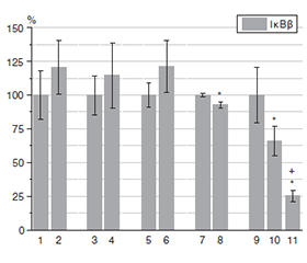
Актуальність. Еукаріотичний транскрипційний фактор NF-κB регулює широкий спектр біологічних процесів, зокрема імунну відповідь й запалення. Дисрегуляція сигнальних механізмів, залежних від NF-κB, пов’язана з тяжкими патологічними станами людини, включно з раком, автоімунними захворюваннями та хронічним запаленням. Інгібітор NF-κB, субодиниця бета (IκBβ), відіграє ключову роль у регуляції активності NF-κB і залучений у розвиток раку щитоподібної залози, особливо в агресивних і метастатичних формах. Мета: порівняти вміст IκBβ у зразках умовно нормальної (УН) тканини щитоподібної залози, тканинах фолікулярної аденоми, зоба, папілярної карциноми щитоподібної залози (ПКЩЗ), метастазів (Мтс), а також у плазмі крові пацієнтів із ПКЩЗ. Матеріали та методи. Післяопераційні зразки тканин і плазми крові були отримані в хірургічному відділенні клініки ДУ «Інститут ендокринології та обміну речовин імені В.П. Комісаренка НАМН України». Кількість IκBβ визначали за допомогою наборів для імуноферментного аналізу. Результати. Статистично значущих відмінностей між рівнями IκBβ у доброякісних новоутвореннях та УН тканині не виявлено. Незначну різницю зафіксовано між УН тканиною та тканиною ПКЩЗ без Мтс. У тканинах ПКЩЗ із метастазами показники IκBβ були більше ніж в 1,5 раза нижчими порівняно з УН тканиною. Найнижчий уміст IκBβ cпостерігався в метастазах. Концентрації IκBβ у плазмі крові осіб із ПКЩЗ були значно нижчими, ніж у контролі, без відмінностей між пацієнтами з Мтс та без них. Висновки. Рівні IκBβ у доброякісних новоутвореннях щитоподібної залози не відрізнялися від таких в УН тканині. У метастатичних тканинах ПКЩЗ та особливо в метастатичних ураженнях концентрації IκBβ були суттєво знижені порівняно з УН тканиною. Зменшення кількості IκBβ у первинних пухлинах і метастатичних ураженнях може свідчити про активацію NF-κB та посилення запального сигналінгу. Зміни експресії IκBβ можуть бути корисними для прогнозування метастатичного потенціалу ПКЩЗ, тоді як рівні IκBβ у плазмі крові розглядаються як перспективний передопераційний діагностичний біомаркер.
Background. The eukaryotic transcription factor NF-κB regulates a variety of biological processes, particularly immune response and inflammatory processes. Dysregulation of NF-κB-dependent signaling mechanisms is associated with serious human diseases, including cancer, autoimmune diseases, and chronic inflammation. NF-κB inhibitor kappa B subunit beta (IκBβ) plays a key role in regulating NF-κB activity and is implicated in thyroid cancer, particularly in aggressive and metastatic forms. The aim of this study was to compare levels of IκBβ in tissue samples of conditionally normal (CN) thyroid tissue, follicular adenoma, goiters, papillary thyroid carcinoma (PTC), metastasis (Mts), and in blood plasma of PTC patients. Materials and methods. Postoperative samples of tissues, blood plasma were obtained from the surgical department of the V.P. Komisarenko Institute of Endocrinology and Metabolism of NAMSU clinic. The amount of IκBβ was determined using enzyme immunoassay kits. Results. There were no significant differences between IκBβ levels in benign neoplasms and CN tissue. A minor difference was detected between CN tissue and PTC tissue without metastases. In PTC tissues with metastases, IκBβ levels were more than 1.5-fold lower than in CN tissue. The lowest IκBβ levels were detected in metastatic tissue. Рlasma IκBβ concentrations in PTC patients were significantly lower than in controls, with no differences between patients with and without metastases. Conclusions. IκBβ levels in benign thyroid neoplasms did not differ from CN tissue. In metastatic PTC tissues and especially in metastatic lesions, IκBβ concentrations were significantly reduced compared to CN tissue. Decreased IκBβ levels in primary tumors and metastatic lesions may indicate NF-κB activation and enhanced inflammatory signaling. Alterations in IκBβ expression may be useful for predicting metastatic potential in PTC, while plasma IκBβ levels could serve as a potential preoperative diagnostic biomarker.
NF-κB; IκBβ; папілярна карцинома щитоподібної залози; метастази; доброякісні новоутворення щитоподібної залози
NF-κB; IκBβ; papillary thyroid carcinoma; metastasis; benign thyroid neoplasms
Для ознакомления с полным содержанием статьи необходимо оформить подписку на журнал.
- Xing M. Molecular pathogenesis and mechanisms of thyroid cancer. Nat Rev Cancer. 2013 Mar;13(3):184-99. doi: 10.1038/nrc3431.
- Guda BB, Pushkarеv VM, Kovalenko AE, Pushkarev VV, Kovzun ОI, Tronko МD. Molecular genetic mechanisms of the pathogenesis of thyroid malignant tumors (review of literature and own data, part 1). Endokrynologia. 2019;24(1):53-65. doi: 10.31793/1680-1466.2019.24-1.53 (in Ukrainian).
- Qian B, Guo S, Zhou J, Qu X, Zhang S. Intraglandular disse–mination is a risk factor for lymph node metastasis in papillary thyroid carcinoma: a propensity score matching analysis. Gland Surg. 2021 Dec;10(12):3169-3180. doi: 10.21037/gs-21-470.
- Li Z, Wang N, Li X, et al. Thyroid cancer: From molecular insights to therapy (Review). Oncol Lett. 2025 Sep 10;30(5):520. doi: 10.3892/ol.2025.15266.
- Niciporuka R, Nazarovs J, Ozolins A, Narbuts Z, Miklasevics E, Gardovskis J. Can We Predict Differentiated Thyroid Cancer Behavior? Role of Genetic and Molecular Markers. Medicina (Kaunas). 2021 Oct 19;57(10):1131. doi: 10.3390/medicina57101131.
- Kobrynska NYa, Pushkarev VM, Levchuk NI, Kovzun OI, Komisarenko ІІ, Tronko MD. Mechanisms and markers of thyroid cancer metastasis. Review of literature and own data (рart 1). Endokrynologia. 2024;29(3):283-93. doi: 10.31793/1680-1466.2024.29-3.283 (in Ukrainian).
- Kobrynska NYa, Pushkarev VM, Levchuk NI, Kovzun OI, Komisarenko ІІ, Tronko MD. Mechanisms and markers of thyroid cancer metastasis. Review of literature and own data (part 2). Endokrynologia. 2024;29(4):372-79. doi: 10.31793/1680-1466.2024.29-4.372 (in Ukrainian).
- Ghosh S. Handbook of transcription factor NF-kappaB. Boca Raton: CRC Press; 2006. 232 р.
- Pushkarev VM, Kovzun OI, Pushkarev VV, Huda BB, Tronko MD. Chronic inflammation and cancer. Role of nuclear factor NF-κB (review of literature and own data). Journal of the National Academy of Medical Sciences of Ukraine. 2015;21(3–4):287-98 (in Ukrainian).
- Mao H, Zhao X, Sun SC. NF-κB in inflammation and cancer. Cell Mol Immunol. 2025 Aug;22(8):811-839. doi: 10.1038/s41423-025-01310-w.
- Cornice J, Verzella D, Arboretto P, et al. NF-κB: Governing Macrophages in Cancer. Genes (Basel). 2024 Jan 31;15(2):197. doi: 10.3390/genes15020197.
- Crescenzi E, Leonardi A, Pacifico F. NF-κB in Thyroid Cancer: An Update. Int J Mol Sci. 2024 Oct 25;25(21):11464. doi: 10.3390/ijms252111464.
- Cormier F, Housni S, Dumont F, et al. NF-κB signaling activation and roles in thyroid cancers: implication of MAP3K14/NIK. Oncogenesis. 2023 Nov 16;12(1):55. doi: 10.1038/s41389-023-00496-w.
- Bradford ММ. A rapid and sensitive methods for the quantitation of microgram quantities of protein utilizing the principle of protein-dye binding. Anal Biochem. 1976;72(1):248-254. doi: 10.1016/0003-2697(76)90527-3.
- Pernot S, Evrard S, Khatib AM. The Give-and-Take Interac–tion Between the Tumor Microenvironment and Immune Cells Regulating Tumor Progression and Repression. Front Immunol. 2022 Apr 13;13:850856. doi: 10.3389/fimmu.2022.850856.
- Garifulin OM, Filonenko VV, Bdzhola AV, et al. Expression of Ribosomal Protein Kinase S6 (S6K1) Isoforms in Different Types of Papillary Thyroid Carcinoma. Cytology and Genetics. 2023;57(4):305-311. doi: 10.3103/S0095452723040059.
- Shi S, Ou X, Liu C, Li R, Zheng Q, Hu L. NF-κB signaling and the tumor microenvironment in osteosarcoma: implications for immune evasion and therapeutic resistance. Front Immunol. 2025 Jan 30;16:1518664. doi: 10.3389/fimmu.2025.1518664.
- Mirzaei S, Saghari S, Bassiri F, et al. NF-κB as a regulator of cancer metastasis and therapy response: A focus on epithelial-–mesenchymal transition. J Cell Physiol. 2022 Jul;237(7):2770-2795. doi: 10.1002/jcp.30759.
- Pozdeyev N, Berlinberg A, Zhou Q, et al. Targeting the NF-κB Pathway as a Combination Therapy for Advanced Thyroid Cancer. PLoS One. 2015 Aug 11;10(8):e0134901. doi: 10.1371/journal.pone.0134901.
- Li X, Abdel-Mageed AB, Mondal D, Kandil E. The nuclear factor kappa-B signaling pathway as a therapeutic target against thyroid cancers. Thyroid. 2013 Feb;23(2):209-18. doi: 10.1089/thy.2012.0237.
- Bilyy OM, Mitriaieva NA, Grebinyk LV, Artiukh SV. The role of NF-kB in carcinogenesis and its connection with inflammation and chemoradioresistance of secondary edematous breast cancer and other malignant tumors (literature review and own research). Ukrainian Journal of Radiology and Oncology. 2024;32(4):459-477. doi: 10.46879/ukroj.4.2024.459-477 (in Ukrainian).
- Bauerle KT, Schweppe RE, Haugen BR. Inhibition of nuclear factor-kappa B differentially affects thyroid cancer cell growth, apoptosis, and invasion. Mol Cancer. 2010 May 21;9:117. doi: 10.1186/1476-4598-9-117.
- Pushkarev VV, Starenki DV, Pushkarev VM, Kovzun OI, Tronko MD. Inhibitor of the transcription factor NF-kB, DHMEQ, enhances the effect of paclitaxel on cells of anaplastic thyroid carcinoma in vitro and in vivo. Ukr Biochem J. 2015 May-Jun;87(3):63-74. doi: 10.15407/ubj87.03.063.
- Pushkarev VM, Kovzun OI, Pushkarev VV, Guda BB, Tronko MD. Biochemical aspects of the combined use of taxanes, irradiation and other antineoplastic agents for the treatment of anaplastic thyroid carcinoma. Ukr Biochem J. 2018 Sep-Oct;90(5):5-19. doi: 10.15407/ubj90.05.005
- Meng Z, Lou S, Tan J, et al. Nuclear factor-kappa B inhibition can enhance therapeutic efficacy of 131I on the in vivo management of differentiated thyroid cancer. Life Sci. 2012 Dec 10;91(23–24):1236-41. doi: 10.1016/j.lfs.2012.09.026.
- Chen F, Yin S, Zhu J, et al. Effects of nuclear factor κB on the uptake of 131iodine and apoptosis of thyroid carcinoma cells. Mol Med Rep. 2018 Apr;17(4):4959-4964. doi: 10.3892/mmr.2018.8481.
- Choi YW, Kim HJ, Kim YH, et al. B-RafV600E inhibits sodium iodide symporter expression via regulation of DNA methyltransferase 1. Exp Mol Med. 2014 Nov 7;46(11):e120. doi: 10.1038/emm.2014.68.