
Інформація призначена тільки для фахівців сфери охорони здоров'я, осіб,
які мають вищу або середню спеціальну медичну освіту.

Підтвердіть, що Ви є фахівцем у сфері охорони здоров'я.


Коморбідний ендокринологічний пацієнт

Журнал «Медицина неотложных состояний» Том 22, №6, 2026

Вернуться к номеру

Математичне та скінченно-елементне моделювання ушкоджень печінки при високошвидкісних осколкових пораненнях
Авторы: Ларін О.О. (1), Негодуйко В.В. (2), Риженко А.П. (3), Скрильников Є.С. (1), Грінченко Є.М. (4)
(1) - Національний технічний університет «Харківський політехнічний інститут», м. Харків, Україна
(2) - Харківський національний медичний університет, м. Харків, Україна
(3) - Військово-медичний клінічний центр Центрального регіону, м. Вінниця, Україна
(4) - Харківський національний університет внутрішніх справ, м. Харків, Україна

Рубрики: Медицина неотложных состояний

Разделы: Клинические исследования

Версия для печати


Резюме

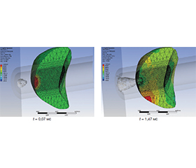
Виконано математичне та на його основі чисельне комп’ютерне моделювання методом скінченних елементів процесів ушкодження печінки при ураженні осколками. Актуальність роботи зумовлена складністю експериментального вивчення динамічного руйнування внутрішніх органів людини, зокрема печінки. Метою дослідження є створення математичної та розробка на її основі скінченно-елементної моделі печінки людини з урахуванням формування в ній ушкоджень від високошвидкісної дії осколків для аналізу динаміки деформаційних процесів, розвитку та впливу ударних хвиль на формування ранового каналу в печінці та оточуючих м’яких тканинах з урахуванням їхніх фізико-механічних властивостей. Виконано моделювання поранення печінки осколками різного розміру у діапазоні швидкостей їх руху, що надало змогу відтворити процес формування ранового каналу та проникнення осколка у тканини. Доведено, що малий осколок може досягти тканин печінки при швидкості руху понад 550 м/с, а великий — вже за швидкості понад 100 м/с. Виконано дослідження напружено-деформованого стану і хвильових процесів у печінці та м’яких тканинах, що її оточують. Максимальні значення надлишкового тиску виникають у зоні проникнення осколка та впливають майже на весь орган, що спричиняє ушкодження навіть за відсутності глибокого проникнення осколка. При цьому для малих осколків переважають сліпі поранення та ударно-хвильові ураження навколишніх тканин, тоді як великі осколки спричиняють як сліпі, так і наскрізні поранення печінки, що характеризуються значно більшою глибиною проникнення та більшим об’ємом руйнування тканин. Результати роботи можуть бути корисними для фахівців і дослідників у біомеханічному моделюванні травмування внутрішніх органів, оцінюванні тяжкості поранень та проведенні судово-медичних експертиз, а також сприятимуть подальшому розвитку моделей на основі методу скінченних елементів для прогнозування ушкоджень внутрішніх органів.

Mathematical and, based on it, numerical computer modeling of liver damage by fragments has been performed with the help of the finite element method. The relevance of the work stems from the complexity of experimental study of dynamic destruction of human internal organs, in particular the liver. The purpose of the study is to create mathematical model and, based on it, to develop finite element model of the human liver taking into account the formation of damage in it from the high-speed action of fragments for analyzing the dynamics of deformation processes, the development and influence of shock waves on the formation of a wound channel in the liver and surrounding soft tissues, considering their physical and mechanical properties. A simulation of a shrapnel injury to the liver by fragments of various sizes and in the range of their movement speeds has been performed, which made it possible to reproduce the process of formation of a wound channel and penetration of a fragment into the tissues. It has been proven that a small fragment can reach the liver tissue at a speed of more than 550 m/s, and a large fragment — at a speed of more than 100 m/s. The stress-strain state and wave processes in the liver and the soft tissues surrounding it were studied. The maximum values of excess pressure arise in the area of fragment penetration and affect almost the entire organ, causing damage even in the absence of deep penetration of the fragment. At the same time, blind wounds and shock-wave lesions of the surrounding tissues prevail in small fragments, and large fragments cause both blind and penetrating liver wounds with significantly greater penetration depth and volume of tissue destruction. The work results will be useful for specialists and researchers in biomechanical modeling of internal organ injuries, assessing the severity of injuries, conducting forensic medical exa­minations, as well as for the further development of finite element models to predict internal organ injuries.


Ключевые слова

математичне моделювання; скінченно-елементний аналіз; печінка; осколкове поранення; руйнування м’яких тканин; рановий канал; ударно-хвильові процеси

mathematical modeling; finite element analysis; liver; shrapnel wound; soft tissue destruction; wound channel; shock wave processes


Для ознакомления с полным содержанием статьи необходимо оформить подписку на журнал.


Список литературы

1. Taddei, L. et al. (2021). Effect of blast loading on injury risk: 3D FEM analysis. Forensic Science International. 326, 110930. https://doi.org/10.1016/j.forsciint.2021.110930.
2. Solomos, G., Larcher, M., Valsamos, G., Karlos, V., Casadei, F. (2020). A survey of computational models for blast induced human injuries for security and defence applications, EUR 30039 EN, Publications Office of the European Union, Luxembourg. https://doi.org/10.2760/685, JRC119310.
3. Chanda, A., Callaway, C. (2018). Computational modeling of blast induced whole-body injury: a review. Journal of medical engineering & technology. 42(2), 88-104. https://doi.org/10.1080/03091902.2018.1432711.
4. Panzer, M.B., Myers, B.S., Capehart, B.P., Bass, C.R. (2012). Development of a finite element model for blast brain injury and the effects of CSF cavitation. Annals of biomedical engineering. 40(7), 1530-1544. https://doi.org/10.1007/s10439-012-0519-2.
5. Seyedpour, S.M., Nabati, M., Lambers, L. (2021). Application of magnetic resonance imaging in liver biomechanics: a systematic review. Frontiers in Physiology. 12, 733393. https://doi.org/10.3389/fphys.2021.733393.
6. Johnson, B., Campbell, S., Campbell-Kyureghyan, N. (2021). Characterizing the Material Properties of the Kidney and Liver in Unconfined Compression and Probing Protocols with Special Reference to Varying Strain Rate. Biomechanics. 1(2), 264-280. https://doi.org/10.3390/biomechanics1020022.
7. Lemine, A.S., Ahmad, Z., Al-Thani, N.J. et al. (2024). Mechanical properties of human hepatic tissues to develop liver-mimicking phantoms for medical applications. Biomech Model Mechanobiol. 23, 373-396. https://doi.org/10.1007/s10237-023-01785-4.
8. Shen, J., Roth, S. (2022). Validation of rib structural responses under dynamic loadings using different material properties: A finite element analysis. Medical engineering & physics. 105, 103820. https://doi.org/10.1016/j.medengphy.2022.103820.
9. Pekedis, M., Ozan, F., Koyuncu, S., Yildiz, H. (2022). The finite element method-based pattern recognition approach for the classification of patient-specific gunshot injury. Proceedings of the Institution of Mechanical Engineers. Part H, Journal of engineering in medicine. 236(5), 665-675. https://doi.org/10.1177/09544119221086397.
10. Menezes, P.D., Gadegaard, N., Natal Jorge, R.M., Pinto, S.I.S. (2021). Modelling human liver microphysiology on a chip through a finite element based design approach. Int J Numer Meth Biomed Engng. 37, e3445. https://doi.org/10.1002/cnm.3445.
11. Zhang, J., Lay, R.J., Roberts, S.K., Chauhan, S. (2021). Towards real-time finite-strain anisotropic thermo-visco-elastodynamic analysis of soft tissues for thermal ablative therapy. Computer methods and programs in biomedicine. 198, 105789. https://doi.org/10.1016/j.cmpb.2020.105789.
12. Yates, K.M., Agnew, A.M., Albert, D.L., Kemper, A.R., Untaroiu, C.D. (2021). Subject-specific rib finite element models with material data derived from coupon tests under bending loading. Journal of the mechanical behavior of biomedical materials. 116, 104358. https://doi.org/10.1016/j.jmbbm.2021.104358.
13. Giudice, J.S., Alshareef, A., Wu, T., Knutsen, A.K., Hiscox, L.V., Johnson, C.L. et al. (2021). Calibration of a Heterogeneous Brain Model Using a Subject-Specific Inverse Finite Element Approach. Frontiers in bioengineering and biotechnology. 9, 664268. https://doi.org/10.3389/fbioe.2021.664268.
14. Avril, S., Goriely, A., Holzapfel, G.A., Kuhl, E., Nordsletten, D., Ogden, R.W. (2026). State-of-the-art and tomorrow’s challenges and opportunities in constitutive modeling of soft biological tissues with a focus on arterial, cardiac and brain biomecha–nics. Acta biomaterialia. 213, 62-87. https://doi.org/10.1016/j.actbio.2026.02.022.
15. Holzapfel, G.A., Humphrey, J.D., Ogden, R.W. (2025). Biomechanics of soft biological tissues and organs, mechanobiology, homeostasis and modelling. Journal of the Royal Society, Interface. 22(222), 20240361. https://doi.org/10.1098/rsif.2024.0361.
16. Pellicer-Valero, O.J., Rupérez, M.J., Martínez-Sanchís, S., Martín-Guerrero, J.D. (2020). Real-time biomechanical modeling of the liver using Machine Learning models trained on Finite Element Method simulations. Expert Syst Appl. 143. https://doi.org/10.1016/j.eswa.2019.113083.
17. Zhu, J., Su, Y., Liu, Z., Liu, B., Sun, Y., Gao, W. et al. (2022). Real-time biomechanical modelling of the liver using LightGBM model. The international journal of medical robotics + computer assisted surgery: MRCAS. 18(6), e2433. https://doi.org/10.1002/rcs.2433.
18. Yang, J., Hu, M., Shi, X., Zhao, D., Yu, L. (2021). Deformation modeling based on mechanical properties of liver tissue for virtuanormal vectors of trianglesl surgical simulation. International journal of computer assisted radiology and surgery. 16(2), 253-267. https://doi.org/10.1007/s11548-020-02297-7.
19. Untaroiu, C.D., Lu, Y.C. (2013). Material characterization of liver parenchyma using specimen-specific finite element models. Journal of the mechanical behavior of biomedical materials. 26, 11-22. https://doi.org/10.1016/j.jmbbm.2013.05.013.
20. Yoganandan, N., Somasundaram, K., Harinathan, B., Banurekha Devaraj, K., Shah, A., Koser, J. et al. (2025). Behind Amor Blunt Trauma Lung and Liver Strains From Indenter Loading Via Finite Element Modeling. Journal of engineering and science in medical diagnostics and therapy. 8(3), 031009. https://doi.org/10.1115/1.4066830.
21. Larin, O., Potopalska, K., Myronenko, M., Polivenok, I. (2025). Computational Modeling of Age-Related Atherosclerotic Plaque Evolution and Mechanical Vulnerability Assessment. In: Sontea, V., Tiginyanu, I., Railean, S. (eds) 7th International Conference on Nanotechnologies and Biomedical Engineering. ICNBME 2025. IFMBE Proceedings, 135. Springer, Cham. https://doi.org/10.1007/978-3-032-06497-4_25.
22. Larin, O., Abdusalamov, R., Itskov, M., Potopalska, K., Fomenko, N. (2025). Thermoplastic Polyurethane Viscoelastic Properties and Usage in Quasi-Zero Stiffness Metastructures: Experimental and Numerical Investigation. Strength of Materials. 57(6), 1197-1208. https://doi.org/10.1007/s11223-026-00848-y.
23. Larin, O., Potopalska, K., Polivanov, O., Nazarenko S., Kalynovskyi, A. (2021). Probabilistic modelling of Container for Discrete Delivery of Extinguishing Agents based on a set of computational simulations. 2021 IEEE 2nd KhPI Week on Advanced Technology. 634-638. https://doi.org/10.1109/KhPIWeek53812.2021.9570100.

Вернуться к номеру