
Журнал «Медицина неотложных состояний» Том 22, №6, 2026
Вернуться к номеру
Ультразвукове дослідження легень порівняно з рутинно призначеною рентгенографією органів грудної клітки при діагностиці пневмонії в дітей: перехресне дослідження в Навчальній лікарні Al-Zahraa (м. Наджаф, Ірак)
Авторы: Ameer Isam Al Aasam (1), Shymaa Abbood Najim Al Wadèes (2), Jasim Mohammed Hashim (1), Raid MR Umran (1), Alaa Jumaah Manji Nasrawi (1)
(1) - Faculty of Medicine, University of Kufa, Najaf, Iraq
(2) - Al-Zahraa Teaching Hospital, Al Najaf Health Directorate, Najaf, Iraq
Рубрики: Медицина неотложных состояний
Разделы: Клинические исследования
Версия для печати
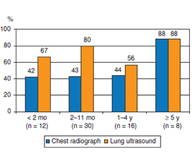
Актуальність. Рентгенографію органів грудної клітки (РОГК) майже автоматично призначають дітям із підозрою на пневмонію, хоча її чутливість щодо виявлення невеликих консолідацій є недостатньою, а сама процедура супроводжується впливом іонізуючого випромінювання на організм дитини. Даних щодо використання ультразвукового дослідження легень (УЗДЛ) як альтернативного методу безпосередньо біля ліжка хворого в рутинній практиці у країнах із низьким і середнім рівнем доходу, зокрема в Іраку, зараз недостатньо. Мета: порівняти діагностичну ефективність УЗДЛ, виконаного біля ліжка хворого, та РОГК, призначеної в рамках стандартного обстеження. Матеріали та методи. Проведено перехресне дослідження за участю дітей, госпіталізованих до педіатричного відділення Навчальної лікарні Al-Zahraa (м. Наджаф) у період із травня до липня 2014 року. Кожній дитині проводили РОГК за призначенням бригади, що здійснювала госпіталізацію, з описом результатів лікарем-рентгенологом, а також УЗДЛ високочастотним лінійним датчиком, виконане педіатром, який пройшов тижневий спеціалізований тренінг і не знав результатів рентгенографії та її опису. Результати обох досліджень фіксували як позитивні або негативні щодо наявності консолідації. Парні пропорції порівнювали за допомогою точного тесту Мак-Немара, узгодженість оцінювали за допомогою κ-коефіцієнта Коена, а чутливість визначали відносно комбінованого візуалізаційного референтного стандарту (наявність консолідації за даними будь-якого з двох методів). Результати. До основного аналізу увійшли 66 дітей; медіана віку становила 6,0 місяця (міжквартильний розмах: 2,5–17,7), при цьому 42 дитини (63,6 %) були віком до одного року. УЗДЛ допомогло виявити консолідацію у 48 дітей (72,7 %; 95% довірчий інтервал (ДІ) 61,0–82,0), РОГК — у 32 (48,5 %; 95% ДІ 36,8–60,3); різниця між показниками становила 24,2 відсоткового пункту (95% ДІ 12,3–36,2) і була статистично значущою (точний тест Мак-Немара, p < 0,001). У 18 дітей позитивний результат було отримано лише за даними ультразвукового дослідження, а у двох — рентгенографії (узгодженість 69,7 %, κ = 0,40, 95% ДІ 0,18–0,62). За комбінованим референтним стандартом чутливість становила 96,0 % (95% ДІ 86,5–98,9) для ультразвукового дослідження та 64,0 % (95% ДІ 50,1–75,9) для рентгенографії. Медіана найбільшого діаметра консолідацій становила 11,0 мм (міжквартильний розмах 8,3–16,0); у 70,8 % випадків він не перевищував 15 мм. Ураження, не виявлені під час рентгенографії, були меншими за ті, що знаходили при використанні цього методу (медіана 9,0 проти 13,5 мм, p = 0,016). Висновки. Серед госпіталізованих пацієнтів, переважно немовлят, УЗДЛ, проведене педіатром після тижневого навчання, виявило консолідацію в більшої кількості дітей (у 1,5 раза), ніж стандартна РОГК; переважно йшлося про дрібні субплевральні осередки, які не візуалізувалися на рентгенограмі. Цей метод є доцільним для первинної візуалізації в таких умовах, тоді як рентгенографію слід проводити в дітей, у яких результати сонографії не пояснюють клінічну картину.
Background. A chest radiograph is ordered almost reflexively for children admitted with suspected pneumonia, although its sensitivity for small consolidations is imperfect and it delivers ionizing radiation to a young chest. Data on lung ultrasound as a bedside alternative in routine practice in low- and middle-income settings, and particularly from Iraq, remain sparse. We compared the yield of bedside lung ultrasound with that of the chest radiograph ordered as part of routine care. Materials and methods. Cross-sectional study of children admitted to the pediatric service of Al-Zahraa Teaching Hospital, Najaf, between May and July 2014. Each child had the chest radiograph requested by the admitting team, reported by a radiologist, and a lung ultrasound performed with a high-frequency linear transducer by a pediatrician who had completed one week of focused training and was blinded to the radiograph and its report. Both were recorded as positive or negative for consolidation. Paired proportions were compared with the exact McNemar test, agreement with Cohen’s kappa, and sensitivity against a composite imaging reference standard (consolidation on either modality). Results. Sixty-six children formed the primary analysis population; median age 6.0 months (interquartile range 2.5–17.7), with 42 (63.6 %) under one year. Lung ultrasound demonstrated consolidation in 48 children (72.7 %, 95% confidence interval (CI) 61.0–82.0) and chest radiography in 32 (48.5 %, 95% CI 36.8–60.3); the paired difference of 24.2 percentage points (95% CI 12.3–36.2) was significant (exact McNemar p < 0.001). Eighteen children were positive on ultrasound alone and two on radiography alone (agreement 69.7 %, κ = 0.40, 95% CI 0.18–0.62). Against the composite reference, sensitivity was 96.0 % (95% CI 86.5–98.9) for ultrasound and 64.0 % (95% CI 50.1–75.9) for radiography. Consolidations had a median largest diameter of 11.0 mm (interquartile range 8.3–16.0); 70.8 % measured 15 mm or less. Lesions missed by radiography were smaller than those it detected (median 9.0 versus 13.5 mm, p = 0.016). Conclusions. In a hospitalized population dominated by infants, lung ultrasound performed by a pediatrician with one week of training identified consolidation in about half again as many children as the routinely ordered chest radiograph, chiefly small subpleural lesions the film did not resolve. It is a reasonable first-line imaging test in this setting, with the radiograph reserved for children whose sonographic findings do not explain the clinical picture.
ультразвукове дослідження легень; рентгенографія органів грудної клітки; пневмонія; діти; ультразвукове дослідження біля ліжка хворого; Ірак
lung ultrasound; chest radiography; pneumonia; children; point-of-care ultrasound; Iraq
Для ознакомления с полным содержанием статьи необходимо оформить подписку на журнал.
- McAllister DA, Liu L, Shi T, Chu Y, Reed C, Burrows J, et al. Global, regional, and national estimates of pneumonia morbidity and mortality in children younger than 5 years between 2000 and 2015: a systematic analysis. Lancet Glob Health. 2019;7(1):e47-e57.
- Harris M, Clark J, Coote N, Fletcher P, Harnden A, –McKean M, et al. British Thoracic Society guidelines for the management of community acquired pneumonia in children: update 2011. Thorax. 2011;66(Suppl 2):ii1-23.
- Bradley JS, Byington CL, Shah SS, Alverson B, Carter ER, Harrison C, et al. The management of community-acquired pneumonia in infants and children older than 3 months of age: clinical practice guidelines by the Pediatric Infectious Diseases Society and the Infectious Diseases Society of America. Clin Infect Dis. 2011;53(7):e25-76.
- Neuman MI, Lee EY, Bixby S, Diperna S, Hellinger J, Markowitz R, et al. Variability in the interpretation of chest radiographs for the diagnosis of pneumonia in children. J Hosp Med. 2012;7(4):294-8.
- Lichtenstein DA, Mezière GA. Relevance of lung ultrasound in the diagnosis of acute respiratory failure: the BLUE protocol. Chest. 2008;134(1):117-25.
- Volpicelli G, Elbarbary M, Blaivas M, Lichtenstein DA, Mathis G, Kirkpatrick AW, et al. International evidence-based re–commendations for point-of-care lung ultrasound. Intensive Care Med. 2012;38(4):577-91.
- Copetti R, Cattarossi L. Ultrasound diagnosis of pneumonia in children. Radiol Med. 2008;113(2):190-8.
- Iuri D, De Candia A, Bazzocchi M. Evaluation of the lung in children with suspected pneumonia: usefulness of ultrasonography. Radiol Med. 2009;114(2):321-30.
- Caiulo VA, Gargani L, Caiulo S, Fisicaro A, Moramarco F, Latini G, et al. Lung ultrasound characteristics of community-acquired pneumonia in hospitalized children. Pediatr Pulmonol. 2013;48(3):280-7.
- Shah VP, Tunik MG, Tsung JW. Prospective evaluation of point-of-care ultrasonography for the diagnosis of pneumonia in children and young adults. JAMA Pediatr. 2013;167(2):119-25.
- Esposito S, Papa SS, Borzani I, Pinzani R, Giannitto C, Consonni D, et al. Performance of lung ultrasonography in children with community-acquired pneumonia. Ital J Pediatr. 2014;40:37.
- Pereda MA, Chavez MA, Hooper-Miele CC, Gilman RH, Steinhoff MC, Ellington LE, et al. Lung ultrasound for the diagnosis of pneumonia in children: a meta-analysis. Pediatrics. 2015;135(4):714-22.
- Urbankowska E, Krenke K, Drobczyński Ł, Korczyński P, Urbankowski T, Krawiec M, et al. Lung ultrasound in the diagnosis and monitoring of community acquired pneumonia in children. Respir Med. 2015;109(9):1207-12.
- Iorio G, Capasso M, De Luca G, Prisco S, Mancusi C, Laga–nà B, et al. Lung ultrasound in the diagnosis of pneumonia in children: proposal for a new diagnostic algorithm. PeerJ. 2015;3:e1374.
- Jones BP, Tay ET, Elikashvili I, Sanders JE, Paul AZ, Nelson BP, et al. Feasibility and safety of substituting lung ultrasonography for chest radiography when diagnosing pneumonia in children: a randomized controlled trial. Chest. 2016;150(1):131-8.
- Ellington LE, Gilman RH, Chavez MA, Pervaiz F, Marin-Concha J, Compen-Chang P, et al. Lung ultrasound as a diagnostic tool for radiographically-confirmed pneumonia in low resource settings. Respir Med. 2017;128:57-64.
- Stadler JAM, Andronikou S, Zar HJ. Lung ultrasound for the diagnosis of community-acquired pneumonia in children. Pediatr Radiol. 2017;47(11):1412-9.
- Balk DS, Lee C, Schafer J, Welwarth J, Hardin J, Novack V, et al. Lung ultrasound compared to chest X-ray for diagnosis of pediatric pneumonia: a meta-analysis. Pediatr Pulmonol. 2018;53(8):1130-9.
- Orso D, Ban A, Guglielmo N. Lung ultrasound in diagnosing pneumonia in childhood: a systematic review and meta-analysis. J Ultrasound. 2018;21(3):183-95.
- Tsou PY, Chen KP, Wang YH, Fishe J, Gillon J, Lee CC, et al. Diagnostic accuracy of lung ultrasound performed by novice versus advanced sonographers for pneumonia in children: a systematic review and meta-analysis. Acad Emerg Med. 2019;26(9):1074-88.
- Wang L, Song W, Wang Y, Han J, Lv K. Lung ultrasonography versus chest radiography for the diagnosis of pediatric community acquired pneumonia in emergency department: a meta-analysis. J Thorac Dis. 2019;11(12):5107-14.
- Najgrodzka P, Buda N, Zamojska A, Marciniewicz E, Lewandowicz-Uszyńska A. Lung ultrasonography in the diagnosis of pneumonia in children — a meta-analysis and a review of pediatric lung imaging. Ultrasound Q. 2019;35(2):157-63.
- Bossuyt PM, Reitsma JB, Bruns DE, Gatsonis CA, Glas–ziou PP, Irwig L, et al. STARD 2015: an updated list of essential items for reporting diagnostic accuracy studies. BMJ. 2015;351:h5527.
- World Health Organization. Revised WHO classification and treatment of childhood pneumonia at health facilities: evidence summaries. Geneva: World Health Organization; 2014.