
Журнал «Здоровье ребенка» Том 21, №5, 2026
Вернуться к номеру
Сироваткові рівні IL-6 та нейтрофільної еластази у дітей з бронхоектатичною хворобо
Авторы: Мазулов О.В., Остренюк Р.С.
Вінницький національний медичний університет імені М.І. Пирогова, м. Вінниця, Україна
Рубрики: Педиатрия/Неонатология
Разделы: Клинические исследования
Версия для печати
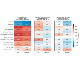
Актуальність. Нейтрофільне запалення дихальних шляхів є провідним патогенетичним механізмом бронхоектатичної хвороби (БЕХ). Прогностична цінність сироваткових біомаркерів запалення у педіатричній популяції залишається невизначеною, а більшість наявних доказів отримано у змішаних за етіологією когортах, а також у дорослих пацієнтів. Мета дослідження: оцінити сироваткові концентрації інтерлейкіну-6 (IL-6) та нейтрофільної еластази (NE/ELA2) у дітей із БЕХ різної етіології, визначити їхню діагностичну цінність та встановити характер зв’язку з ключовими точками контролю перебігу — частотою загострень, що потребували антибіотикотерапії, частотою госпіталізацій, об’ємом форсованого видиху за першу секунду та якістю життя, оціненою за допомогою опитувальника Bronchiectasis Child-specific Parent-proxy Quality of Life. Матеріали та методи. Обстежено 150 дітей віком 2–17 років, яких розподілили на три групи: з бронхоектазами, не пов’язаними з муковісцидозом (non-CF, n = 50), з бронхоектазами, пов’язаними з муковісцидозом (CF, n = 50), та практично здорові діти (n = 50). IL-6 та NE/ELA2 визначали методом імуноферментного аналізу. Для статистичного аналізу застосовували критерії Краскела — Уолліса та Манна — Уїтні, ROC-аналіз із порівнянням площ під ROC-кривими за методом Делонга, кореляційний аналіз за Спірменом, часткову рангову кореляцію та логістичну регресію Фірта. Результати. Обидва біомаркери формували ступінчастий градієнт: контроль < non-CF < CF (усі попарні p < 0,001), а їхні розподіли у хворих і здорових не перекривалися (AUC = 1,000). Для клінічно змістовного розмежування етіологічних груп дискримінативна здатність була помірною: IL-6 — AUC = 0,884; NE/ELA2 — AUC = 0,865 (p = 0,653). В об’єднаній когорті виявлено 21 значущу кореляцію із 34 та 13 значущих відношень шансів із 18 (3,6–10,2). Висновки. Сироваткові рівні IL-6 та NE/ELA2 надійно відображають наявність та нозологічну категорію захворювання. Водночас у межах діагностично однорідних груп їхній зв’язок з індивідуальним клінічним перебігом не досягав статистичної значущості, що з урахуванням поперечного дизайну свідчить про обмежену прогностичну цінність для моніторингу і потребує підтвердження у проспективних дослідженнях. За наявних даних моніторинг перебігу доцільно ґрунтувати передусім на клінічних показниках та якості життя.
Background. Neutrophilic airway inflammation is a central pathogenic mechanism in bronchiectasis. However, the prognostic value of serum inflammatory biomarkers in the paediatric population remains uncertain, and most available evidence derives from mixed-etiology cohorts and adult patients. The aim of the study was to evaluate serum interleukin-6 (IL-6) and neutrophil elastase (NE/ELA2) concentrations in children with bronchiectasis of different etiologies, determine their diagnostic utility, and assess associations with key disease-control outcomes, including the rate of exacerbations requiring antibiotic therapy, hospitalisation frequency, forced expiratory volume in 1 second, and quality of life measured using bronchiectasis child-specific parent-proxy QoL instrument. Materials and methods. A total of 150 children aged 2–17 years were enrolled and divided into three groups: non-cystic fibrosis bronchiectasis (non-CF, n = 50), cystic fibrosis-associated bronchiectasis (CF, n = 50), and apparently healthy controls (n = 50). IL-6 and NE/ELA2 were measured by enzyme-linked immunosorbent assay. Kruskal-Wallis and Mann-Whitney tests, ROC analysis with area-under-the-curve (AUC) comparison using DeLong’s method, Spearman correlation, partial rank correlation, and Firth logistic regression were applied. Results. Both biomarkers exhibited a stepwise gradient: controls < non-CF < CF (all pairwise p < 0.001), and their distributions in patients and healthy controls did not overlap (AUC = 1.000). For clinically meaningful discrimination between etiological groups, performance was moderate: IL-6, AUC = 0.884; NE/ELA2, AUC = 0.865 (DeLong p = 0.653). In the pooled cohort, 21 significant correlations among 34 tested associations were identified, as well as 13 significant odds ratios among 18 evaluated comparisons (3.6–10.2). Conclusions. Serum IL-6 and NE/ELA2 reliably reflect disease presence and etiologic category. Within diagnostically homogeneous groups, however, their association with individual clinical course did not reach statistical significance, which — given the cross-sectional design — indicates limited prognostic value for monitoring and warrants confirmation in prospective studies. Based on the present data, disease monitoring should rely primarily on clinical outcomes and quality-of-life measures.
бронхоектатична хвороба; діти; інтерлейкін-6; нейтрофільна еластаза; ROC-аналіз; якість життя
bronchiectasis; children; interleukin-6; neutrophil elastase; ROC analysis; quality of life
Для ознакомления с полным содержанием статьи необходимо оформить подписку на журнал.
- Chang AB, Bush A, Grimwood K. Bronchiectasis in children: diagnosis and treatment. Lancet. 2018;392(10150):866-879. doi: 10.1016/S0140-6736(18)31554-X.
- Gaillard EA, Carty H, Heaf D, Smyth RL. Reversible bronchial dilatation in children: comparison of serial high-resolution computed tomography scans of the lungs. Eur J Radiol. 2003;47(3):215-220. doi: 10.1016/S0720-048X(02)00122-5.
- Chalmers JD, Chang AB, Chotirmall SH, Dhar R, McShane PJ. Bronchiectasis. Nat Rev Dis Primers. 2018;4(1):45. doi: 10.1038/s41572-018-0042-3.
- Zhang J, Wang S, Shao C. Reversible bronchial dilatation in adults. Clin Exp Pharmacol Physiol. 2021;48(7):966-970. doi: 10.1111/1440-1681.13503.
- Chang AB, Kantar A, Redding GJ, Hill AT. Is bronchiectasis a rever–sible disorder? Arch Bronconeumol. 2023. doi: 10.1016/j.arbres.2022.11.007.
- Burgel PR, Bush A. Reversal of bronchiectasis in children: early intervention may stop the vicious circle! Am J Respir Crit Care Med. 2024. doi: 10.1164/rccm.202404-0764ED.
- Mills DR, et al. Radiographic outcomes in pediatric bronchiectasis and factors associated with reversibility. Am J Respir Crit Care Med. 2024;210(1):97-107. doi: 10.1164/rccm.202402-0411OC.
- Silvestre-Roig C, Hidalgo A, Soehnlein O. Neutrophil he–terogeneity: implications for homeostasis and pathogenesis. Blood. 2016;127(18):2173-2181. doi: 10.1182/blood-2016-01-688887.
- Amitani R, et al. Effects of human neutrophil elastase and Pseudomonas aeruginosa proteinases on human respiratory epithelium. Am J Respir Cell Mol Biol. 1991;4(1):26-32. doi: 10.1165/ajrcmb/4.1.26.
- Sandhaus RA, Turino G. Neutrophil elastase-mediated lung disease. COPD. 2013;10(Suppl 1):60-63. doi: 10.3109/15412555.2013.764403.
- Sly PD, et al. Lung disease at diagnosis in infants with cystic fibrosis detected by newborn screening. Am J Respir Crit Care Med. 2009;180(2):146-152. doi: 10.1164/rccm.200901-0069OC.
- Dente FL, et al. Neutrophilic bronchial inflammation correlates with clinical and functional findings in patients with noncystic fibrosis bronchiectasis. Mediators Inflamm. 2015;2015:642503. doi: 10.1155/2015/642503.
- Lin HC, et al. IL-6 and TIMP-1 correlated to airway pathogen colonization and predict disease severity in patients with non-cystic fibrosis bronchiectasis. J Inflamm Res. 2024;17:5701-5709. doi: 10.2147/JIR.S465413.
- Pizzutto SJ, Upham JW, Yerkovich ST, Chang AB. High pulmonary levels of IL-6 and IL-1β in children with chronic suppurative lung disease are associated with low systemic IFN-γ production in response to non-typeable Haemophilus influenzae. PLoS One. 2015;10(6):e0129517. doi: 10.1371/journal.pone.0129517.
- Barbour V, Clark J, Peiperl L, Veitch E, Yamey G. Better reporting, better research: guidelines and guidance in PLoS Medicine. PLoS Med. 2008;5(4):e97. doi: 10.1371/journal.pmed.0050099.
- Bossuyt PM, et al. STARD 2015: an updated list of essential items for reporting diagnostic accuracy studies. BMJ. 2015;351:h5527. doi: 10.1136/bmj.h5527.
- Kekeç H, Şişmanlar Eyüboğlu T, Aslan AT, Duman Y, Medeni V, et al. Bronchiectasis in children: a comparative analysis of cystic fibrosis and non-cystic fibrosis etiologies using the Bhalla score. Balkan Med J. 2025;42:429-439. doi: 10.4274/balkanmedj.galenos.2025.2025-6-247.
- Prentice BJ, Wales S, Doumit M, Owens L, Widger J. Children with bronchiectasis have poorer lung function than those with cystic fibrosis and do not receive the same standard of care. Pediatr Pulmonol. 2019;54:1921-1926. doi: 10.1002/ppul.24491.
- Chang AB, Fortescue R, Grimwood K, Alexopoulou E, Bell L, Boyd J, et al. European Respiratory Society guidelines for the management of children and adolescents with bronchiectasis. Eur Respir J. 2021;58(2):2002990. doi: 10.1183/13993003.02990-2020.
- Chalmers JD, Mall MA, Nielsen KG, Chang AB, Aliberti S, Blasi F, et al. Neutrophil-derived biomarkers in bronchiectasis: identifying a common therapeutic target. Eur Respir J. 2025;66(3):2500081. doi: 10.1183/13993003.00081-2025.
- Keam SJ. Brensocatib: First Approval. Drugs. 2026;86(2):265-270. doi: 10.1007/s40265-025-02255-0.
- Liang CC, Zhao J, Zhang YQ, Chen J, Wang Y. Research progress on the effect of neutrophil elastase and its inhibitors in respiratory diseases. J Int Med Res. 2025;53(6):3000605251352789. doi: 10.1177/03000605251352789.
- Cant E, Bottier M, Shuttleworth M, Stobo J, Perea L, Finch S, et al. Inflammatory and mucociliary dysfunction-based endotypes across the spectrum of chronic airway diseases. Chest. 2025. doi: 10.1016/j.chest.2025.07.4087.
- Ghezzi M, Di Cicco M, Canali G, Lamberti R, Bertolucci G, Zirpoli S, et al. Early Diagnosis of Non-Cystic Fibrosis Bronchiectasis in Childhood: Shedding Light on Barriers and Opportunities. Pediatr Pul–monol. 2025;60(11):e71365. doi: 10.1002/ppul.71365.
- Tsouprou M, Koumpagioti D, Botsa E, Douros K, Moriki D. Utilization of inhaled antibiotics in pediatric non-cystic fibrosis bronchiectasis: a comprehensive review. Antibiotics (Basel). 2025;14(2):165. doi: 10.3390/antibiotics14020165.