
Международный эндокринологический журнал Том 22, №7, 2026
Вернуться к номеру
Переваги застосування натурального мікронізованого прогестерону у формі песаріїв для лікування гіперплазії ендометрія без атипії
Авторы: N. Karimova (1), G. Subanova (1), G. Karimova (2), V. Tursunova (1), A. Egemkulova (1)
(1) - Osh State University, Osh, Kyrgyz Republic
(2) - Bukhara State Medical Institute, Bukhara, Uzbekistan
Рубрики: Эндокринология
Разделы: Клинические исследования
Версия для печати
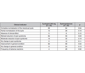
Актуальність. Гіперплазія ендометрія без атипії пов’язана з естроген-прогестероновим дисбалансом, порушеннями менструального циклу та патологічними кровотечами, тоді як використання натурального мікронізованого прогестерону може запропонувати фізіологічний терапевтичний підхід. У той же час його клінічні, морфологічні й гормональні ефекти потребують подальшого вивчення. У попередніх дослідженнях розглядалися фармакологія прогестерону, фактори ризику гіперплазії, результати лікування та прогресування захворювання, але дані щодо клінічного, морфологічного, ультразвукового й гормонального впливу натурального мікронізованого прогестерону у формі песарію залишалися обмеженими. Мета: визначити порівняльну клінічну ефективність та безпеку натурального мікронізованого прогестерону в пацієнток із гіперплазією ендометрія без атипії. Матеріали та методи. Це проспективне контрольоване клінічне спостереження було проведено в Національному центрі охорони здоров’я матері та дитини м. Бішкек (Киргизстан). Дані було зібрано на основі клінічних спостережень за 50 жінками віком 30–50 років із гістологічно підтвердженою гіперплазією ендометрія без атипії. Пацієнтки були розділені на дві нерандомізовані групи (основну й контрольну) по 25 осіб відповідно до призначеної терапії, що відображає рутинну клінічну практику. Результати. Встановлено, що застосування натурального мікронізованого прогестерону сприяло швидшій нормалізації клінічних симптомів, скороченню часу до настання ремісії та вищому показнику повного клінічного одужання. Морфологічну нормалізацію ендометрія було досягнуто у 84 % випадків порівняно із 60 % (p = 0,02). Зменшення товщини ендометрія з 14,2 ± 1,3 мм до 5,8 ± 0,7 мм супроводжувалося відновленням його структури та чіткості контурів. Гормональні зміни включали зниження рівня естрогенів до 98 ± 15 пг/мл і підвищення вмісту прогестерону до 14,8 ± 1,5 нг/мл, що привело до нормалізації їх співвідношення. У контрольній групі ці зміни були менш вираженими. Частота побічних реакцій становила 4 проти 28 % (p < 0,05), потреба в коригуванні лікування — 0 проти 16 %, а частота припинення терапії — 0 проти 12 %, що свідчить про ліпшу переносимість лікування. Висновки. Натуральний мікронізований прогестерон забезпечив більш повну клінічну, морфологічну й гормональну відповідь і продемонстрував сприятливий профіль безпеки.
Background. Endometrial hyperplasia without atypia is associated with oestrogen-progesterone imbalance, menstrual disturbances, and abnormal bleeding, while natural micronised progesterone may offer a physiological therapeutic approach, although its clinical, morphological, and hormonal effects require further investigation. The reviewed studies addressed progesterone pharmacology, hyperplasia risk factors, treatment outcomes, and disease progression, but evidence on the clinical, morphological, ultrasound, hormonal, and tolerability effects of natural micronised progesterone in pessary form remained limited. This study purposed to determine the comparative clinical efficacy and safety of natural micronised progesterone in patients with endometrial hyperplasia without atypia. Materials and methods. This prospective controlled clinical observation study was conducted at the National Centre for Maternal and Child Health in Bishkek, Kyrgyzstan. Data were collected based on clinical observations of 50 patients aged 30–50 years with histologically confirmed endometrial hyperplasia without atypia. The patients were divided into two non-randomised groups (main and control) of 25 each according to the prescribed therapy, reflecting routine clinical practice. Results. The study established that natural micronised progesterone was associated with a faster normalisation of clinical symptoms, shorter time to remission and a higher rate of complete clinical recovery. Morphological normalisation of the endometrium was achieved in 84 % of cases, compared to 60 % (p = 0.02). A reduction in endometrial thickness from 14.2 ± 1.3 mm to 5.8 ± 0.7 mm was accompanied by the restoration of its structure and contour definition. Hormonal changes included a reduction in oestrogen to 98 ± 15 pg/ml and an increase in progesterone to 14.8 ± 1.5 ng/ml, normalising their ratio. These changes were less pronounced in the control group. The incidence of adverse reactions was 4 versus 28 % (p < 0.05), the need for treatment adjustment was 0 versus 16 %, and therapy discontinuation was 0 versus 12 %, reflecting the treatment’s better tolerability. Conclusions. Natural micronised progesterone provided a more complete clinical, morphological and hormonal response, with a favourable safety profile.
естроген-прогестероновий дисбаланс; гіперплазія ендометрія; морфологічне регресування; ультразвукове дослідження; клінічна ремісія; аномальні маткові кровотечі; секреторна трансформація; натуральний мікронізований прогестерон
oestrogen-progesterone imbalance; endometrial hyperplasia; morphological regression; ultrasound imaging; clinical remission; abnormal uterine bleeding; secretory transformation; natural micronised progesterone
Для ознакомления с полным содержанием статьи необходимо оформить подписку на журнал.
- Dhillon-Smith RK, Khairy M, Bamford T, et al. A comparison of progesterone via vaginal oil capsules versus pessaries for luteal phase support in assisted reproduction treatment: A multicentre cohort study of 42 291 cycles. Hum Reprod. 2026 Jan;41(1):59-68. doi: 10.1093/humrep/deaf219.
- Labarta E, Rodríguez-Varela C, Salvaleda-Mateu M, Kohls G, Bosch E. Luteal phase support using micronized vaginal progesterone as pessaries or capsules in artificial cycles: Is there any difference? Reprod Biomed Online. 2024 May;48(5):103638. doi: 10.1016/j.rbmo.2023.103638.
- Herencia A, Bernabeu A, Pitas A, et al. Progesterone levels on the day of embryo transfer using a single pessary of 400 mg of vaginal progesterone vs. 200 mg × 2 pessaries in hormonal replacement cycles. JBRA Assist Reprod. 2023 Nov;27(4):651-654. doi: 10.5935/1518-0557.20230021.
- Shafi O. Engineering for Obstetrics and Gynaecology: Fabrication of the Transdermal Progesterone Patch. London: University College London; 2024.
- Abdirasulova ZA, Begum LJ. Comparative study of prevalence in uterine cancer: Kyrgyzstan and India. Int J Novel Res Dev. 2023 Apr;8(4):492-498.
- Kibets EA, Akmatbekova ZS, Abaeva TA, Muzaparov MS. Assessment of risk factors for endometrial hyperplasia of women at reproductive age. Health Care Kyrgyzstan. 2022 Jan;(4):75-81.
- Sebok P, Keszthelyi M, Vida B, et al. Oncologic and reproductive outcomes after fertility-sparing treatments for endometrial hyperplasia with atypia: A systematic review and meta-analysis. Cancers. 2025 Dec;17(24):3966. doi: 10.3390/cancers17243966.
- Prip CM, Stentebjerg M, Bennetsen MH, Petersen LK, Bor P. Risk of atypical hyperplasia and endometrial carcinoma after initial diagnosis of non-atypical endometrial hyperplasia: A long-term follow-up study. PLoS One. 2022 Apr;17(4):e0266339. doi: 10.1371/journal.pone.0266339.
- World Medical Association. WMA Declaration of Helsinki — Ethi–cal Principles for Medical Research Involving Human Participants. 2024.
- Shi WJ, Fadare O. The significance of papillary architecture in the follow-up biopsies of patients with progestin-treated atypical endometrial hyperplasia. J Pathol Transl Med. 2026 Jan;60(1):58-68. doi: 10.4132/jptm.2025.09.12.
- Kaliyamoorthy S, Radhakrishnan V, Arasu M, Ramasamy J. A study on histopathological association of endometrial hyperplasia with estrogen receptor, progesterone receptor status. Acta Sci Med Sci. 2022 Jan;6(1):226-232. doi: 10.31080/ASMS.2022.06.1143.
- Ring KL, Mills AM, Modesitt SC. Endometrial hyperplasia. Obstet Gynecol. 2022 Dec;140(6):1061-1075. doi: 10.1097/aog.0000000000004989.
- Nawaz A, Javaid A, Waheed SA, Raza MW, Haq EU, Ullah I. Duration-dependent histomorphological alterations in human endometrium following progestin therapy: Evidence from a tertiary care center. Int J Pharm Res Technol. 2026 Mar;16(1):836-844.
- Chou AJ, Bing RS, Ding DC. Endometrial atypical hyperplasia and risk of endometrial cancer. Diagnostics. 2024 Nov;14(22):2471. doi: 10.3390/diagnostics14222471.
- Hermann C, Williams K, Ko EM. Endometrial hyperplasia. In: Shoupe D, ed. Handbook of Gynecology. Cham: Springer; 2023. 1029-1047. doi: 10.1007/978-3-031-14881-1_3.
- Li JJX, Ho JYY, Lee CHC, et al. Digital image analysis of gland-to-stroma ratio by CD10 objectively differentiates between low-grade endometrioid carcinoma and atypical hyperplasia on endometrial biopsy. Pathology. 2025 Jun;57(4):455-460. doi: 10.1016/j.pathol.2024.11.012.
- Mohan R, Mohanraj A, Ramdas A, Padma A. Spectrum of histopathological changes in abnormal uterine bleeding with estrogen and progesterone receptor expression in endometrial hyperplasia and carcinoma. J Midlife Health. 2025 Oct-Dec;16(4):395-401. doi: 10.4103/jmh.jmh_33_25.
- Harmsen MJ, Trommelen LM, de Leeuw RA, et al. Uterine junctional zone and adenomyosis: Comparison of MRI, transvaginal ultrasound and histology. Ultrasound Obstet Gynecol. 2023 Jul;62(1):42-60. doi: 10.1002/uog.26117.
- Capasso I, Cucinella G, Wright DE, et al. Artificial intelligence model for enhancing the accuracy of transvaginal ultrasound in detecting endometrial cancer and endometrial atypical hyperplasia. Int J Gynecol Cancer. 2024 Oct;34(10):1547-1555. doi: 10.1136/ijgc-2024-005652.
- Lv M, Chen P, Bai M, et al. Progestin resistance and corresponding management of abnormal endometrial hyperplasia and endometrial carcinoma. Cancers. 2022 Dec;14(24):6210. doi: 10.3390/cancers14246210.
- Van Weelden WJ, Birkendahl PB, Lalisang RI, IntHout J, Kruitwagen RFPM, Romano A, Pijnenborg JMA. The effect of progestin therapy in advanced and recurrent endometrial cancer: A systematic review and meta-analysis. BJOG. 2023 Jan;130(2):143-152. doi: 10.1111/1471-0528.17331.
- Shao F, Li Y, Zhao Y. Progestin plus metformin improves outcomes in patients with endometrial hyperplasia and early endometrial cancer more than progestin alone: A meta-analysis. Front Endocrinol. 2023 Jun;14:1139858. doi: 10.3389/fendo.2023.1139858.
- Elassall GM, Sayed EG, Abdallah NA, et al. Levonorgestrel-releasing intrauterine system versus systemic progestins in management of endometrial hyperplasia: A systemic review and meta-analysis. J Gynecol Obstet Hum Reprod. 2022 Oct;51(8):102432. doi: 10.1016/j.jogoh.2022.102432.
- Kaemmle LM, Stadler A, Janka H, von Wolff M, Stute P. The impact of micronized progesterone on cardiovascular events — A systematic review. Climacteric. 2022 Aug;25(4):327-336. doi: 10.1080/13697137.2021.2022644.
- Ermakhanova T, Bazarbekova RB, Svyatova GS, Dossanova AK. Genotype-phenotype association in congenital adrenal hyperplasia due to 21-hydroxylase deficiency in children. Clin Endocrinol. 2023 May;98(5):654-661. doi: 10.1111/cen.14859.
- Dyndar O, Lypko I. Predictors of the development of benign proliferative uterine diseases in women of reproductive age. Med Sci Ukr. 2025 Jun;21(2):3-15. doi: 10.32345/2664-4738.2.2025.01.
- Kachailo IA, Guz IA, Kuzmina OO. State of the hormonal profile in endometrial hyperplasia. World Med Biol. 2023 Nov;19(86):69-73. doi: 10.26724/2079-8334-2023-4-86-69-73.
- Wang L, Wei W, Cai M. A review of the risk factors associated with endometrial hyperplasia during perimenopause. Int J Womens Health. 2024 Sep;16:1475-1482. doi: 10.2147/IJWH.S481509.
- Yıldız P, Keles E, Aydın E, et al. Efficacy of vaginal microni–zed progesterone versus oral micronized progesterone in the treatment of abnormal uterine bleeding: A prospective randomized controlled trial. South Clin Istanb Eurasia. 2023 Mar;34(1):25-30. doi: 10.14744/scie.2022.57255.
- Ilmuratova S, Manzhuova L, Bazarbayeva A, et al. Focus on the endocrine system of children born after reproductive technologies in Kazakhstan. Qatar Med J. 2025 Jan;2025(1):9. doi: 10.5339/qmj.2025.9.
- Rossi A, Simeoli C, Salerno M, et al. Imbalanced cortisol concentrations in glycogen storage disease type I: Evidence for a possible link between endocrine regulation and metabolic derangement. Orphanet J Rare Dis. 2020 Apr;15(1):99. doi: 10.1186/s13023-020-01377-w.
- Foschi M, Groccia G, Rusce ML, et al. Estradiol and micronized progesterone: A narrative review about their use as hormone replacement therapy. J Clin Med. 2025 Oct;14(20):7328. doi: 10.3390/jcm14207328.
- Aslanova RA, Rzaguliyeva LM. Hormonal microenvironment of the follicle as a predictor of oocyte nuclear maturity and normal fertilization in IVF/ICSI cycles: A prospective observational study. Reprod Med Cent Asia. 2026 Mar;2026(1):131-135. doi: 10.37800/RM.1.2026.681.
- Memi E, Pavli P, Papagianni M, Vrachnis N, Mastorakos G. Diagnostic and therapeutic use of oral micronized progesterone in endocrinology. Rev Endocr Metab Disord. 2024 Aug;25:751-772. doi: 10.1007/s11154-024-09882-0.
- Ferraro GA, Molle M, Gesuete FP, et al. Extremely rare case of unilateral gigantomastia after hormonal stimulation treated with a modified wise pattern technique: A case report. Int J Surg Case Rep. 2024 Feb;115:109258. doi: 10.1016/j.ijscr.2024.109258.
- Susak K, Kurochka V. Complex therapy of adenomyosis in women of reproductive age. Med Sci Ukr. 2025 Dec;21(4):13-22. doi: 10.32345/2664-4738.4.2025.02.
- Löfqvist H. Menopause treatments with hormones. In: Mo–dern Menopausal Hormone Treatment: Facts and Myths About Sex Hormones. Cham: Springer; 2022. 27-45. doi: 10.1007/978-3-030-92735-6_3.
- Liu Y, Yue Q, Wang L, Wang M, Huang Y. Using 2D/3D ultrasound observation of endometrial thickness, endometrial vo–lume, and blood flow changes to predict the outcome of frozen embryo transfer cycles: A prospective study. Quant Imaging Med Surg. 2023 Jun;13(6):3915-3927. doi: 10.21037/qims-22-705.
- Nocita E, Martire FG, Paladino C, et al. Ultrasound follow-up in young women with severe dysmenorrhea predicts early onset of endometriosis. J Gynecol Obstet Hum Reprod. 2025 Oct;54(8):103003. doi: 10.1016/j.jogoh.2025.103003.
- Ciancio FF, Insalaco G, Millan S, et al. Stage II endometrial cancer: The diagnostic power of hysteroscopic excisional biopsy and MRI in the pre-operative cervical stroma assessment. Eur J Obstet Gynecol Reprod Bio. 2024 Jul;298:140-145. doi: 10.1016/j.ejogrb.2024.05.015.
- Svyatova GS, Mirzakhmetova DD, Berezina GM, Murtazaliyeva AV. Genetic factors of idiopathic recurrent miscarriage in Kazakh population. J Reprod Infertil. 2022 Jan-Mar;23(1):39-45. doi: 10.18502/jri.v23i1.8451.
- Guo S, Li W, Chen X, Liu M. Exploring the application value of ultrasound in animal studies of stem cell therapy for intrauterine adhesions. Sci Rep. 2025 Aug;15:29319. doi: 10.1038/s41598-025-14996-9.