Резюме
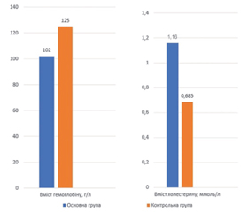
Актуальність. Експериментальні дослідження, що вивчають консервацію постекстракційної лунки в умовах атеросклерозу у кролів, значно обмежені. Мета: вивчення даних літератури щодо моделювання атеросклерозу та стоматологічних втручань на кролях; перевірка запропонованої експериментальної моделі. Матеріали та методи. Було проведено наративний огляд літератури у базі даних PubMed. Експериментальне дослідження передбачало щоденне введення кролям основної групи 95% холестерину (ХС), розчиненого у соняшниковій олії та змішаного зі 100 г тертої моркви, по 0,5 г/кг маси тіла 12 тижнів. Кролі контрольної групи отримували стандартний раціон. Через 12 тижнів здійснювався забір венозної крові з проведенням загального аналізу крові та визначенням загального ХС і тригліцеридів. Результати опрацьовано статистично (Statistica 14.0, TIBCO Software Inc, США). Дані подано як медіану (нижній; верхній квартилі). За поріг значущості прийнято р < 0,05. Результати. Для моделювання атеросклерозу запропоновано застосовувати ХС, розчинений в олеїновій олії, пальмову та кокосову олію, смалець, ХС у гранулах із додаванням сіна, люцерни посівної та зелені тощо. Єдиного стандартизованого протоколу дієтичної індукції атеросклеротичних змін не існує. Кролів активно використовують для вивчення процедур і матеріалів для постекстракційної кісткової пластики (застосовували оброблений дентинний матрикс з багатим на тромбоцити фібрином, звичайний чи перехресно-зв’язаний колаген, полімерну мікроволоконну матрицю). За результатами власного експерименту, через 12 тижнів атерогенної дієти в особин дослідної групи спостерігалися візуальні відмінності (масна шерсть, гіперемована слизова оболонка, рихлі ясна). В основній групі спостерігався вірогідно нижчий рівень гемоглобіну порівняно з контрольною групою: 102,00 (77,00; 113,00) г/л проти 125,00 (124,00; 128,00) г/л, а також менший показник ширини розподілу еритроцитів, обидва р < 0,05. Вміст ХС у групах дослідження вірогідно відрізнявся і становив 0,685 (0,650; 0,960) ммоль/л у контрольній групі та 1,16 (0,93; 1,63) ммоль/л у дослідній, р < 0,05. Висновки. Кролі є зручним біологічним видом для моделювання стоматологічних досліджень на тлі атеросклерозу. Описана вище дієтологічна модель атеросклерозу на кролях дозволяє досягти проатерогенного профілю крові. Отримана в експерименті гіперхолестеринемія асоціювалася з анемією та зниженням ширини розподілу еритроцитів у кролів.
Background. Experimental studies investigating tooth extraction socket preservation in rabbits with atherosclerosis are scarce. Objective: to review the current literature on experimental models of atherosclerosis and dental interventions in rabbits and to validate the proposed experimental model. Materials and methods. A narrative literature review was conducted using the PubMed database. In the experimental study, rabbits in the main group received 95% cholesterol dissolved in sunflower oil and mixed with 100 g of grated carrots at a dose of 0.5 g/kg body weight daily for 12 weeks. Animals in the control group were maintained on a standard diet. After 12 weeks, venous blood samples were collected for a complete blood count and determination of total cholesterol and triglyceride levels. Statistical analysis was performed using Statistica 14.0 (TIBCO Software Inc., USA). Data are presented as median (interquartile range). Statistical significance was defined as p < 0.05. Results. Various dietary approaches have been proposed for experimental induction of atherosclerosis, including cholesterol dissolved in vegetable oil, palm and coconut oil, lard, and cholesterol granules supplemented with hay, alfalfa, or fresh greens. However, no standardized dietary protocol for inducing atherosclerotic changes has yet been established. Rabbits are widely used as an experimental model for evaluating techniques and biomaterials for bone grafting after tooth extraction, including treated dentin matrix combined with platelet-rich fibrin, native and cross-linked collagen, and polymeric microfiber matrices. In our study, rabbits fed an atherogenic diet for 12 weeks demonstrated visible clinical changes, including poor coat condition, hyperemic oral mucosa, and friable gingival tissues. The experimental group showed significantly lower hemoglobin levels than the control group — 102.00 (77.00; 113.00) g/L vs. 125.00 (124.00; 128.00) g/L, as well as a significantly lower red cell distribution width, both p < 0.05. Total cholesterol concentrations differed significantly between the groups, reaching 0.685 (0.650; 0.960) mmol/L in the control group and 1.16 (0.93; 1.63) mmol/L in the experimental group (p < 0.05). Conclusions. Rabbits represent a suitable experimental model for dental research in the setting of atherosclerosis. The proposed dietary protocol successfully induced a proatherogenic blood profile. Experimentally induced hypercholesterolemia was associated with anemia and a reduced red cell distribution width in rabbits.
Список литературы
1. Al Otaibi NM, Dunne M, Ayoub AF, Naudi KB. A novel surgical model for the preclinical assessment of the osseointegration of dental implants: a surgical protocol and pilot study results. J Transl Med. 2021;19(1):276. doi: 10.1186/s12967-021-02944-w.
2. Van Erk M, Van Luijk J, Yang F, Leeuwenburgh SCG, Sn-chez-Fernndez MJ, Hermans E, et al. A systematic review and meta-analyses on animal models used in bone adhesive research. J Orthop Res. 2022;40(3):624-633. doi: 10.1002/jor.25057.
3. Blanc-Sylvestre N, Bouchard P, Chaussain C, Bardet C. Pre-Clinical Models in Implant Dentistry: Past, Present, Future. Biomedicines. 2021;9(11):1538. doi: 10.3390/biomedicines9111538.
4. Cheng J, Huang H, Chen Y, Wu R. Nanomedicine for Diagnosis and Treatment of Atherosclerosis. Adv Sci (Weinh). 2023;10(36):e2304294. doi: 10.1002/advs.202304294.
5. Liang L, Xiao F, Li M, Xu F, Peng F, Luo C, et al. Quantification of Cardiac Iron Overload at 3 TMRI in a Rabbit Model Utilizing ME-GRE T2* Sequence. J Magn Res on Imaging. 2024. doi: 10.1002/jmri.29534.
6. Hao Q, Lv S, Zhang J, Liu H. Effects of cardiac contractility modulation on autophagy and apoptosis of cardiac myocytes in rabbits with chronic heart failure. PLoS One. 2024;19(12):e0306242. doi: 10.1371/journal.pone.0306242.
7. Kar R, Mukhopadhyay D, Angom RS. Progress in Disease Modeling for Myocardial Infarction and Coronary Artery Disease: Bridging In Vivo and In Vitro Approaches. Hearts. 2024;5(4):429-447. doi: 10.3390/hearts5040031.
8. Hong JY, Herr Y, Naenni N, Thoma DS, Buranawat B, Shin SI, et al. Comparison of Healing Outcomes Between Simultaneous and Staged Implant Placement With Sinus Floor Elevation: A Preclinical Study Using a Rabbit Sinus Model. J Clin Periodontol. 2025;52(12):1802-1812. doi: 10.1111/jcpe.70025.
9. Al Thani NA, Hasan M, Yalcin HC. Use of Animal Models for Investigating Cardioprotective Roles of SGLT2 Inhibitors. J Cardiovasc Transl Res. 2023;16(5):975-986. doi: 10.1007/s12265-023-10379-5.
10. Genovs P, Arias-Mutis J, Parra G, Such-Miquel L, Zarzoso M, Del Canto I, et al. Development and Long-Term Follow-Up of an Experimental Model of Myocardial Infarction in Rabbits. Animals (Basel). 2020;10(9):1576. doi: 10.3390/ani10091576.
11. Reed SD. Histologic characterization of spontaneous catecholamine-induced cardiomyopathy in laboratory New Zealand White rabbits. J Vet Diagn Invest. 2024;36(5):759-764. doi: 10.1177/1040638724124474.
12. International BR. Retracted: Cardiac Contractility Modulation Attenuates Chronic Heart Failurein a Rabbit Model via the PI3K/AKT Pathway. Biomed Res Int. 2024;2024:9760278. doi: 10.1155/2024/9760278.
13. Freeman M, Huethorst E, Boland E, Dunne M, Burton F, Denning C, et al. A novel method for the percutaneous induction of myocardial infarction by occlusion of small coronary arteries in the rabbit. Am J Physiol Heart Circ Physiol. 2024;326(3):H735-H751. doi: 10.1152/ajpheart.00657.2023.
14. Zhang Y, Fatima M, Hou S, Bai L, Zhao S, Liu E. Research methods for animal models of atherosclerosis (Review). Mol Med Rep. 2021;24(6):871. doi: 10.3892/mmr.2021.12511.
15. Bonaterra GA, Bender K, Wilhelm B, Schwarzbach H, Metz S, Kelber O, et al. Effect of cholesterol re-supplementation and atorvastatin on plaque composition in the thoracic aorta of New Zealand white rabbits. BMC Cardiovasc Disord. 2020;20(1):420. doi: 10.1186/s12872-020-01703-x.
16. Abou-Khalil S, Poulsen R, Stemerman MB, Moore S, Alavi MZ. Vitamin B12 Inhibits Progression of Hypercholesterolemia and Atherosclerosis in Rabbits Fed a High Cholesterol Diet. McGill J Med [Internet]. 2020 [cited 2026 Jul. 8];3(1). https://mjm.mcgill.ca/article/view/584.
17. Donis N, D’Emal C, Hulin A, Gustin M, Dulgheru R, Lancellotti P, et al. Effect of cholesterol- and palmoil-enriched diets on rabbit aorta and aortic valve calcification. Archives of Cardiovascular Diseases Supplements. 2020;12(2–4):251. doi: 10.1016/j.acvdsp.2020.03.124.
18. Donis N, Jiang Z, D’Emal C, Dulgheru R, Giera M, Blomberg N, et al. Regular Dietary Intake of Palmitate Causes Vascular and Valvular Calcificationin a Rabbit Model. Front Cardiovas Med. 2021;8:692184. doi: 10.3389/fcvm.2021.692184.
19. Rahim INA, Omar E, Muid SA, Kasim NAM. Antiatherogenic and plaque stabilizing effects of saffron ethanolic extract in athe–rosclerotic rabbits. BMC Complement Med Ther. 2025;25(1):187. doi: 10.1186/s12906-025-04927-6.
20. Zabriskie MS, Wang S, Kim SE, McNally JS, de Havenon AH, Parker DL, et al. New Zealand White Rabbits Fed High Cholesterol Diets Develop Morbid Systemic Diseases before Intracranial Atherosclerosisis Detected. J Vet Sci Med Diagn. 2019;8(3):273.
21. Tekin MB, Toker H. The effect of hyperlipidemia on bone graft regeneration of peri-implantal created defects in rabbits. Int J Implant Dent. 2019;5(1):18. doi: 10.1186/s40729-019-0170-x.
22. Assmann AK, Buschmann J, Reimers S, Karakas A, Weber E, Aubin H, et al. Development of an atherosclerosis rabbit model to evaluate the hemodynamic impact of extracorporeal circulation. Animal Model Exp Med. 2025;8(3):523-533. doi: 10.1002/ame2.12556.
23. Papavasileiou DA, Leventis M, Agrogiannis G, Kalyvas D. Effect of Isolating the Periosteum with a Resorbable Barrier Membrane on Neoangiogenesis in Guided Bone Regeneration: An Experimental Study. Cureus. 2025;17(3):e81069. doi: 10.7759/cureus.81069.
24. Sedek EM, Khalil NM, Abdul-Monem MM. Histomorphometric and immunohistochemical assessment of treated dentin matrix delivered by platelet-rich fibrin for socket preservation in rabbit model. BMC OralHealth. 2025;25:225. doi: 10.1186/s12903-025-05569-3.
25. Li H, Yang C, Chen G, Wang B, Li J, Xu L. Effect of radiation cross-linked collagen scaffold in alveolar ridge preservation of extraction socket. J Biomed Mater Res A. 2024;112(10):1699-1711. doi: 10.1002/jbm.a.37723.
26. Covani U, Giammarinaro E, Panetta D, Salvadori PA, Cosola S, Marconcini S. Alveolar Bone Remodeling with or without Collagen Filling of the Extraction Socket: A High-Resolution X-ray Tomography Animal Study. J Clin Med. 2022;11(9):2493. doi: 10.3390/jcm11092493.
27. Soldatyuk V, Rozhko M, Pantus A. Clinical-morphological substantiation of the fibrous matrix with biogel cenobone application for preservation of the alveolar process of the jaw safter the teeth removal. Georgian Med News. 2022;(330):74-80.
28. Scarano A, Khater AGA, Gehrke SA, Inchingolo F, Tari SR. Animal Models for Investigating Osseointegration: An Overview of Implant Research over the Last Three Decades. J Funct Biomater. 2024;15(4):83. doi: 10.3390/jfb15040083.
29. Motschnig T. Establishment of Haematologic Reference Intervals for New Zealand White Rabbits with the Siemens Advia 2120i™. Diplomar beit. Veterinr medizinische Universitt Wien, 2020. https://phaidra.vetmeduni.ac.at/api/object/o:2839/get.
30. Tanczos B, Somogyi V, Bombicz M, Juhasz B, Nemeth N, Deak A. Changes of Hematological and Hemorheological Parameters in Rabbits with Hypercholesterolemia. Metabolites. 2021;11(4):249. doi: 10.3390/metabo11040249.
31. Abdelhalim MA, Moussa SA. Biochemical changes of hemoglobin and osmotic fragility of red blood cells in high fat diet rabbits. Pak J Biol Sci. 2010;13(2):73-7.
32. Oliveira JA, de Oliveira Alves R, Nascimento IM, et al. Pro- and anti-inflammatory cytokines and osteoclastogenesis-related factors in peri-implant diseases: systematic review and meta-analysis. BMC Oral Health. 2023;23:420. doi: 10.1186/s12903-023-03072-1.
33. Sun H, Meng S, Chen J, Wan Q. Effects of Hyperlipidemia on Osseointegration of Dental Implants and Its Strategies. J Funct Biomater. 2023;14(4):194. doi: 10.3390/jfb14040194.
34. Li H, Chen X, Wang B, Wu C, Li J, Xu L. Radiation cross-linked collagen scaffolds facilitatero otcoverage and keratinized gingival regeneration. Bioact Mater. 2025;51:138-149. doi: 10.1016/j.bioactmat.2025.04.023.
35. Ergnol E, Akgn Orkun R, emsi R, Ekim O, Uludamar A, Dinel Sepici A. Novel Method for Tooth Extractionin Rabbits: Advantages and Considerations. Indian Journal of Animal Research. 2026;60(1):157-162. doi: 10.18805/IJAR.BF-1901.
36. Imoto K, Hoshi K, Odashima K, Nogami S, Unuma H, Ya–mauchi K. Static and dynamic guided bone regeneration using a shape-memory polyethylen eterephthalate membrane: An experimental study in rabbit mandible. Clin Implant Dent Relat Res. 2024;26(4):734-741. doi: 10.1111/cid.13337.