
Журнал «Травма» Том 27, №5, 2026
Вернуться к номеру
Зв’язок передопераційних імунозапальних показників із порушенням консолідації переломів стегнової кістки в пацієнтів похилого та старечого віку
Авторы: A.A. Ablakulov (1), V.R. Akramov (1), B.Z. Khamdamov (1), A.O. Okhunov (2), J.P. Radjabov (3)
(1) - Bukhara State Medical Institute named after Abu Ali ibn Sino, Bukhara, Republic of Uzbekistan
(2) - Tashkent State Medical University, Tashkent, Republic of Uzbekistan
(3) - Samarkand State Medical University, Samarkand, Republic of Uzbekistan
Рубрики: Травматология и ортопедия
Разделы: Клинические исследования
Версия для печати
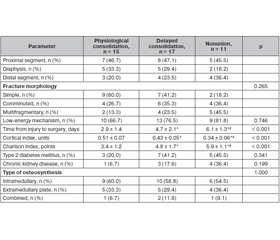
Актуальність. Порушення консолідації переломів стегнової кістки в пацієнтів літнього віку є клінічно значущою проблемою, що визначається не лише механічними, а й біологічними чинниками. Розуміння ролі передопераційних імунозапальних та остеометаболічних маркерів у формуванні результату зрощення залишається недостатнім. Мета: оцінити зв’язок передопераційних показників системного запалення, цитокінової регуляції та кісткового ремоделювання з наступним порушенням консолідації переломів стегнової кістки в пацієнтів похилого і старечого віку. Матеріали та методи. Одноцентрове ретроспективне когортне дослідження включало 43 осіб, прооперованих з приводу переломів стегнової кістки (2020–2022). Кожному пацієнту присвоювали категорію на основі клінічної та радіологічної оцінки через 6–12 місяців або під час ревізійної операції: фізіологічна консолідація (n = 15), уповільнена консолідація (n = 17), незрощення (n = 11). Основним лабораторним показником було співвідношення RANKL/OPG (ліганд рецептора активатора ядерного фактора каппа
B/остеопротегерин). Додатково оцінювали нейтрофільно-лімфоцитарний індекс (NLR), рівні С-реактивного білка (CRP), інтерлейкіну-6 (IL-6), фактора некрозу пухлини (TNF-), IL-10, трансформуючого фактора росту (TGF-), RANKL та OPG. Застосовано дисперсійний аналіз Велча, точний критерій Фішера — Фрімена — Холтона, корекцію Бенджаміні — Хохберга й Холма. Результати. При погіршенні консолідації NLR (2,2 ± 0,7 3,5 ± 1,1 5,1 ± 1,8), IL-6 (7,8 ± 2,6 15,4 ± ± 5,1 26,7 ± 8,9 пг/мл), TNF- (9,6 ± 3,4 17,8 ± 5,7 29,3 ± 9,1 пг/мл) та співвідношення RANKL/OPG (0,29 ± 0,08 0,66 ± 0,19 1,49 ± 0,44; p < 0,001 для всіх) послідовно зростали (p < 0,001). Рівні IL-10 та TGF- знижувалися. При порушеній консолідації співвідношення RANKL/OPG було вищим на 0,70 од. (95% довiрчий iнтервал 0,49–0,90; Hedges g = 1,63), IL-6 — на 12,04 пг/мл, TNF- — на 12,72 пг/мл. Усі додаткові лабораторні відмінності, за винятком рівня моноцитів, залишалися вірогідними після корекції на множинні порівняння. Висновки. Порушення консолідації переломів стегнової кістки в пацієнтів літнього віку асоційоване з передопераційним прозапальним і резорбтивним профілем. Найбільші та найстійкіші відмінності виявлено для співвідношення RANKL/OPG, IL-6 і TNF-. Результати відображають нескориговані клінічні асоціації та потребують підтвердження в проспективному дослідженні на незалежній вибірці.
Background. Impaired consolidation of femoral fractures in elderly patients is a clinically significant problem determined not only by mechanical but also by biological factors. Understanding of the role of preoperative immuno-inflammatory and osteometabolic markers in shaping the union outcome remains insufficient. The purpose was to assess the relationship between preoperative post-traumatic indicators of systemic inflammation, cytokine regulation, and bone remodeling with subsequent impaired consolidation
of femoral fractures in elderly and senile patients. Materials and methods. A single-center retrospective cohort study included 43 patients operated on for femoral fractures (2020–2022). Each patient was assigned one mutually exclusive category based on clinical and radiological assessment at 6–12 months or at the time of revision surgery: physiological consolidation (n = 15), delayed consolidation (n = 17), nonunion (n = 11). The primary laboratory indicator was the RANKL/OPG (receptor activator of nuclear factor-kappa B ligand/osteo-protegerin) ratio. Additionally, the neutrophil-to-lymphocyte ratio (NLR), C-reactive protein (CRP), interleukin-6 (IL-6), tumor necrosis factor (TNF-), IL-10, transforming growth factor (TGF-), RANKL and OPG were studied. Welch’s analysis of variance, the Fisher-Freeman-Halton exact test, and the Benjamini-Hochberg and Holm corrections were applied. Results. As the outcome worsened, the NLR (2.2 ± 0.7 3.5 ± 1.1 5.1 ± 1.8), IL-6 (7.8 ± 2.6 15.4 ± 5.1 26.7 ± 8.9 pg/mL), TNF- (9.6 ± 3.4 17.8 ± 5.7 29.3 ± 9.1 pg/mL) and the RANKL/OPG ratio (0.29 ± 0.08 0.66 ± 0.19 1.49 ± 0.44; p < 0.001 for all) increased consistently. IL-10 and TGF- levels decreased. When delayed consolidation and nonunion were combined, the RANKL/OPG ratio was higher by 0.70 units (95% confidence interval 0.49–0.90; Hedges’ g = 1.63), IL-6 by 12.04 pg/mL, and TNF- by 12.72 pg/mL. All additional laboratory differences, except for monocyte levels, remained significant after correction for multiple comparisons. Conclusions. Impaired consolidation of femoral fractures in elderly patients was associated with a preoperative pro-inflammatory and resorptive profile. The largest and most consistent differences were found for RANKL/OPG, IL-6 and TNF-. The findings reflect unadjusted clinical associations and require confirmation in a prospective study on an independent sample.
перелом стегнової кістки; уповільнена консолідація; незрощення; RANKL/OPG; інтерлейкін-6; фактор некрозу пухлини ; нейтрофільно-лімфоцитарний індекс; літній вік
femoral fracture; delayed consolidation; nonunion; RANKL/OPG; interleukin-6; tumor necrosis factor ; neutrophil-to-lymphocyte ratio; elderly age