Інформація призначена тільки для фахівців сфери охорони здоров'я, осіб,
які мають вищу або середню спеціальну медичну освіту.

Підтвердіть, що Ви є фахівцем у сфері охорони здоров'я.


Всесвітній день боротьби із запальними захворюваннями кишечника
день перший
день другий

Коморбідний ендокринологічний пацієнт

Всесвітній день боротьби із запальними захворюваннями кишечника
день перший
день другий

Коморбідний ендокринологічний пацієнт

Международный эндокринологический журнал Том 16, №1, 2020

Вернуться к номеру

Інсулініндукований набряк: рідкісне ускладнення в пацієнта з уперше діагностованим цукровим діабетом 1-го типу (клінічний випадок)

Авторы: H.Y. Kostek(1), N. Köroğlu(2), F.Ö. Çömlek(1), E. Dilek(1), F. Tutunculer(1)
(1) — Department of Pediatrics, Division of Pediatric Endocrinology, Trakya University Faculty of Medicine, Edirne, Turkey
(2) — Department of Pediatrics, Trakya University Faculty of Medicine, Edirne, Turkey

Рубрики: Эндокринология

Разделы: Справочник специалиста

Версия для печати


Резюме

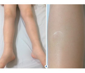
Спричинений введенням інсуліну набряк належить до рідкісних ускладнень інсулінотерапії. Він виникає невдовзі після початку інтенсивної терапії інсуліном у пацієнтів з уперше діагностованим цукровим діабетом (ЦД) 1-го типу або в пацієнтів із погано контрольованим ЦД 2-го типу після початку терапії інсуліном, а також у пацієнтів із низькою масою тіла при введеннi великих доз інсуліну. Процес характеризується розвитком набряку нижніх кінцівок або менш поширеним набряком після введення інсуліну з подальшим спонтанним розсмоктуванням протягом декількох тижнів. Автори повідомляють про випадок розвитку набряку нижніх кінцівок та мошонки в 9-річного хлопчика з уперше діагностованим ЦД 1-го типу через декілька днів після початку лікування інсуліном.

Вызванный введением инсулина отек относится к редким осложнениям инсулинотерапии. Он возникает вскоре после начала интенсивной терапии инсулином у пациентов с впервые диагностированным сахарным диабетом (СД) 1-го типа или у пациентов с плохо контролируемым СД 2-го типа после начала терапии инсулином, а также у пациентов с низкой массой тела при введении больших доз инсулина. Процесс характеризуется развитием отека нижних конечностей или менее распространенным отеком после введения инсулина с последующим самопроизвольным рассасыванием в течение нескольких недель. Авторы сообщают о случае развития отека нижних конечностей и мошонки у 9-летнего мальчика с впервые диагностированным СД 1-го типа через несколько дней после начала лечения инсулином.

Insulin-induced oedema is a rare complication of insulin therapy and occurs shortly after the initiation of intensive insulin therapy in patients with newly diagnosed type 1 diabetes mellitus (DM) or in patients with poorly controlled type 2 DM following the initiation of insulin therapy and also in underweight patients on large doses of insulin. It is characterized by the development of lower extremity oedema or less common generalized oedema after administration of insulin and resolves spontaneously within a few weeks. We report a case of a 9-year-old boy with newly diagnosed type 1 DM, who developed insulin oedema of lower extremities and scrotum within a few days after the initiation of insulin treatment.


Ключевые слова

набряк; цукровий діабет 1-го типу; інсулін

отек; сахарный диабет 1-го типа; инсулин

oedema; type 1 diabetes mellitus; insulin

Introduction

Peripheral or generalized oedema is an unusual complication of insulin therapy in the absence of heart, liver or renal diseases [1]. It has been reported in newly diagnosed type 1 diabetes mellitus (DM), in poorly controlled type 2 DM following the initiation of insulin therapy and in underweight patients on large doses of inslulin [2]. It can present with a spectrum of severity ranging from mild peripheral oedema to cardiac failure and massive serous effusions [3]. Its nature is commonly transient and benign and frequently is a self-limited condition that resolves spontaneously without any treatment or with diuretic therapy [3–7].
Although multiple potential mechanisms underlying insulin oedema have been proposed [5, 8], the pathogenetic mechanisms of insulin-induced oedema remain to be clarified [4]. The first pediatric report was described in 1979 and since then around 16 cases have been described [9, 10].
We report a 9-year-old boy with newly diagnosed type 1 DM, who presented with oedema of the lower extremities and scrotum within a few days after the initiation of insulin therapy.

Case presentation

A 9-year-old boy was admitted to our hospital with a 2-month history of polyuria and polydipsia and with a recent weight loss of 6 kg. He was diagnosed with type 1 DM. He had no family history of type 1 DM or autoimmune diseases. On admission, his height was 130 cm (–0.88 SD) and his weight was 27 kg (–0.75 SD), body mass index was 15.9 kg/m2. He was on pubertal Tanner stage 2. Physical examination was normal except for clinical signs of mild dehydration. Laboratory investigation revealed a blood glucose level of 476 mg/dl, ketonuria without acidosis, venous blood pH of 7.33 and elevated glycosylated haemoglobin concentration of 19.6 % (normal value 3.6–5.8). He was initially treated with subcutaneous regular insulin for determination of daily insulin requirement and was managed on basal-bolus regimen, consisting of once-daily insulin glargine and 3 times daily pre-meal insulin lispro (total 31 units daily, 1.6 units/kg/day). On the 4th day of the therapy, the insulin dosage had to be increased to 2.2 units/kg/day to achieve blood glucose level within the target range. Mild bilateral pitting ankle oedema was first noted on day 6 of the therapy and progressed over the tibiae and scrotum next day (Fig. 1). At this time, patient’s weight increased from 27 (on admission) to 32 kg. His blood pressure and vital signs were normal. The rest of his physical examination parameters were unremarkable. Laboratory investigations revealed renal and kidney function tests to be normal as well as serum albumin level. Urine analysis was normal. Chest X-ray and abdominal ultrasonography were normal. Doppler ultrasonography of the lower leg veins and arteries showed no evidence of thrombosis. Based on the clinical and laboratory findings, the patient was diagnosed with insulin oedema.
He was treated conservatively with salt and fluid restriction. Five days later, oedema started improving when his insulin requirement decreased to 1.1 units/kg/day with excellent glycemic control. Oedema completely disappeared within 10 days and no relapse occurred so far.
In addition, because he had Tanner stage 2 testicular development on physical examination, we performed a GnRH stimulation test due to suspicion of central precocious puberty (CPP). GnRH stimulation test revealed a pubertal luteinizing hormone peak of 5.08 mIU/L, confirmed the activation of the gonadotropic axis. Bone age was 10 year and 6 months according to the Greulich and Pyle method. A contrast enhanced brain magnetic resonance imaging was normal. A diagnosis of idiopathic CPP was performed and the patient started treatment with GnRH agonists.

Discussion

Insulin-induced oedema is a rare and probably underreported complication appearing after initiating or intensifying insulin therapy [11]. Its nature is generally transient and benign and it is usually a self-limiting condition that resolves spontaneously without any treatment or with diuretic therapy; however, it can present in a variety of ways, from mild peripheral oedema to cardiac failure and massive serous effusions [3, 5–7].
The diagnosis of insulin oedema is based on the exclusion of all other known causes of oedema. Therefore, it should be considered after excluding cardiac, renal and hepatic causes of oedema in a diabetic child [12]. Our patient was diagnosed with insulin oedema after other identifiable causes of oedema, such as cardiac dysfunction, liver failure, nephrotic syndrome and thrombosis were all excluded by ultrasonography and the results of blood and urine examinations.
The majority of the pediatric patients reported so far were recently diagnosed with type 1 DM [1, 6, 10, 12, 13]. The other several predisposition factors including the use of large dose insulin, underweight, hypoalbuminemia, recurrent hypoglycemia under poor diabetic control, current treatment for ketoacidosis and 3243 mitochondrial tRNALeu (UUR) mutation have been proposed in the literature [14].
Our patient had many risk factors for the development of insulin oedema. He had remarkably weight loss during the previous 2 months. He had been currently treated for diabetic ketosis and was taking large doses of insulin (> 1 units/kg/day). Despite all of these described predisposition factors mentioned in the literature the questions as to why insulin oedema is rare and whether any individual susceptibility or particular physiological conditions provoking oedema exist remain unanswered [14].
Multiple pathogenic mechanisms for insulin oedema are described. Insulin treatment has mainly two effects including antinatriuresis and increased capillary permeability. Insulin has a direct antinatriuretic effect by enhancing renal tubular sodium reabsorption by stimulating the Na+/K+ exchanger 3 in the proximal tubule, and it causes vasodilation and increases vascular permeability [4]. It has also an indirect antinatriuresis effect via suppressing secretion of glucagon which causes natriuresis [15]. Transient inappropriate hyperaldosteronism has been suggested as the other contributing factor to develop fluid retention [4]. Rarely, mutation of the 3243 mitochondrial tRNA has been reported in peripheral oedema and hepatic dysfunction, suggesting a further role of mitochondrial function in vasomotor function in the periphery but the majority of the patients with this mutation had type 2 DM [16].
Treatment approaches for insulin oedema are various depending on oedema severity. As insulin oedema is generally a transient condition, it may resolve with fluid and salt restriction and reduction in insulin dose.
Diuretic therapy may be indicated in more severe decompensated cases. An aldosterone antagonist such as spironolactone may be chosen in cases of severe insulin oedema associated with inappropriate hyperaldosteronism [4]. Specific enzyme therapy like coenzyme Q is used in selected patients with mitochondrial mutation [16]. Our patient was treated conservatively with fluid and salt restriction. Oedema started improving within 5 days and completely disappeared over 15 days. In most of the reported cases in literature, oedema resolved within 7–10 days with only fluid and salt restriction, without necessity of diuretics [1, 6, 10], similar to our case.
In conclusion, insulin-induced oedema should be considered during introduction of insulin therapy in children and adolescents with newly diagnosed type 1 DM especially on high-dose daily insulin therapy.
Conflicts of interests. Authors declare the absence of any conflicts of interests and their own financial interest that might be construed to influence the results or interpretation of their manuscript.

Список литературы

1. Mamoulakis D., Bitsori M., Galanakis E., Raissaki M., Kalmanti M. Insulin induced edema in children and adolescents. J. Paediatr. Child Health. 2006. 42. 655-657.

2. Juliusson P.B., Bjerknes R., Sovik O., Kvistad P.H. Generalized edema following insulin treatment of newly diagnosed diabetes mellitus. Tidsskr. Nor. Laegeforen. 2001. 212. 919-20.

3. Lee P., Kinsella J., Borkman M., Carter J. Bilateral pleural effusions, ascites, and facial and peripheral oedema in a 19-year-old woman 2 weeks following commencement of insulin lispro and detemir — an unusual presentation of insulin oedema. Diabet. Med. 2007. 24. 1282-85.

4. Kalambokis G., Tsatsoulis A., Economou G., Tsianos E.V. A case of insulin edema with inappropriate hyperaldosteronism. J. Endocrinol. Invest. 2004. 27. 957-960.

5. Evans D.J. Pritchard-Jones K., Trotman-Dikenson B. Insulin edema. Postgrad. Med. J. 1986. 62. 665-8.

6. Baş V.N., Cetinkaya S., Agladioglu S.Y., Kendirci H.V.P., Bilgili H., Yıldırım N., Aycan Z. Insulin oedema in newly diagnosed type 1 diabetes mellitus. J. Clin. Res. Pediatr. Endocrinol. 2010. 2. 46-48.

7. Adamo L., Thoelke M. Generalized insulin edema after intensification of treatment with insulin analogues. BMJ Case Report. 2013. doi: 10.1136/bcr-2012-007037.

8. Kalambokis G.N., Tsatsoulis A.A., Tsianos E.V. The edematogenic properties of insulin. Am. J. Kidney Dis. 2004. 44. 579-90.

9. Lawrence J.R., Dunnigan M.G. Diabetic (insulin) oedema. Br. Med. J. 1979. 2. 445.

10. Bulus A.D., Andiran N., Köksal A.O. Insulin edema in type 1 diabetes mellitus: Report of a case and brief review of the literature. Iran J. Pediatr. 2016. 26. e5077.

11. Hopkins D.F., Cotton S.J., Williams G. Effective treatment of insulin-induced edema using ephedrine. Diabetes Care. 1993. 16. 1026-1028.

12. Goturu A., Jain N., Lewis I. Bilateral cataracts and insulin oedema in a child with type 1 diabetes. BMJ Case Reports. 2013. doi: 10.1136/bcr-2012-008235.

13. Aravamudhan A., Gardner C., Smith C., Senniappan S. Insulin oedema in a child with newly diagnosed diabetes mellitus. Eur. J. Pediatr. 2014. 173. 685-687.

14. Zenda T., Murase Y., Yoshida I., Muramoto H., Okada T., Yagi K. Does the use of insulin in a patient with liver dysfunction increase water retention in the body, i.e. cause insulin oedema? Eur. J. Gastroenterol. Hepatol. 2003. 15. 545-549.

15. Spark R.F., Arky R.A., Boulter P.R., Saudek C.D., O’Brian J.T. Renin, aldosterone ad glucagon in the natriuresis of fasting. N. Eng. J. Med. 1975. 292. 1335-1340.

16. Suzuki Y., Kadowaki H., Taniyama M., Kadowaki T., Katagiri H. et al. Insulin edema in diabetes mellitus associated with the 3243 mitochondrial tRNA Leu (UUR) mutation: a case report. Diabetes Res. Clin. Pract. 1995. 29. 137-142.


Вернуться к номеру