Інформація призначена тільки для фахівців сфери охорони здоров'я, осіб,
які мають вищу або середню спеціальну медичну освіту.

Підтвердіть, що Ви є фахівцем у сфері охорони здоров'я.


Всесвітній день боротьби із запальними захворюваннями кишечника
день перший
день другий

Коморбідний ендокринологічний пацієнт

Всесвітній день боротьби із запальними захворюваннями кишечника
день перший
день другий

Коморбідний ендокринологічний пацієнт

Международный эндокринологический журнал Том 16, №6, 2020

Вернуться к номеру

Can metastasis and recurrence be detected with Endocan and Vascular Endothelial Growth Factor in thyroid papillary cancer?

Авторы: Hande Peynirci(1), Canan Ersoy(2), Pınar Sisman(3), Ozlem Saraydaroglu(4), Coskun Ozer Demirtas(5), Ozen Oz Gul(2)
(1) — Health Sciences University Kanuni Sultan Suleyman Training and Research Hospital, Department of Endocrinology and Metabolism Diseases, Istanbul, Turkey
(2) — Uludag University Faculty of Medicine, Department of Internal Medicine, Division of Endocrinology and Metabolism Diseases, Bursa, Turkey
(3) — Private Medicana Hospital, Department of Endocrinology and Metabolism Diseases, Bursa, Turkey
(4) — Uludag University Faculty of Medicine, Department of Pathology, Bursa, Turkey
(5) — Marmara University Faculty of Medicine, Department of Internal Medicine, Division of Gastroenterology, Istanbul, Turkey

Рубрики: Эндокринология

Разделы: Клинические исследования

Версия для печати


Резюме

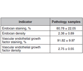
Актуальність. Відомо, що ендокан асоціюється з різними типами злоякісних утворень. Установлено, що фактор росту судинного ендотелію (VEGF) підвищує експресію ендокану. Метою проведеного дослідження було виявлення наявності рецидиву захворювання та/або метастазування за допомогою змін ендокану та VEGF у пацієнтів із папілярним раком щитоподібної залози. Матеріали та методи. Ретроспективне дослідження проводилося від січня 2005 року до грудня 2015 року. Реєстрували стать, вік, а також вік пацієнта на час діагностики тиреоїдного раку, тривалість спостереження. Дослідну групу було розподілено на дві групи, які становили пацієнти з післяопераційними рецидивами і/або метастазами та без них. Патологічні зразки обробляли Anti-ESM-1 та Anti-VEGFA, оцінювали відсоток забарвлення та щільність. Результати. Під спостереженням перебували 59 пацієнтів (43 жінки та 16 чоловіків). Середній вік становив 52,39 ± 13,75 року. Установлено, що в середньому найдовший діаметр пухлини становив 21,31 ± 20,20 мм, а тривалість спостереження — 37,24 ± 32,68 місяця. Серед пацієнтів 54,2 % мали рецидив та/або метастазування, тоді як у 45,8 % рецидиву чи метастазування не спостерігалося. Відсоток щільності ендокану становив 84,26 ± 20,32 та 2,56 ± 0,75 у групі рецидивів та/або метастазів, 75,56 ± 24,06 та 2,11 ± 1,02 — відповідно у групі без них. Щільність ендокану була вищою в пацієнтів із рецидивом та/або метастазами, але не досягла статистичної значущості (р = 0,077, р = 0,136 відповідно). Істотної різниці між двома групами щодо забарвлення та щільності VEGF не виявлено. Висновки. Необхідні подальші дослідження для оцінки ролі ендокану та фактора росту судинного ендотелію для прогнозування рецидиву та/або метастазування папілярної карциноми щитоподібної залози.

Актуальность. Известно, что эндокан ассоци­ируется с различными типами злокачественных образований. Установлено, что фактор роста сосудистого эндотелия (VEGF) повышает экспрессию эндокана. Целью проведенного исследования было выявление наличия рецидива заболевания и/или метастазирования с помощью изменений эндокана и VEGF у пациентов с папиллярным раком щитовидной железы. Материалы и методы. Ретроспективное исследование проводилось с января 2005 года по декабрь 2015 года. Регистрировали пол, возраст, а также возраст пациента на момент диагностики тиреоидного рака, продолжительность наблюдения. Исследовательская группа была разделена на две подгруппы, которые составили пациенты с послеоперационными рецидивами и/или метастазами и без них. Патологические образцы обрабатывали Anti-ESM-1 и Anti-VEGFA, оценивали процент окрашивания и плотность. Результаты. Под наблюдением находились 59 пациентов (43 женщины и 16 мужчин). Средний возраст составил 52,39 ± 13,75 года. Установлено, что наиболее длинный диаметр опухоли составил 21,31 ± 20,20 мм, а продолжительность наблюдения — 37,24 ± 32,68 месяца. Среди пациентов 54,2 % имели рецидив и/или метастазирование, тогда как у 45,8 % рецидива или метастазирования не наблюдалось. Процент плотности эндокана составил 84,26 ± 20,32 и 2,56 ± 0,75 в группе рецидивов и/или метастазов, 75,56 ± 24,06 и 2,11 ± 1,02 — соответственно в группе без них. Плотность эндокана была выше у пациентов с рецидивом и/или метастазами, но не достигла статистической значимости (р = 0,077, р = 0,136 соответственно). Существенной разницы между двумя группами по окраске и плотности VEGF не обнаружено. Выводы. Необходимы дальнейшие исследования для оценки роли эндокана и фактора роста сосудистого эндотелия для прогнозирования рецидива и/или метастазирования папиллярной карциномы щитовидной железы.

Background. Endocan is known to be associated with different type of malignancies and vascular endothelial growth factor (VEGF) has been shown to upregulate endocan expression. We purposed to determine whether the presence of disease recurrence and/or metastasis can be detected with pathological evaluation of endocan and VEGF in patients with thyroid papillary cancer. Materials and methods. This study was performed retrospectively between January 2005 and December 2015. Patients’ gender, age, also age at diagnosis, and duration of follow-up were recorded. The study group was divided into two groups comprised of patients with and without postoperative recurrence and/or metastasis. Pathological samples were treated with Anti-ESM-1 and Anti-VEGFA, staining percentage and density were evaluated. Results. A total of 59 patients (43 female and 16 male) were included. The mean age was 52.39 ± 13.75 years. Mean longest tumor diameter was found to be 21.31 ± 20.20 mm, and follow-up duration was 37.24 ± 32.68 months. Among the patients, 54.2 % had recurrence and/or metastasis, while 45.8 % did not have either. The percentage of endocan staining and density was 84.26 ± 20.32 and 2.56 ± 0.75 in the recurrence and/or metastases group, 75.56 ± 24.06 and 2.11 ± 1.02, respectively in the group without. Endocan staining and density was higher in the patients with recurrence and/or metastasis but not statistically significant (p = 0.077, p = 0.136, respectively). No significant difference was found between two groups in terms of VEGF staining and density. Conclusions. These markers might be further evaluated for determination their role in recurrence and/or metastasis of thyroid papillary carcinoma.


Ключевые слова

папілярний рак щитоподібної залози; ендокан; фактор росту судинного ендотелію; рецидив; метастазування

папиллярный рак щитовидной железы; эндокан; фактор роста сосудистого эндотелия; рецидив; метастазирование

thyroid papillary cancer; endocan; vascular endothelial growth factor; recurrence; metastasis

Introduction

Papillary thyroid carcinomas are responsible for more than 75–85 % of all thyroid cancers. They are more than twice as frequent in females and the incidence of papillary cancer has been found to increase from 3.4 to 12.5 per 100,000 individuals in the last 50 years; however, death rates from the disease have remained stable (around 0.5 per 100,000 individuals) [1]. Various clinical and pathologic features of the disease that have been identified as risk factors for tumor recurrence and mortality are as follows: age at diagnosis, primary tumor size, presence of soft tissue invasion, and distant metastases. In fact, improved outcomes have been associated with the extent of initial surgery and the use of radioiodine therapy in those with advanced disease [2]. Whereas new serological markers with adequate efficiency and specificity are expected to identify for preventing unnecessary treatments with adverse effects, negative impact of the disease on the patients quality of life due to the fear of recurrences, and higher health costs.
Endocan is a soluble proteoglycan (50 kDa) which is constituted of a mature polypeptide of 165 amino acids and a single dermatan sulphate chain covalently attached to the serine residue at position 137. Previously known as endothelial-cell-specific molecule-1 (ESM-1) is secreted by human vascular endothelial cells. Endocan, as a proteoglycan (PG) function in the regulation of important endothelial processes, including proliferation, differentiation, migration, and cell adhesion [3]. Additionally, it has also been demonstrated to play a role in inflammation, shock, vascular disorders, and angiogenesis [4, 5]. The formation of new blood vessels from pre-existing ones is defined as angiogenesis. It is a crucial process in normal physiology as well as pathological conditions such as inflammation, abnormal tumor growth and metastasis. Growth factors role in promoting angiogenesis like endocan is also well-known. Vascular endothelial growth factor (VEGF) is one of the most important proangiogenic molecules which is specific for endothelial cells and it has critical effects [6]. VEGF, a member of the Platelet Derived Growth Factor Superfamily, is a 34- to 45-kDa heparin binding glycoprotein and plays an important role in thyroid malignancies [6, 7]. Besides, VEGF’s critical control over the synthesis and secretion of endocan is also revealed [8]. 
There is an increasing data that had demonstrated an association between overexpression of endocan levels and lung, cardiovascular, kidney, and autotoimmune disorders, sepsis and preeclampsia as well as different types of malignancies [3, 4, 9, 10]. An overwhelming majority of researchers consider endocan to be a valid therapeutic target in cancer. In this study, we aimed to determine the usefulness of pathological endocan and VEGF staining in identifying the presence of recurrence and/or metastasis in thyroid papillary cancer.

Materials and methods

This was a retrospective study performed between January 2005 and December 2015 by collaboration of the Endocrinology and Metabolism Diseases and Pathology Departments of Uludag University Medical Faculty, Bursa, Turkey. Patients older than 18 years of age who had confirmed diagnosis of thyroid papillary cancer due to the histopathological examination with a complete medical record were included in the study. Patients with other organs malignancies, cardiovascular, lung and kidney diseases that can affect endocan expression were excluded. During the conduct of the study, Good Clinical Practices Guidelines and the Declaration of Helsinki were followed. The study was approved by Uludağ University Ethics Committee (Reference number: 2019.10/12).
Patients’ gender, age, also age at diagnosis, and duration of follow-up were assessed and recorded from the files. The study group was divided into two groups comprised of patients with and without postoperative recurrence and/or metastasis. The tumor diameter recorded with preoperative USG and tumor diameter in the pathological examination were reevaluated, and the largest of these was defined as the final tumor diameter.
Immunohistochemical staining with Anti-ESM-1 (Polyclonal, ab224591, Abcam, 1/200 dilution) and Anti VEGFA (Polyclonal, ab39250, Abcam, 1/200 dilution) were performed on pathology samples. Density and staining rates were compared among the groups.
Statistical analysis was performed using the Statistical Package for Social Sciences (SPSS) software version 20 for Windows (IBM SPSS Inc., Chicago, IL). Data are presented as mean ± standard error of mean (SEM). Statistical comparisons between the means of two groups were performed using the Student’s t-test (two-tailed). P values lower or equal to 0.05 were considered to be statistically significant.

Results

In this study, a total of 59 patients diagnosed as thyroid papillary cancer were included. Forty-three (72.9 %) were female and 16 (27.1 %) were male and were aged between 24–78 (52.39 ± 13.75) years. The mean age at diagnosis was 47.08 ± 14.38 (16–74) years. Mean longest tumor diameter was found to be 21.31 ± 20.20 mm and follow-up duration was 37.24 ± 32.68 months. Endocan and VEGF staining percentages and densities were shown in Table 1. 
Of the 27 (45.8 %) patients without recurrence and/or metastasis, 21 (77.8 %) were female and 6 (22.2 %) were male. The mean age of these patients was 55.41 ± 10.65 (34–76) years, while mean age at the time of diagnosis was 50.81 ± 10.79 (28–74) years. The mean longest tumor diameter was 15.74 ± 17.65 mm and follow-up period was 45.96 ± 26.78 months. The percentage and density of endocan was 75.56 ± 24.06 % and 2.11 ± 1.02, respectively. VEGF staining percentage was 92.08 ± 9.66 % and density was 2.79 ± 0.51 in this group. 
Of the 32 (54.2 %) patients with recurrence and/or metastasis, 22 (68.8 %) were female and 10 (31.3 %) were male. The patients aged between 24 and 78 (49.84 ± 15.62) years and the mean age at diagnosis was 43.94 ± 16.35 (16–72) years. The mean longest tumor diameter was 26.16 ± 21.28 mm and follow-up period was 29.88 ± 35.70 months. The percentage of endocan staining was 84.26 ± 20.32 % and density was 2.56 ± 0.75. The percentage and density of VEGF was 91.61 ± 10.36 % and 2.71 ± 0.59, respectively with recurrence and/or metastasis.
There was a statistically significant difference in terms of tumor diameter and follow-up duration between the patients with or without recurrence and/or metastasis. No significant difference was noted between the two groups with respect to endocan staining/density and VEGF staining/density. The datas of the two groups were shown in Table 2. 

Discussion

The most common cancer is thyroid cancer amongst endocrine gland tumors and the most common histotype is papillary cancer. In the literature, a study involving patients with a median follow-up of 16 years found that the cancer-related mortality in patients with papillary cancer in the absence of metastases was only 6 percent [11]. Although the majority of patients with papillary cancer are not expected to die directly due to their disease; early recognition of the metastasis and/or recurrence of papillary cancer will positively affect the prognosis of a small but significant group of patients and also health expenditure [2, 11]. Despite extensive studies in determining biomarkers for the prognosis of thyroid cancers, there is little evidence and few real candidates for specific biomarkers [12–14]. Therefore, there is a requirement for the determination of markers that can predict which patients have a higher risk of metastasis and recurrence or to identify patients with poor outcome in papillary thyroid cancers.
Endocan was first described by Lassalle et al. in 1996 after cloning from human umbilical vein endothelial cell complementary DNA library [15]. It is a circulating PG that has been shown to have a role in inflammation and tumor progression; therefore, recently it has been studied with increasing interest as an important contributor to cancer and inflammation, both structurally and functionally [16, 17]. Various studies employing semi-quantitative and quantitative measurements have identified endocan to be associated with various characteristics of cancer and cancer cells, including tumor development [18], angiogenesis and invasion [19], cell survival and metastasis development [20], and also patient survival [21]. The normal function of endocan may shed some light to these associations with cancer, as its expression is known to be increased with activation/presence of inflammation and various angiogenic factors that play a role in tumor development and progression [9]. For instance, its role in the adhesion of leukocytes and endothelial cells may also be effective in the adhesion of circulating cancerous cells to the endothelial vasculature, which are essential processes for invasion [22]. Due to these properties, endocan is regarded as a potential endothelial cell marker and a new target for therapy in recent years [5]. The association between endocan levels and several tumors such as hepatocellular carcinoma, colorectal cancer, gastric cancer, ovarian cancer, glioblastoma, lung cancer, nasopharyngeal carcinoma, and bladder cancer were shown in some studies but we could not find any data regarding the levels of endocan in patients with thyoid papillary cancer [9, 10, 19–21]. To our knowledge, this is the first study to investigate the endocan staining in the determination of recurrence and/or metastasis in papillary cancer. Additionally, there is little information about the relationship between benign thyroid diseases and endocan levels. Arpacı et al, showed that subclinical hypothyroidism is associated with increased levels of serum endocan due to the its endothelial dysfunction [23].
Various inflammatory cytokines such as tumor necrosis factor-α (TNF-α) and interleukin-1 (IL-1) induce secretion of endocan. Moreover, growth factors, especially VEGF have been shown to upregulate endocan expression [8]. The association of VEGF expression and thyroid carcinoma has been demonstrated by various studies. The regulation of VEGF production in thyroid cancer is unclear. Hypoxia, low pH, inflammatory cytokines such as IL6, growth factors, sex hormones, and chemokines are established factors that increase VEGF production [24]. As it is seen in physiological events and tumor growth, its role in metastasis or recurrence of different kind of tumors such as head and neck cancers, breast cancer, lung cancer, oesophageal cancer, colon cancer, hepatocellular carcinoma, gastric cancers, kidney cancer, ovarian cancer, bladder cancer and melanoma is also revealed [6, 25, 26]. Increased expression of VEGF on determinating recurrence and/or metastasis of thyroid papillary carcinoma has not yet been clearly elucidated. Indeed, inconsistency exists in the literature concerning the effect of increased VEGF expression on increased risk of metastasis or recurrence. Some studies have shown that elevation of VEGF in thyroid papillary cancer is associated with recurrence and poor prognosis [25, 27, 28]. Lack of association between the risk of recurrence and/or metastasis and increased VEGF expression in the previous study is in line with the data from several studies such as another Turkish study done by Karaca et al. They demonstrated that VEGF expression was higher in differantiated thyroid carcinoma than the nodular goiter but they did not found a correlation between the expression of VEGF and the size of tumor, capsule invasion, lymph node metastasis or progression-free survival [29]. In another study conducted by Soh et al. no difference was reported in VEGF staining between primary and metastatic thyroid tumors [7]. In another recent study, no significant difference was revealed in VEGF, VEGFR-1 and VEGFR-2 expression in terms of lymph node involvement and extra-thyroidal extension [30]. 
The main limitation of our study is a relatively small sample size. Also, the retrospective single-center design is likely to cause bias. We also did not evaluate other factors/mediators which are known to be important in the activation of endocan expression.

Conclusions

In the current study, endocan staining and density was higher in the patients with recurrence and/or metastasis but not statistically significant. No differences were found between patients with and without metastasis/recurrence of papillary thyroid cancer in terms of VEGF staining and density. These findings may be related to the relatively small number of patients included. Further prospective researches with higher number of patients is warrented to predict their role in recurrence and/or metastasis.

Additional information

The authors state that this manuscript is original and has not been previously published and is not under consideration in the same or substantially similar form in any other journals. All authors listed have contributed sufficiently to the project to be included as authors, and all those who are qualified to be authors are listed in the author by line.
Conflicts of interests. Authors declare the absence of any conflicts of interests and their own financial interest that might be construed to influence the results or interpretation of their manuscript.
Funding Source Declaration
This research did not receive any specific grant from any funding agency in the public, commercial, or not-for-profit sector.
Authors contribution: Hande Peynirci — concept and design of the study, collection and processing of the material, analysis of the data, text writing, final approval of the version; Canan Ersoy — concept and design of the study, text writing, final approval of the version; Pınar Sisman — collection and processing of the material, analysis of the data, final approval of the version; Ozlem Saraydaroglu — collection and processing of the material, analysis of the data, final approval of the version; Coskun Ozer Demirtas — collection and processing of the material, final approval of the version; Ozen Oz Gul — analysis of the data, final approval of the version.

Список литературы

  1. Davies L., Welch H.G. Current thyroid trends in the United States. JAMA Otolaryngol Head Neck Surg. 2014. 140(4). 317-322. doi: 10.1001/jamaoto.2014.1.
  2. Eustatia-Rutten C.F., Corssmit E.P., Biermasz N.R., Pereira A.M., Romijn J.A., Smit J.W. Survival and death causes in differentiated thyroid carcinoma. J. Clin. Endocrinol. Metab. 2006. 91(1). 313-319. doi: 10.1210/jc.2005-1322. 
  3. Kali A., Shetty K.S. Endocan: a novel circulating proteoglycan. Indian J. Pharmacol. 2014. 46(6). 579-583. doi: 10.4103/0253-7613.144891.
  4. Balta S., Mikhailidis D.P., Demirkol S., Ozturk C., Celik T., Iyisoy A. Endocan: A novel inflammatory indicator in cardiovascular disease? Atherosclerosis. 2015. 243(1). 339-343. doi: 10.1016/ j.atherosclerosis. 2015.09.030.
  5. Sarrazin S., Adam E., Lyon M. et al. Endocan or endothelial cell specific molecule-1 (ESM-1): a potential novel endothelial cell marker and a new target for cancer therapy. Biochim. Biophys. Acta. 2006. 1765(1). 25-37. doi: 10.1016/j.bbcan.2005.08.004.
  6. Ferrara N. Vascular endothelial growth factor: basic science and clinical progress. Endocr. Rev. 2004. 25(4). 581-611. doi: 10.1210/er.2003-0027.
  7. Soh E.Y., Duh Q.Y., Sobhi S.A. et al. Vascular endothelial growth factor expression is higher in differentiated thyroid cancer than in normal or benign thyroid. J. Clin. Endocrinol. Metab. 1997. 82(11). 3741-3747. doi: 10.1210/jcem.82.11.4340.
  8. Rennel E., Mellberg S., Dimberg A. et al. Endocan is a VEGF-A and PI3K regulated gene with increased expression in human renal cancer. Exp. Cell. Res. 2007. 313(7). 1285-1294. doi: 10.1016/j.yexcr.2007.01.021.
  9. Delehedde M., Devenyns L., Maurage C.A., Vivès R.R. Endocan in Cancers: A Lesson from a Circulating Dermatan Sulfate Proteoglycan. Int. J. Cell. Biol. 2013. 2013. 705027. doi: 10.1155/2013/705027.
  10. Yang J., Yang Q., Yu S., Zhang X. Endocan: A new marker for cancer and a target for cancer therapy. Biomed. Rep. 2015. 3(3). 279-283. doi: 10.3892/br.2015.438.
  11. Mazzaferri E.L., Jhiang S.M. Long-term impact of initial surgical and medical therapy on papillary and follicular thyroid cancer. Am. J. Med. 1994. 97(5). 418-428. doi: 10.1016/0002-9343(94)90321-2.
  12. Soares P., Celestino R., Melo M., Fonseca E., Sobrinho-Simões M. Prognostic biomarkers in thyroid cancer. Virchows Arch. 2014. 464(4). 333-346. doi: 10.1007/s00428-013-1521-2.
  13. Shi Y., Su C., Hu H. et al. Serum MMP-2 as a potential predictive marker for papillary thyroid carcinoma. PLoS One. 2018. 13(6). e0198896. doi: 10.1371/journal.pone.0198896.
  14. Zarkesh M., Zadeh-Vakili A., Akbarzadeh M., Fanaei S.A., Hedayati M., Azizi F. The role of matrix metalloproteinase-9 as a prognostic biomarker in papillary thyroid cancer. BMC Cancer. 2018. 18(1). 1199. doi: 10.1186/s12885-018-5112-0.
  15. Lassalle P., Molet S., Janin A. et al. ESM-1 is a novel human endothelial cell-specific molecule expressed in lung and regulated by cytokines. J. Biol. Chem. 1996. 271(34). 20458-20464. doi: 10.1074/jbc.271.34.20458.
  16. Tı́már J., Lapis K., Dudás J., Sebestyén A., Kopper L., Kovalszky I. Proteoglycans and tumor progression: Janus-faced molecules with contradictory functions in cancer. Semin. Cancer Biol. 2002. 12(3). 173-186. doi: 10.1016/S1044-579X(02)00021-4.
  17. Delehedde M., Lortat-Jacob H., Gallagher J.T. et al. Proteoglycan involvement in inflammatory diseases. new developments in GAG-based therapies. Medicinal Chemistry Reviews-Online. 2005. 2. 345-357. doi: 10.2174/1567203054637560.
  18. Scherpereel A., Gentina T., Grigoriu B. et al. Overexpression of endocan induces tumor formation. Cancer Res. 2003. 63(18). 6084-6089.
  19. Chen L.Y., Liu X., Wang S.L., Qin C.Y. Over-expression of the Endocan gene in endothelial cells from hepatocellular carcinoma is associated with angiogenesis and tumour invasion. J. Int. Med. Res. 2010. 38(2). 498-510. doi: 10.1177/147323001003800213.
  20. Kang Y.H., Ji N.Y., Han S.R. et al. ESM-1 regulates cell growth and metastatic process through activation of NF-kappaB in colorectal cancer. Cell. Signal. 2012. 24(10). 1940-1949. doi: 10.1016/j.cellsig.2012.06.004.
  21. Liu N., Zhang L.H., Du H. et al. Overexpression of endothelial cell specific molecule-1 (ESM-1) in gastric cancer. Ann. Surg. Oncol. 2010. 17(10). 2628-2639. doi: 10.1245/s10434-010-1037-9.
  22. Béchard D., Scherpereel A., Hammad H. et al. Human endothelial-cell specific molecule-1 binds directly to the integrin CD11a/CD18 (LFA-1) and blocks binding to intercellular adhesion molecule-1. J. Immunol. 2001. 167(6). 3099-3106. doi: 10.4049/jimmunol.167.6.3099.
  23. Arpaci D., Karakece E., Tocoğlu A.G. et al. Endocan, TGF-beta, and ADMA as risk factors for endothelial dysfunction and possible vascular disease in patients with subclinical hypothyroidism. Ann. Clin. Lab. Sci. 2016. 46(6). 601-607. 
  24. Shibuya M., Claesson-Welsh L. Signal transduction by VEGF receptors in regulation of angiogenesis and lymphangiogenesis. Exp. Cell. Res. 2006. 312(5). 549-560. doi: 10.1016/j.yexcr.2005.11.012.
  25. Klein M., Vignaud J.M., Hennequin V. et al. Increased expression of the vascular endothelial growth factor is a pejorative prognosis marker in papillary thyroid carcinoma. J. Clin. Endocrinol. Metab. 2001. 86(2). 656-658. doi: 10.1016/j.yexcr.2005.11.012.
  26. Ferrara N., Davis-Smyth T. The biology of vascular endothelial growth factor. Endocr. Rev. 1997. 18(1). 4-25. doi: 10.1210/edrv.18.1.0287.
  27. Lennard C.M., Patel A., Wilson J. et al. Intensity of vascular endothelial growth factor expression is associated with increased risk of recurrence and decreased disease-free survival in papillary thyroid cancer. Surgery. 2001. 129(5). 552-558. doi: 10.1067/msy.2001.112592.
  28. Kilicarslan A.B., Ogus M., Arici C., Pestereli H.E., Cakir M., Karpuzoglu G. Clinical importance of vascular endothelial growth factor (VEGF) for papillary thyroid carcinomas APMIS. 2003. 111(3). 439-443. doi: 10.1034/j.1600-0463.2003.t01-1-1110209.x.
  29. Karaca Z., Tanriverdi F., Unluhizarci K. et al. VEGFR1 expression is related to lymph node metastasis and serum VEGF may be a marker of progression in the follow-up of patients with differentiated thyroid carcinoma. Eur. J. Endocrinol. 2011. 164(2). 277-284. doi: 10.1530/EJE-10-0967. 
  30. Mohamad Pakarul Razy N.H., Wan Abdul Rahman W.F., Win T.T. Expression of Vascular Endothelial Growth Factor and Its Receptors in Thyroid Nodular Hyperplasia and Papillary Thyroid Carcinoma: A Tertiary Health Care Centre Based Study. Asian Pac. J. Cancer Prev. 2019. 20(1). 277-282. doi: 10.31557/APJCP.2019.20.1.277.

Вернуться к номеру