Інформація призначена тільки для фахівців сфери охорони здоров'я, осіб,
які мають вищу або середню спеціальну медичну освіту.

Підтвердіть, що Ви є фахівцем у сфері охорони здоров'я.


Всесвітній день боротьби із запальними захворюваннями кишечника
день перший
день другий

Коморбідний ендокринологічний пацієнт

Всесвітній день боротьби із запальними захворюваннями кишечника
день перший
день другий

Коморбідний ендокринологічний пацієнт

Международный эндокринологический журнал Том 16, №8, 2020

Вернуться к номеру

Автоімунний тиреоїдит і харчові чинники

Авторы: Chandanvir Saini
Warnrnambool Medical Clinic, Lyndoch, Australia

Рубрики: Эндокринология

Разделы: Справочник специалиста

Версия для печати


Резюме

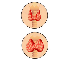
Автоімунний тиреоїдит (АІТ) вважається найпоширенішим автоімунним захворюванням. На сьогодні визнано, що генетична сприйнятливість, фактори навколишнього середовища та імунні розлади сприяють його розвитку. Щодо харчових чинників, дані свідчать про високий рівень споживання йоду, дефіцит селену та заліза з потенційною значимістю стану вітаміну D. Для з’ясування ролі факторів харчування у ризику, патогенезі та лікуванні АІТ використано джерела PubMed стосовно йоду, заліза, селену, вітаміну D, міо-інозитолу та лікування АІТ. Хронічний вплив надмірного споживання йоду індукує автоімунний тиреоїдит частково через те, що йодований тиреоглобулін (Tg) є більш імуногенним. Селенопротеїни необхідні для підтримання функціонального стану щитоподібної залози. Існують дані рандомізованих конт­рольованих досліджень, що селен/селенопротеїни можуть зменшити титри антитіл до тиреоїдної пероксидази (ТПО) у хворих на гіпотиреоз та післяпологовий тиреоїдит. У хворих на АІТ часто спостерігається дефіцит заліза, оскільки автоімунний гастрит, який погіршує всмоктування заліза, є доволі частим супутнім захворюванням. Останнім часом виникли докази, які вказують на позитивну роль вітаміну D, включаючи проліферацію та диференціацію нормальних та ракових клітин, серцево-судинну функцію та імуномодуляцію. Дефіцит вітаміну D особливо продемонстрований у хворих на АІТ. У пацієнтів з АІТ виявлено нижчий рівень вітаміну D, ніж у осіб з контрольної групи, і повідомляється про зворотну залежність вмісту сироваткового вітаміну D від титру антитіл до ТПО/Tg. Адекватне споживання селену є життєво важливим у районах дефіциту/надлишку йоду, а в регіонах із низьким споживанням селену може бути доцільним додавання 50–100 мкг/день селену. Міо-інозитол та селен здатні відновити еутиреоїдний стан, а також покращити самопочуття у хворих на АІТ із субклінічним гіпотиреозом. З огляду також на безпеку використання цих двох молекул, відсутність побічних ефектів комбінацію міо-інозитолу та селену можна вважати дуже ефективною та безпечною терапією для лікування АІТ.

Аутоиммунный тиреоидит (АИТ) считается самым распространенным аутоиммунным заболеванием. В настоящее время признано, что генетическая восприимчивость, факторы окружающей среды и иммунные расстройства способствуют его развитию. Что касается пищевых факторов, данные свидетельствуют о высоком уровне потребления йода, дефиците селена и железа с потенциальной значимостью статуса витамина D. Для выяснения роли факторов питания в риске, патогенезе и лечении АИТ использованы источники PubMed относительно йода, железа, селена, витамина D, мио-инозитола и лечения АИТ. Хроническое воздействие чрезмерного потребления йода индуцирует аутоиммунный тиреоидит отчасти потому, что йодированный тиреоглобулин (Tg) более иммуногенный. Селенопротеины необходимы для поддержания функционального состояния щитовидной железы. Существуют данные рандомизированных конт­ролируемых исследований, что селен/селенопротеины могут уменьшить титры антител к тиреоидной пероксидазе (ТПО) у больных гипотиреозом и послеродовым тиреоидитом. У больных АИТ часто наблюдается дефицит железа, поскольку аутоиммунный гастрит, который ухудшает всасывание железа, является довольно частым сопутствующим заболеванием. В последние годы возникли доказательства, указывающие на положительную роль витамина D, включая пролиферацию и дифференциацию нормальных и раковых клеток, сердечно-сосудистую функцию и иммуномодуляцию. Дефицит витамина D особенно продемонстрирован у больных АИТ. У пациентов с АИТ обнаружен более низкий уровень витамина D, чем у лиц из контрольной группы, и сообщается об обратной зависимости содержания сывороточного витамина D от титра антител к ТПО/Tg. Адекватное потребление селена является жизненно важным в районах дефицита/избытка йода, а в регионах с низким потреблением селена может быть целесо­образным добавление 50–100 мкг/день селена. Мио-инозитол и селен способны восстановить эутиреоидное состояние, а также улучшить самочувствие у больных АИТ с субклиническим гипотиреозом. Принимая во внимание также безопасность использования двух молекул, отсутствие побочных эффектов, комбинацию мио-инозитола и селена можно считать очень эффективной и безопасной терапией для лечения АИТ.

Autoimmune thyroiditis (AT) is considered to be the most common autoimmune disease. It is currently accepted that genetic susceptibility, environmental factors, and immune disorders contribute to its development. With regard to nutritional factors, evidence implicates high iodine intake and deficiencies of selenium and iron with a potential relevance of vitamin D status. To elucidate the role of nutritional factors in the risk, pathogenesis, and treatment of AT, PubMed and the Cochrane Library were searched for publications on iodine, iron, selenium, and vitamin D and risk/treatment of AT. Chronic exposure to excess iodine intake induces autoimmune thyroiditis, partly because highly iodinated thyroglobulin (Tg) is more immunogenic. Selenoproteins are essential to thyroid action. There is evidence from observational studies and randomized controlled trials that selenium/selenoproteins can reduce thyroid peroxidase (TPO)-antibody titers, hypothyroidism, and postpartum thyroiditis. AT patients are frequently iron deficient, since autoimmune gastritis, which impairs iron absorption, is a common co-morbidity. In recent years, evidence has emerged pointing to various roles for vitamin D, including, proliferation and differentiation of normal and cancer cells, cardiovascular function, and immunomodulation. Vitamin D deficiency has been especially demonstrated in AT patients. Lower vitamin D status has been found in AT patients than in controls, and inverse relationships of serum vitamin D with TPO/Tg antibodies have been reported. Adequate selenium intake is vital in areas of iodine deficiency/excess, and in regions of low selenium intake a supplement of 50–100 μg/day of selenium may be appropriate. Myo-inositol and selenium are able to restore the euthyroid state as well as improve the wellbeing of AT with subclinical hypothyroidism. Bearing in mind also the safety of these two molecules’ usage, accentuated by the absence of side effects, the Myo-Ins-Se combination can be considered a very efficacious and safe therapy for AT treatment.


Ключевые слова

автоімунний тиреоїдит; йод; залізо; селен; вітамін D; міо-інозитол

аутоиммунный тиреоидит; йод; железо; селен; витамин D; мио-инозитол

autoimmune thyroiditis; iodine; iron; selenium; vitamin D; myo-inositol

Autoimmune thyroiditis (AT) is the most prevalent autoimmune disorder characterized by the destruction of thyroid cells caused by leukocytes and antibody-mediated immune processes accompanied by hypothyroidism [1]. In recent years, evidence has emerged pointing to various roles for vitamin D, including, proliferation and differentiation of normal and cancer cells, cardiovascular function, and immunomodulation. Vitamin D deficiency has been especially demonstrated in AT patients [2]. 
Chronic exposure to excess iodine intake induces autoimmune thyroiditis, partly because highly iodinated thyroglobulin (Tg) is more immunogenic. Recent introduction of universal salt iodization can have a similar, though transient, effect. Selenoproteins are essential to thyroid action. In particular, the glutathione peroxidases protect the thyroid by removing excessive hydrogen peroxide produced for Tg iodination. Genetic data implicate the anti-inflammatory selenoprotein S in AT risk. Antibodies to thyroid peroxidase (TPO), the enzyme that catalyses thyroid-hormone production and antibodies to the receptor for the thyroid-stimulating hormone, are characteristic of AT. It is presently accepted that genetic susceptibility, environmental factors, including nutritional factors and immune disorders contribute to the development of AT. There is evidence from observational studies and randomized controlled trials that selenium/selenoproteins can reduce TPO-antibody titers, hypothyroidism, and postpartum thyroiditis. Iron deficiency impairs thyroid metabolism. TPO, the enzyme responsible for the production of thyroid hormones, is a heme (iron-containing) enzyme which becomes active at the apical surface of thyrocytes only after binding heme [3]. 
AT patients are frequently iron deficient, since autoimmune gastritis, which impairs iron absorption, is a common co-morbidity. Treatment of anemic women with impaired thyroid function with iron improves thyroid-hormone concentrations, while thyroxine and iron together are more effective in improving iron status [4]. Lower vitamin D status has been found in HT patients than in controls, and inverse relationships of serum vitamin D with TPO/Tg antibodies have been reported. However, other data and the lack of trial evidence suggest that low vitamin D status is more likely the result of autoimmune disease processes that include vitamin D receptor dysfunction. Clinicians should check patients’ iron (particularly in menstruating women) and vitamin D status to correct any deficiency. Adequate selenium intake is vital in areas of iodine deficiency/excess, and in regions of low selenium intake a supplement of 50–100 μg/day of selenium may be appropriate.
Chronic exposure to excess iodine intake induces AT, partly because highly-iodinated Tg is more immunogenic. The recent introduction of universal salt iodisation can have a similar, although transient, effect. Iron deficiency impairs thyroid metabolism. TPO is a haem enzyme that becomes active only after binding haem. AT patients are frequently iron-deficient since autoimmune gastritis, which reduces iron absorption and coeliac disease which causes iron loss, are frequent co-morbidities. In two-thirds of women with persistent symptoms of hypothyroidism despite appropriate levothyroxine therapy, restoration of serum ferritin above 100 µg/l ameliorated symptoms. Selenoproteins are essential to thyroid action. In particular, the glutathione peroxidases remove excessive hydrogen peroxide produced there for the iodination of Tg to form thyroid hormones. There is evidence from observational studies and randomised controlled trials that selenium, probably as selenoproteins, can reduce TPO-antibody concentration, hypothyroidism and postpartum thyroiditis. Appropriate status of iodine, iron and selenium is crucial to thyroid health.
Much research suggests that Mediterranean eating habits and lifestyle contribute to counteract the risk of chronic diseases while promoting longevity, but little information is available on the effects of the Mediterranean diet (Med-Diet) on thyroid function, particularly among overweight/obese subjects. Nevertheless, consistent data reported a slight increase in serum levels of the thyroid-stimulating hormone (TSH) and a higher rate of conversion of thyroxine (T4) to triiodothyronine (T3) in obesity. In cross-sectional study authors [5] investigated the relationship between adherence to the Med-Diet and circulating thyroid hormones in a cohort of overweight/obese subjects Southern Italy. 324 consecutive outpatient subjects (228 women and 96 men, age range 14–72 years) taking no drug therapy and showing normal levels of thyroid hormones, but complicated by overweight and obesity were involved in the study. Higher adherence to Med-Diet was found to be inversely related to free T3 (p < 0.01) and T4 (p < 0.01) serum levels. People consuming at least four spoonful of extra-virgin olive oil (EVOO) per day, as well as those consuming at least two servings of vegetables per day, had lower free T3 levels (p = 0.033 and p = 0.021, respectively). Furthermore, consuming at least four spoonfuls of EVOO per day was found to be associated to lower free T4 serum concentrations (p = 0.011). Multinomial logistic regression models, performed on tertiles of thyroid hormones to further investigate the relationship with Med-Diet, corroborated the significance only for free T4. Increased adherence to the Med-Diet was independently associated to a slightly reduced thyroid function, but still within the reference range for free T3 and T4 serum levels. This first finding in this field opens up a research line on any underlying biological interplay.
The aim of other study [6] was to investigate the effect of vitamin D on circulating thyroid autoantibodies and thyroid hormones profile (T4, T3, and TSH) in females with AT. Forty-two women with AT disease were enrolled in this randomized clinical trial study and divided into vitamin D and placebo groups. Patients in the vitamin D and placebo groups received 50 000 IU vitamin D and placebo pearls, weekly for 3 months, respectively. The serum levels of 25-hydroxy vitamin D [25(OH) D], Ca++ ion, anti-TPO Ab, anti-thyroglobulin antibody (anti-Tg Ab), T4, T3, and TSH were measured at the baseline and at the end of the study. The results of this study showed a significant reduction of anti-Tg Ab and TSH hormone in the Vitamin D group compared to the start of the study; however, there was a no significant reduction of anti-TPO Ab in the Vitamin D group compared to the placebo group (p = 0.08). No significant changes were observed in the serum levels of T3 and T4 hormones. Therefore, vitamin D supplementation can be helpful for alleviation of the disease activity in AT patients.
Vitamin D is a steroid hormone traditionally connected to phosphocalcium metabolism. The main functions of vitamin D are the regulation of phosphocalcium metabolism and the promotion of bone homeostasis [7]. The discovery of pleiotropic expression of its receptor (VDR) and of the enzymes involved in its metabolism has led to the exploration of the other roles of this vitamin. The influence of vitamin D on autoimmune disease-namely, on autoimmune thyroid disease-has been widely studied. Most of the existing data support a relationship between vitamin D deficiency and a greater tendency for development and/or higher titers of antibodies linked to AT, Graves’ disease, and/or postpartum thyroiditis [8]. However, there have also been some reports contradicting such relationships, thus making it difficult to establish a unanimous conclusion. Even if the existence of an association between vitamin D and autoimmune thyroid disease is assumed, it is still unclear whether it reflects a pathological mechanism, a causal relationship, or a consequence of the autoimmune process. The relationship between vitamin D polymorphisms and this group of diseases has also been the subject of study, often with divergent results [9]. 
Previous studies have reported the presence of VDR in murine thyrotropic cells [10]. A strong molecular homology between VDR and thyroid hormone has been demonstrated, as well as the presence of VDR in murine follicular thyroid cells. The incubation of these cells with 1,25(OH)2D inhibited the uptake of iodine and cell growth [11].
A higher risk of AT in subjects born in summer [12] and no relationship between month of birth and Graves’ disease [13] were described in Danish register-based studies. A higher frequency of birth in spring was noticed in Greek children with AT [14]. Seasonality of birth month may be related to Vitamin D levels (higher frequency of deficiency in the end of winter, beginning of spring), but also may relate to viral exposure and other factors which vary in different regions and years [14].
A study performed in Korea revealed that iodine excess was associated with thyroid dysfunction only in vitamin D-deficient individuals [15].
In patients with AT, K. Vondra et al. found a positive relationship between 25(OH)D levels and the fT4/fT3 ratio, which disappeared after supplementation with cholecalciferol. The authors speculated that the decreased ratio may be a compensatory adaptation to Vitamin D deficiency [16].
S. Kivity et al. reported an association between Vitamin D deficiency, defined as 25(OH)D < 10 ng/ml, and a higher frequency of AT and the presence of thyroid antibodies, in general [17]. A.D. Unal et al. found lower levels of 25(OH)D in individuals with autoimmune thyroid pathology, with the Graves’ disease group registering lower levels than those with AT and an inverse correlation between the levels of 25(OH)D and antithyroid antibody titers [18]. In a meta-analysis in 2015, J. Wang et al. reported lower levels of 25(OH)D and higher prevalence of deficiency in individuals with AT vs. controls [19].
There is evidence supporting a relationship between vitamin D and AT. G. Tamer et al. identified lower 25(OH)D levels in individuals with AT versus control subjects, with a tendency for a higher prevalence of deficiency in patients with hypothyroidism than in those in euthyroidism [20]. 
There are also data supporting this relationship at age extremes. A higher prevalence of AT and anti-TPO titers in association with 25(OH)D < 20 ng/ml was found in individuals over 65 years of age. It should be noted, however, that the AT group was older and had higher creatinine levels [21]. In pediatric patients with AT vs. healthy controls, a higher prevalence of Vitamin D deficiency was also found [22]. However, in an analysis of pediatric patients with type 1 diabetes mellitus with vs. without AT, 25(OH)D levels < 20 ng/ml were found in both groups, with no difference between the two [23].
In a systematic review and meta-analysis, S. Wang et al. concluded that supplementation with Vitamin D appeared to significantly reduce levels of anti-TPO (for treatments ≥ 6 months) and anti-Tg, with no reported serious adverse effects [24]. More recently, V.F. Koehler et al. retrospectively analyzed 933 patients with AT and found a greater reduction in anti-TPO levels in a 58-patient sub-group that had an improvement in their initially insufficient Vitamin D level (< 30 ng/ml) vs a control group that maintained a Vitamin D level below the threshold. The difference between the groups, however, was not statistically significant [25].
Other factors may influence the effect of Vitamin D supplementation on AT. Testosterone replacement in testosterone-deficient men has been associated with a more pronounced reduction in anti-TPO/-Tg titers and increased thyroid secretory capacity (SPINA-GT index) with Vitamin D supplementation (vs. testosterone-naïve men). Selenomethionine supplementation has also been shown to enhance the effect of Vitamin D on these parameters in 47 AT women [26].
Supplementation may also have a preventive component. A group of 11,017 participants in a wellness program were supplemented with Vitamin D for over a year, aiming to reach physiological levels defined as 25(OH)D > 40 ng/ml. It was found that concentrations of 25(OH)D ≥ 50 ng/ml reduced the risk of hypothyroidism by 30 % (from 0.4 % — 44 cases/11,017 participants to 0.28 % — 31 cases) and elevated antibody titers by 32 %. Increased levels of 25(OH)D in patients with hypothyroidism have been associated with improved thyroid function [27].
Several questions can be raised regarding the relationship between Vitamin D and AT, the first one being whether such a relationship actually exists. With respect to this matter, although there is some inconsistency in the results of the studies carried out to date, most of the data point toward an association between lower Vitamin D levels and increased risk of developing the disease and/or higher antibody titers and/or more difficulty in its treatment, especially for vitamin D deficiency. Polymorphisms in genes associated with Vitamin D function/metabolism also appear to have some influence on the risk of AT.
Myo-Inositol is an isomer of a C6 sugar alcohol. Several studies suggested that Myo-Inositol plays an important role in several cellular processes. In particular, it has been demonstrated that Myo-Inositol is the precursor for the synthesis of phosphoinositides, which are part of the phosphatidylinositol (PtdIns) signal transduction pathway. PtdIns is responsible for signal transduction across the plasma membrane, via second messenger: inositol 1,4,5-triphosphate that modulates intracellular Ca2+ release or by being a docking site for several signal transduction proteins [28].
TSH signaling is rather complex; indeed, two different signal cascades are generated. One branch of the signal cascade involves as second messenger cyclic AMP (cAMP), while another branch is inositol dependent [29]. Indeed, while the cAMP is more involved in cell growth differentiation and the T3–T4 secretion, the inositol-dependent branch regulates H2O2-mediated iodination. In particular, it has been shown that relatively low TSH concentrations are able to stimulate cAMP mediated signal cascade, while only 100-fold higher TSH concentrations are able to stimulate the inositol-mediated signal cascade [29].
M. Nordio and R. Pajalich [30] evaluated whether the association between Myo-Inositol and Selenium can ensure euthyroidism in subclinical hypothyroidism patients with AT. This study was performed as a prospective randomized double-blinded, controlled study in women (mean age 38 years) with AT and TPOAb; inclusion criteria were TgAb and/or TPOAb above 350 IU/mL, TSH levels between 4,01 mIU/l and 9,99 mIU/l, and a normal free-thyroxine level (0.6–1.8 ng/dl) as well as typical hypoechogenicity of the thyroid in high-resolution sonography. The primary endpoint of the study was restoration of TSH levels (lower than 4 mIU/l). Secondary end points were decreased in serum TPOAb and TgAb concentrations, free thyroid hormone levels and improvement of the thyroid and quality of life estimation. All patients enrolled signed an informed consent. Patients were randomized into 2 groups according to their initial TPOAb concentrations. group A consisted of 24 patients who received orally 83 g selenomethionine/day, in a soft gel capsule; group B consisted of 24 patients who received a combined treatment plus Myo-Inositol 600 mg also in 83 g selenomethionine, soft gel capsule, orally, for 6 months. The patients were asked to take the medication with water about 2 h before or after a meal. They were not given further treatment, such as over-the-counter vitamins or trace elements. All patients were otherwise healthy. No patients were substituted with LT4. TPOAb, TgAb, TSH, and free thyroid hormones were determined by commercial assays. The echogenicity of the thyroid was monitored with high-resolution ultrasound. This article demonstrated that the beneficial effects obtained by selenomethionine treatment on patients affected by subclinical hypothyroidism, likely due to the presence of autoantibody (TPOAb and TgAb), are further improved by cotreatment with Myo-Inositol. Indeed, due to its action as TSH second messenger, Myo-Inositol treatment reduces TSH levels closer to physiological concentrations.
S.M. Ferrari et al. evaluated the immune-modulating effect of myo-inositol in association with seleno-methionine in patients with euthyroid AT [31]. Twenty-one consecutive Caucasian patients with newly diagnosed euthyroid chronic AT were evaluated. All subjects were treated with myo-inositol in association with selenium (600 mg/83 mg) tablets, twice per day, for six months. A complete thyroid assessment was done before the treatment, and after six months. After the treatment TSH levels significantly declined with respect to basal values, overall, in patients with an initial TSH value in the high normal range (2.1 < TSH < 4.0), suggesting that the combined treatment can reduce the risk of a progression to hypothyroidism in subjects with autoimmune thyroid diseases. Authors found that after the treatment antithyroid autoantibodies levels declined. They first show an immune-modulatory effect of myo-inositol in association with seleno-methionine in patients with euthyroid AT. Further studies are needed to extend the observations in a large population, to evaluate the effect on the quality of life, and to study the mechanism of the effect on chemokines.
Myo-inositol and selenium are able to restore the euthyroid state as well as improve the wellbeing of AT with subclinical hypothyroidism. Bearing in mind also the safety of these two molecules’ usage, accentuated by the absence of side effects, the Myo-inositol — Selenium combination can be considered a very efficacious and safe therapy for AT treatment.
Conflicts of interests. Author declares the absence of any conflicts of interests and their own financial interest that might be construed to influence the results or interpretation of their manuscript.

Список литературы

  1. Flores-Rebollar A., Moreno-Castañeda L., Vega-Servín N.S., López-Carrasco G., Ruiz-Juvera A. Prevalence of autoimmune thyroiditis and thyroid dysfunction in healthy adult mexicans with a slightly excessive iodine intake. Nutr. Hosp. 2015. 32(2). 918-924. doi: 10.3305/nh.2015.32.2.9246. 
  2. Hu S., Rayman M.P. Multiple Nutritional Factors and the Risk of Hashimoto’s Thyroiditis. Thyroid. 2017. 27(5). 597-610. doi: 10.1089/thy.2016.0635. 
  3. Rayman M.P. Multiple nutritional factors and thyroid disease, with particular reference to autoimmune thyroid disease. Proc. Nutr. Soc. 2019. 78(1). 34-44. doi: 10.1017/S0029665118001192. 
  4. Harris Rosenzweig P., Volpe S.L. Effect of iron supplementation on thyroid hormone levels and resting metabolic rate in two college female athletes: a case study. Int. J. Sport Nutr. Exerc. Metab. 2000. 10(4). 434-443. doi: 10.1123/ijsnem.10.4.434. 
  5. Zupo R., Castellana F., Panza F, Lampignano L., Murro I., Di Noia C., Triggiani V. et al. Adherence to a Mediterranean Diet and Thyroid Function in Obesity: A Cross-Sectional Apulian Survey. Nutrients. 2020, Oct 16. 12(10). 3173. doi: 10.3390/nu12103173. 
  6. Chahardoli R., Saboor-Yaraghi A.A., Amouzegar A., Khalili D., Vakili A.Z., Azizi F. Can Supplementation with Vitamin D Modify Thyroid Autoantibodies (Anti-TPO Ab, Anti-Tg Ab) and Thyroid Profile (T3, T4, TSH) in Hashimoto’s Thyroiditis? A Double Blind, Randomized Clinical Trial. Horm. Metab. Res. 2019. 51(5). 296-301. doi: 10.1055/a-0856-1044.
  7. Vieira I.H., Rodrigues D., Paiva I. Vitamin D and Autoimmune Thyroid Disease-Cause, Consequence, or a Vicious Cycle? Nutrients. 2020. 12(9). 2791. doi: 10.3390/nu12092791.
  8. Kmieć P., Sworczak K. Vitamin D in thyroid disorders. Exp. Clin. Endocrinol. Diabetes. 2015. 123(7). 386-393. doi: 10.1055/s-0035-1554714. 
  9. Kim D. The Role of Vitamin D in Thyroid Diseases. Int. J. Mol. Sci. 2017. 18(9). 1949. doi: 10.3390/ijms18091949. 
  10. Barchetta I., Baroni M., Leonetti F., De Bernardinis M., Bertoccini L., Fontana M., Mazzei E. et al. TSH levels are associated with vitamin D status and seasonality in an adult population of euthyroid adults. Clin. Exp. Med. 2015. 15. 389-396. doi: 10.1007/s10238-014-0290-9.
  11. Muscogiuri G., Tirabassi G., Bizzaro G., Orio F., Paschou S.A., Vryonidou A., Balercia G. et al. Vitamin D and thyroid disease: To D or not to D? Eur. J. Clin. Nutr. 2015. 69. 291-296. doi: 10.1038/ejcn.2014.265.
  12. Thvilum M., Brandt F., Brix T.H., Hegedüs L. Month of birth is associated with the subsequent diagnosis of autoimmune hypothyroidism. A nationwide Danish register-based study. Clin. Endocrinol. (Oxf.). 2017. 87(6). 832-837. doi: 10.1111/cen.13425. 
  13. Sivalingam S., Thvilum M., Brix T.H., Hegedüs L., Brandt F. No link between season of birth and subsequent development of Graves’ disease or toxic nodular goitre: a nationwide Danish register-based study. Endocr. Connect. 2018. 7(10). 1090-1095. doi: 10.1530/EC-18-0185.
  14. Kyrgios I., Giza S., Tsinopoulou V.R., Maggana I., Haidich A.B., Galli-Tsinopoulou A. Seasonality of month of birth in children and adolescents with autoimmune thyroiditis: a continuing conundrum. J. Pediatr. Endocrinol. Metab. 2018. 31(10). 1123-1131. doi: 10.1515/jpem-2018-0051. 
  15. Kim M., Song E., Oh H.S., Park S., Kwon H., Jeon M.J., Kim W.G., Kim W.B., Shong Y.K., Kim T.Y. Vitamin D deficiency affects thyroid autoimmunity and dysfunction in iodine-replete area: Korea national health and nutrition examination survey. Endocrine. 2017. 58(2). 332-339. doi: 10.1007/s12020-017-1425-z. 
  16. Vondra K., Bílek R., Matucha P., Salátová M., Vosátková M., Stárka L., Hampl R. Vitamin D Supplementation Changed Relationships, Not Levels of Metabolic-Hormonal Parameters in Autoimmune Thyroiditis. Physiol. Res. 2017. 26. 409-417. doi: 10.33549/physiolres.933727.
  17. Kivity S., Agmon-Levin N., Zisappl M., Shapira Y., Nagy E.V., Dankó K., Szekanecz Z. et al. Vitamin D and autoimmune thyroid diseases. Cell. Mol. Immunol. 2011. 8(3). 243-247. doi: 10.1038/cmi.2010.73.
  18. Unal A.D., Tarcin O., Parildar H., Cigerli O., Eroglu H., Demirag N.G. Vitamin D deficiency is related to thyroid antibodies in autoimmune thyroiditis. Cent. Eur. J. Immunol. 2014. 39(4). 493-497. doi: 10.5114/ceji.2014.47735. 
  19. Wang J., Lv S., Chen G., Gao C., He J., Zhong H., Xu Y. Meta-analysis of the association between vitamin D and autoimmune thyroid disease. Nutrients. 2015. 7(4). 2485-2498. doi: 10.3390/nu7042485. 
  20. Tamer G., Arik S., Tamer I., Coksert D. Relative vitamin D insufficiency in Hashimoto’s thyroiditis. Thyroid. 2011. 21(8). 891-896. doi: 10.1089/thy.2009.0200. 
  21. Muscogiuri G., Mari D., Prolo S., Fatti L.M., Cantone M.C., Garagnani P., Arosio B. et al. 25 Hydroxyvitamin D Deficiency and Its Relationship to Autoimmune Thyroid Disease in the Elderly. Int. J. Environ. Res. Public Health. 2016. 13(9). 850. doi: 10.3390/ijerph13090850. 
  22. Metwalley K.A., Farghaly H.S., Sherief T., Hussein A. Vitamin D status in children and adolescents with autoimmune thyroiditis. J. Endocrinol. Invest. 2016. 39(7). 793-797. doi: 10.1007/s40618-016-0432-x. 
  23. Demir K., Keskin M., Kör Y., Karaoğlan M., Bülbül Ö.G. Autoimmune thyroiditis in children and adolescents with type 1 diabetes mellitus is associated with elevated IgG4 but not with low vitamin D. Hormones (Athens). 2014. 13(3). 361-368. doi: 10.14310/horm.2002.1481. 
  24. Wang S., Wu Y., Zuo Z., Zhao Y., Wang K. The effect of vitamin D supplementation on thyroid autoantibody levels in the treatment of autoimmune thyroiditis: a systematic review and a meta-analysis. Endocrine. 2018. 59(3). 499-505. doi: 10.1007/s12020-018-1532-5. 
  25. Koehler V.F., Filmann N., Mann W.A. Vitamin D Status and Thyroid Autoantibodies in Autoimmune Thyroiditis. Horm. Metab. Res. 2019. 51(12). 792-797. doi: 10.1055/a-1023-4181. 
  26. Krysiak R., Kowalcze K., Okopień B. Selenomethionine potentiates the impact of vitamin D on thyroid autoimmunity in euthyroid women with Hashimoto’s thyroiditis and low vitamin D status. Pharmacol. Rep. 2019. 71(2). 367-373. doi: 10.1016/j.pharep.2018.12.006. 
  27. Mirhosseini N., Brunel L., Muscogiuri G., Kimball S. Physiological serum 25-hydroxyvitamin D concentrations are associated with improved thyroid function-observations from a community-based program. Endocrine. 2017. 58(3). 563-573. doi: 10.1007/s12020-017-1450-y.
  28. Kutateladze T. Translation of the phosphoinositide code by PI effectors. Nat. Chem. Biol. 2010. 6. 507-513. https://doi.org/10.1038/nchembio.390.
  29. Ohye H., Sugawara M. Dual oxidase, hydrogen peroxide and thyroid diseases. Exp. Biol. Med. (Maywood). 2010. 235(4). 424-433. doi: 10.1258/ebm.2009.009241. 
  30. Nordio M., Pajalich R. Combined treatment with Myo-inositol and selenium ensures euthyroidism in subclinical hypothyroidism patients with autoimmune thyroiditis. J. Thyroid Res. 2013. 2013. 424163. doi: 10.1155/2013/424163. 
  31. Ferrari S.M., Fallahi P., Di Bari F., Vita R., Benvenga S., Antonelli A. Myo-inositol and selenium reduce the risk of developing overt hypothyroidism in patients with autoimmune thyroiditis. Eur. Rev. Med. Pharmacol. Sci. 2017. 21(2 Suppl). 36-42. PMID: 28724175.

Вернуться к номеру