Інформація призначена тільки для фахівців сфери охорони здоров'я, осіб,
які мають вищу або середню спеціальну медичну освіту.

Підтвердіть, що Ви є фахівцем у сфері охорони здоров'я.


Всесвітній день боротьби із запальними захворюваннями кишечника
день перший
день другий

Коморбідний ендокринологічний пацієнт

Всесвітній день боротьби із запальними захворюваннями кишечника
день перший
день другий

Коморбідний ендокринологічний пацієнт

Международный эндокринологический журнал Том 17, №3, 2021

Вернуться к номеру

Чи належать метаболічний синдром і його компоненти до факторів ризику виникнення поліпів жовчного міхура?

Авторы: Emine Duygu Boz(1), Refik Demirtunç(2), Mehmet Sözen(3)
(1) — Department of İnternal Medicine, Silivri State Hospital, Istanbul, Turkey
(2) — Department of Internal Medicine, Haydarpaşa Numune Training and Research Hospital, Istanbul, Turkey
(3) — Department of Endocrinology and Metabolism, Kocaeli University Faculty of Medicine, Kocaeli, Turkey

Рубрики: Эндокринология

Разделы: Клинические исследования

Версия для печати


Резюме

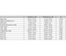
Актуальність. Поліпи жовчного міхура, як правило, є доброякісними утвореннями, що походять із слизової оболонки, і зазвичай виявляються випадково під час рентгенологічних досліджень або після холецистектомії. Поліпи жовчного міхура доволі поширені і можуть мати ризик злоякісного переродження. У цьому дослідженні було встановлено, чи є метаболічний синдром (МС) фактором ризику для виникнення поліпів жовчного міхура. Мета дослідження — визначити поширеність МС та його компонентів у пацієнтів з поліпами жовчного міхура. Матеріали та методи. Проведено ретроспективне перехресне дослідження. Автори дослідили вік, стать та анамнез хвороби у 90 дорослих осіб (45 — з поліпами жовчного міхура, 45 — без поліпів). Ріст та маса тіла, індекс маси тіла, окружність талії та лабораторні дані були отримані з лікарняної системи обробки даних. Для діагностики МС використовували діагностичний критерій МС NCEP-ATP III та Міжнародної діабетичної федерації (IDF). Результати. У дослідженні під спостереженням перебувало 51,1 % (n = 46) жінок і 48,8 % (n = 44) чоловіків. Середній вік становив 58,79 ± 15,70 року. МС виявлено в 56,7 % (n = 51) випадків за критеріями NCEP-ATP III та в 64,4 % (n = 58) випадків за критеріями IDF. У пацієнтів з поліпом жовчного міхура МС виявлено у 55,55 % за критеріями NCEP-ATP III та у 66,66 % за критеріями IDF. Частота МС не була однаковою у групі осіб з поліпами жовчного міхура та контрольній групі (р > 0,01). Встановлено, що абдомінальне ожиріння є фактором ризику розвитку поліпа жовчного міхура (співвідношення шансів 14,23; 95% ДІ: 1,751–15,722; р < 0,01). Хоча показники не досягнули рівня статистичної значущості, низький рівень ліпопротеїнів високої щільності та артеріальна гіпертензія були виявлені приблизно удвічі частіше у пацієнтів з поліпами жовчного міхура, ніж у групі контролю. Висновки. Хоча МС не асоціюється з розвитком поліпа жовчного міхура, ожиріння розглядається як єдиний вірогідний фактор ризику.

Background. Gallbladder polyps are usually benign lesions originating from the mucosa and are usually detected incidentally during radiological examinations or after cholecystectomy. Gallbladder polyps are common and may have malignant risk. In this study, it was investigated whether metabolic syndrome (MS) is a risk factor for gallbladder polyps. This study aimed to determine the prevalence of MS and its components in patients with gallbladder polyps. Materials and methods. We conducted a retrospective, cross-sectional study. We investigated the age, gender and past medical history of 90 adults (45 with polyps, 45 without polyps). Body height and weight, body mass index, waist circumference and laboratory data were obtained from the hospital data processing system. National Cholesterol Education Program Adult Treatment Panel III (NCEP-ATP III) and International Diabetes Foundation (IDF) MS diagnostic criterion were used for the diagnosis of MS. Results. 51.1 % (n = 46) of the subjects participating in the study were female and 48.8 % (n = 44) were male. The mean age was 58.79 ± 15.70 years. MS was found in 56.7 % (n = 51) of the cases according to the criteria of NCEP-ATP III and, in 64.4 % (n = 58) of the cases according to the IDF criteria. In patients with a gallbladder polyp, MS was detected in 55.55 % according to the criteria of NCEP-ATP III and in 66.66 % according to the IDF criteria. The rates of MS were not similar in the gallbladder polyp group and control group (p > 0.01). Abdominal obesity was found to be a risk factor for the development of gallbladder polyp (odds ratio: 14.23, 95% CI: 1.751–15.722; p < 0.01). Although it was not statistically significant, low HDL and hypertension were detected approximately 2 times higher in patients with gallbladder polyps than in the control group. Conclusions. While MS is not associated with the development of gallbladder polyp, obesity is seen as a sole risk factor.


Ключевые слова

поліп жовчного міхура; метаболічний синдром; абдомінальне ожиріння; фактори ризику

gallbladder polyp; metabolic syndrome; abdominal obesity; risk factors

Introduction

Gallbladder polyps (GP) are usually benign lesions originating from the mucosa and are usually detected incidentally during radiological examinations or after cholecystectomy [1]. Detection rates of GP have gradually increased since ultrasonography (USG) became widely used. GP is seen in 1.5–4.5 % of USG-performed patients, 0.5–13.8 % of those undergoing cholecystectomy, and 4–8 % in patients with gallstones [2]. GP is generally classified as true polyps and pseudopolyps. Pseudopolyps include cholesterol polyps, inflammatory polyps, and hyperplastic polyps, while true polyps include adenomas and adenocarcinomas. While pseudopolyps are benign and do not require any intervention, true GP should be surgically removed as they carry the risk of malignancy [3].
It is important to know the risk factors that contribute to the formation of gallbladder polyp. These risk factors include male gender, high body mass index (BMI), old age, chronic hepatitis B and C, hyperlipidemia, obesity, glucose intolerance and metabolic syndrome (MS) [4, 5]. MS is a combination of diseases which one’s obesity, high triglyceride (Tg), low high density lipoprotein (HDL) cholesterol, hyperglycemia and hypertension [6]. MS is associated with gallstone disease [7]. However, there are very few literature studies investigating the relationship between MS and GP. This study aimed to determine the prevalence of MS and its components in patients with GP.

Materials and methods

We conducted a retrospective, cross-sectional study on individuals who admitted to XXXX Hospital Internal Medicine and Gastroenterology Clinic. Informed consent and institutional review board approval were obtained. To evaluate the prevalence of GP in individuals with MS, the data of the subjects. Forty five patients with GP were included in the study and 45 people without GP were taken as the control group. Patients aged 18 and over were included in the study, while, pregnant women, congenital polyposis syndromes, chronic viral hepatitis, primary sclerosing cholangitis, and patients with cholangio cellular carcinoma were excluded. 
The height and weight of the patients were measured with calibrated devices. BMI was calculated by dividing the weight (kg) by the square of the height (m2). Waist circumference (WC) was measured in the horizontal plane from the midpoint of the distance between the arcus costarium and spina iliaca anterior superior. Blood pressure was measured on the right arm with a standard mercury sphygmomanometer.
Physical examination findings of the patients were screened retrospectively from anamnesis forms. Fasting blood glucose (FPG), liver and kidney function tests, total cholesterol, triglyceride (Tg), high HDL, low density lipoprotein (LDL) and viral hepatitis markers were documented from the hospital information processing system. National Cholesterol Education Program Adult Treatment Panel III (NCEP-ATP III) and International Diabetes Foundation (IDF) MS diagnostic criteria were used for the diagnosis of MS. According to the NCEP-ATP III diagnostic criteria, MS is defined if three or more of the following criteria are met: 1) Abdominal obesity; WC, males > 102 cm, females > 88 cm; 2) Hypertriglyceridemia; 150 mg/dL; 3) Low HDL cholesterol; < 40 mg/dL in men and < 50 mg/dL in women; 4) Hypertension; ≥ 130/85 mm Hg; 5) High fasting plasma glucose; > 110 mg/dL [8]. According to the IDF diagnostic criteria, MS is defined together with central obesity (WC; 94 cm in men or ≥ 80 cm in women) and two of the following. 1) Tg > 150 mg/dL; 2) Low HDL cholesterol; < 40 mg/dL in men and < 50 mg/dL in women; 3) Blood pressure ≥ 130/85 mm Hg; 4) Fasting hyperglycemia (glucose level > 100 mg/dL) or a prior diagnosis of diabetes or impaired glucose tolerance [9].
GP were detected by previous abdominal USG or pathology examinations after cholecystectomy.
Ethical considerations
This study was approved by the Clinical Research Ethics Committee of XXXX Hospital with the decision number HNEAH-KAEK 2017/97 dated 09.10.2017.
Statistical analysis
SPSS 21 (Statistical Package for the Social Sciences) program was used for statistical analysis. Statistical analysis was done for 2 × 2 tables according to the expected and observed cell status by fisher, yates corrected chi-square or pearson chi-square; and for the tables that were not in 2 × 2 order according to the expected and observed cells by Pearson’s chi-square or likelihood ratio. Significance was evaluated at p < 0.01 and p < 0.05 levels.

Results

A total of 90 patients, 45 patients with GP and 45 subjects without GP, were included in our retrospective study; 51.1 % (n = 46) of the patients were female, 48.8 % (n = 44) were male, and the average age of all subjects was 58.79 ± 15.70. Average of all participants weight was 76.96 ± 17.30 kg, height 165.0 ± 10.1 cm, BMI 27.88 ± 5.23 kg/m2, WC 100.43 ± 11.30 cm.
When the demographic data of those with and without GP were compared with each other, the mean age of those in the control group was found to be higher. However, height, body weight, BMI and WC measurements were similar. GP were detected in 48.8 % of women (n = 22) and 51.1 % of men (n = 23). When the laboratory data of both groups were compared, parameters other than FPG and HDL were found to be similar. While FPG was found to be higher in the control group, HDL was higher in the group with gall bladder (Tabl. 1).
MS was found in 56.7 % (n = 51) of the cases according to NCEP-ATP III, and in 64.4 % (n = 58), according to IDF criteria. When the GP group and control group were compared, no statistically significant difference was found between the rates of MS (p > 0.01). According to the NCEP-ATP III criteria, MS was detected in 57.7 % (n = 26) of the patients in the control group and in 55.5 % (n = 25) of those with GP. According to the IDF criteria, MS was found in 62.2 % (n = 28) of the patients in the control group and in 66.6 % (n = 30) of those with GP. The distribution of patient characteristics in the GP and control group according to both NCEP-ATP III and IDF criteria are given in tabl. 2.
When the GP group and the control group were compared according to the NCEP-ATP III MS criteria, a statistically significant difference was found between the cases with high blood glucose (FPG ≥ 110 mg/dl) (p < 0.01). FPG was found above 110 mg/dl in 62.2 % (n = 28) of the control group cases and 33.3 % (n = 15) of the GP cases.
When the GP group and control group were compared according to IDF MS criteria, a significant difference was found in terms of abdominal obesity (p < 0.01). WC was found above the criteria in 75.5 % (n = 34) of the control group cases and in 97.7 % (n = 44) of the cases of the GP group. According to the IDF MS criteria, abdominal obesity is a risk factor for the development of GP (odds ratio: 14.23, 95% CI: 1.751–15.722).
According to both NCEP-ATP III and IDF MS criteria, no statistical difference was found between the two groups in terms of low HDL, high Tg and high blood pressure criteria (p > 0.01). However, although it was not statistically significant, low HDL and hypertension were detected approximately 2 times higher in patients with GP than in the control group.

Discussion

GP is usually detected in 5–6 % of adults. True GPs are uncommon, but many other lesions may display GP-like appearances on radiological imaging. GP are usually asymptomatic and are usually detected incidentally [10]. While most benign GPs are usually composed of cholesterol polyps, the main concern in clinical practice is true polyps with malignant risk. The pathophysiological process between true polyps and gallbladder cancer has not been clearly revealed yet. However, given the low survival rates of gallbladder cancer, the follow-up and proper management of GP gives clinicians the opportunity to successfully eliminate this cancer risk [11].
The malignant risk of true GPs raises a number of questions regarding the timing of prophylactic cholecystectomy and determination of patients suitable for surveillance. In previous studies, it was shown that all malignant gallbladder tumors were found in polyps larger than 6 mm, gallbladder cancer was observed in 43–77 % of polyps with a size of 10–20 mm, and polyps larger than 20 mm were pathognomonic for gallbladder cancer [12]. Therefore, current guidelines for the management of GP have largely focused on their size. If there is no risk factor for gallbladder malignancy and polyp is 6–9 mm or if there is a risk factor for gallbladder malignancy and polyp is < 5 mm, follow-up ultrasound of the gallbladder is recommended at 6th month, 1st year and then up to 5 years. If the patient has no risk factors for malignancy and polyp is < 5 mm, follow-up is recommended at 1st year, 3rd year, and 5th year. If GP are > 10 mm, cholecystectomy is recommended [13].
A review of the literature on risk factors for GP revealed conflicting results. Although many studies show that the prevalence of gallbladder polyp is higher in men [14], there are also data indicating that the frequency of polyp is not related to gender [15]. In our study, a relationship between the frequency of GP and gender could not be shown. Studies have shown that the frequency of gallbladder polyp increases with age [14]. In our study, in accordance with the literature, the mean age of the GP group was found to be 51.44 ± 12.33 years.
The most common type of GP is cholesterol polyps. According to some hypotheses polyps form when cholesterol accumulates directly in the gallbladder, similar to plaque formation in atherosclerosis [16]. Y. Lee et al. in their study, showed that low HDL and high Tg levels contributed to the development of GP [17]. On the contrary, there are literature data indicating that dyslipidemia is not associated with the development of GP [18]. In our study, no significant relationship was found between lipid levels and GP (p > 0.01).This finding may be due to the small size of our study group and the fact that it is a cross-sectional study.
Studies have shown that there are conflicting results between GP and obesity. In previous studies, parameters such as BMI, WC or waist-to-hip ratio were used in the evaluation of obesity. However, these markers do not accurately reflect visceral fat volume, which can play a crucial role in the pathogenesis of obesity-related diseases [19]. The pathogenesis of GP are not fully understood. S.H. Lim et al. concluded that obesity, together with MS and insulin resistance, was a possible risk for GP [5]. Studies suggest that obesity contributes to the formation of gallbladder cholesterol polyps [20]. However, obesity is not the only risk factor for the development of GP because the relationship is not consistent. Many studies have not shown a relationship between obesity and GP [15, 21]. 
In our study, abdominal obesity was detected in 97.7 % of the GP group according to the IDF criteria. In multivariate analysis, abdominal obesity was found to be a risk factor for the development of GP (odds ratio: 14.23, 95% CI: 1.751–15.722). In a recent study that evaluated the relationship of GP with visceral adipose tissue (VAD) and total adipose tissue (TAD) calculated by using computed tomography (CT), VAD and TAD were found to be independent risk factors for GP. In the same study, BMI and WC were not detected as significant variables. These findings suggest that visceral adipose tissue may be more important than BMI and WC parameters in the pathogenesis of GP [19].
A limited number of recent studies have advocated the role of MS in the pathogenesis of GP [5, 18]. However, in other studies, MS was not associated with the development of GP [22, 23]. In our study, MS was detected in 55.5 % (n = 25) of cases with GP according to NCEP-ATP III criteria and in 66.6 % (n = 30), according to IDF criteria. There was no significant difference in the incidence of MS according to both NCEP ATP III and IDF criteria (p > 0.01). In studies, gallstones, hepatitis virus infection, and cholecystitis were strong risk factors for the formation of GP [23, 24]. These data suggest that local factors such as local inflammation, rather than systemic factors, may play a key role in the formation of GP.
This study has a few limitations. First, because this study was retrospective and had a cross-sectional design, the duration of MS was not predicted and the temporal relationship between MS and GP could not be evaluated. Second, the histology of the polyps was confirmed in only a very small proportion of subjects, making subgroup analysis not possible by histology. Third is the limited number of patients. 

Conclusions

MS was seen more frequently in those with GP, however no statistically significant relationship was found due to the small sample size. Neverthleless, abdominal obesity has been found to be a risk factor for the development of GP. Because GP carry malignant potential, preventing abdominal obesity, which is a risk factor for the development of polyp, can reduce the development of rapidly progressing gallbladder cancer. However, prospective studies with large series are needed to explain the relationship between MS and GP.
 
Received 10.03.2021
Revised 07.04.2021
Accepted 20.04.2021

Список литературы

  1. Bhatt N.R., Gillis A., Smoothey C.O., Awan F.N., Ridgway P.F. Evidence based management of polyps of the gall bladder: a systematic review of the risk factors of malignancy. Surgeon. 2016. 14. 278-286. doi: 10.1016/j.surge.2015.12.001. 
  2. Heitz L., Kratzer W., Gräter T. Gallbladder polyps — a follow-up study after 11  years. BMC Gastroenterol. 2019. 19. 42. https://doi.org/10.1186/s12876-019-0959-3. 
  3. McCain R.S., Diamond A., Jones C., Coleman H.G. Current practices and future prospects for the management of gallbladder polyps: A topical review. World J. Gastroenterol. 2018. 24(26). 2844-2852. doi: 10.3748/wjg.v24.i26.2844. 
  4. Choi Y.S., Do J.H., Seo S.W., Lee S.E., Oh H.C., Min Y.J., Kang H. Prevalence and Risk Factors of Gallbladder Polypoid Lesions in a Healthy Population. Yonsei Med. J. 2016. 57(6). 1370-5. doi: 10.3349/ymj.2016.57.6.1370.
  5. Lim S.H., Kim D.H., Park M.J., Kim Y.S., Kim C.H., Yim J.Y., Cho K.R., et al. Is Metabolic Syndrome One of the Risk Factors for Gallbladder Polyps Found by Ultrasonography during Health Screening? Gut Liver. 2007. 1(2). 138-44. doi: 10.5009/gnl.2007.1.2.138.
  6. Huang P.L. A comprehensive definition for metabolic syndrome. Dis. Models Mech. 2009. 2(5–6). 231-7. doi: 10.1242/dmm.001180. 
  7. Méndez-Sánchez N., Chavez-Tapia N.C., Motola-Kuba D., Sanchez-Lara K., Ponciano-Rodríguez G., Baptista H., Ramos M.H., Uribe M. Metabolic syndrome as a risk factor for gallstone disease. World J. Gastroenterol. 2005. 11(11). 1653-7. doi: 10.3748/wjg.v11.i11.1653. 
  8. Expert Panel on Detection, Evaluation, and Treatment of High Blood Cholesterol in Adults. Executive Summary of The Third Report of The National Cholesterol Education Program (NCEP) Expert Panel on Detection, Evaluation, And Treatment of High Blood Cholesterol In Adults (Adult Treatment Panel III). JAMA. 2001. 285(19). 2486-97. doi: 10.1001/jama.285.19.2486.
  9. Alberti K.G., Zimmet P., Shaw J. Metabolic syndrome — a new world-wide definition. A Consensus Statement from the International Diabetes Federation. Diabet. Med. 2006. 23(5). 469-80. doi: 10.1111/j.1464-5491.2006.01858.x. 
  10. Pickering O., Pucher P.H., Toale C., Hand F., Anand E., Cassidy S., McEntee G., Toh S.K.C. Prevalence and Sonographic Detection of Gallbladder Polyps in a Western European Population. J. Surg. Res. 2020. 250. 226-231. doi: 10.1016/j.jss.2020.01.003. 
  11. Babu B.I., Dennison A.R., Garcea G. Management and diagnosis of gallbladder polyps: a systematic review. Langenbecks Arch. Surg. 2015. 400. 455-462. doi: 10.1007/s00423-015-1302-2.
  12. Gurusamy K.S., Abu-Amara M., Farouk M., Davidson B.R. Cholecystectomy for gallbladder polyp. Cochrane Database Syst. Rev. 2009. 2009(1). CD007052. doi: 10.1002/14651858.CD007052.pub2. 
  13. Wiles R., Thoeni R.F., Barbu S.T., Vashist Y.K., Rafaelsen S.R., Dewhurst C., Arvanitakis M. et al. Management and follow-up of gallbladder polyps : Joint guidelines between the European Society of Gastrointestinal and Abdominal Radiology (ESGAR), European Association for Endoscopic Surgery and other Interventional Techniques (EAES), International Society of Digestive Surgery - European Federation (EFISDS) and European Society of Gastrointestinal Endoscopy (ESGE). Eur. Radiol. 2017. 27(9). 3856-3866. doi: 10.1007/s00330-017-4742-y. 
  14. Park J.K., Yoon Y.B., Kim Y.T., Ryu J.K., Yoon W.J., Lee S.H., Yu S.J. et al. Management strategies for gallbladder polyps: is it possible to predict malignant gallbladder polyps? Gut Liver. 2008. 2(2). 88-94. doi: 10.5009/gnl.2008.2.2.88. 
  15. Ahn D.W., Jeong J.B., Kang J., Kim S.H., Kim J.W., Kim B.G., Lee K.L., et al. Fatty liver is an independent risk factor for gallbladder polyps. World J. Gastroenterol. 2020. 26(44). 6979-6992. doi: 10.3748/wjg.v26.i44.6979. 
  16. Dairi S., Demeusy A., Sill A.M., Patel S.T., Kowdley G.C., Cunningham S.C. Implications of gallbladder cholesterolosis and cholesterol polyps? J. Surg. Res. 2016. 200(2). 467-72. doi: 10.1016/j.jss.2015.08.037. 
  17. Lee Y.J., Park K.S., Cho K.B., Kim E.S., Jang B.K., Chung W.J., Hwang J.S. Shifting prevalence of gallbladder polyps in Korea. J. Korean Med. Sci. 2014. 29(9). 1247-52. doi: 10.3346/jkms.2014.29.9.1247. 
  18. Oh J.S., Lee H.M., Lin T.C., Seo H.N., Kong H.R., Cho S.A., Choi B.G. Relationship between Gallbladder Polyps and Metabolic Syndrome Components in Korean Male Adults. Korean J. Fam. Pract. 2018. 8(1). 21-24. https://doi.org/10.21215/kjfp.2018.8.1.21.
  19. Lee J.K., Hahn S.J., Kang H.W., Jung J.G., Choi H.S., Lee J.H., Han I.W. et al. Visceral Obesity Is Associated with Gallbladder Polyps. Gut Liver. 2016. 10(1). 133-9. doi: 10.5009/gnl14506. 
  20. Yamin Z., Xuesong B., Guibin Y., Liwei L., Fei L. Risk factors of gallbladder polyps formation in East Asian population: A meta-analysis and systematic review. Asian Journal of Surgery. 2020. 43(1). 52-59. https://doi.org/10.1016/j.asjsur.2019.03.015. 
  21. Li Y., Tejirian T., Collins J.C. Gallbladder Polyps: Real or Imagined? Am. Surg. 2018. 84(10). 1670-1674. PMID: 30747692. 
  22. Lim S.H., Kim D., Kang J.H., Song J.H., Yang S.Y., Yim J.Y., Chung S.J. et al. Hepatic fat, not visceral fat, is associated with gallbladder polyps: a study of 2643 healthy subjects. J. Gastroenterol. Hepatol. 2015. 30(4). 767-74. doi: 10.1111/jgh.12841. 
  23. Stinton L.M., Shaffer E.A. Epidemiology of gallbladder disease: cholelithiasis and cancer. Gut Liver. 2012. 6(2). 172-87. doi: 10.5009/gnl.2012.6.2.172. 
  24. Shabanzadeh D.M. Incidence of gallstone disease and complications. Curr. Opin. Gastroenterol. 2018. 34(2). 81-89. doi: 10.1097/MOG.0000000000000418.

Вернуться к номеру