
Журнал «Здоровье ребенка» 8 (59) 2014
Вернуться к номеру
Isolated pulmonary artery stenosis: early neonatal and postnatal diagnostics, clinical manifestations, treatment and prognosis
Авторы: Kalashnikova K.A., Nikitina N.O. - Odessa National Medical University, Department of Propedeutics of Pediatrics, Odessa, Ukraine
Рубрики: Педиатрия/Неонатология
Разделы: Справочник специалиста
Версия для печати
Summury. Isolated pulmonary artery stenosis is a congenital anomaly, which is characterized with the presence of a right ventricle outflow tract obstruction and a disorder of the blood outflow passages from it in the pulmonary circuit. According to the International Disease Classification -10, the following is defined: Q25.6 Pulmonary artery stenosis.
The malformation Rate, according to the sectional and clinical data, ranges among all the congenital heart malformations from 2,4 to 12 %.
Interaction with other defects. Atresia and pulmonary artery stenosis are described in case of multiple chromosome anomalies, gene disorders and 60 syndromes of multiple development malformations.
Clinical picture. Pulmonary artery stenosis can be valvular (90% cases), subvalvular (infundibular) and supravalvular. The clinical picture of the malformation depends on the degree of the stenosis, compensatory hypertrophy, heart’s right ventricle potency, condition of the pulmonary circuit circulation. The clinical picture varies from the asymptomatic forms to the severe manifestations with the marked dyspnea under the slightest physical exertion, heartache of the anginal character and cyanosis.
Early neonatal and postnatal diagnostics. The malformation flows without significant complications in the first months of life. A child doesn’t have any complaints with the slight and moderate forms of stenosis, has no lagging in the development, acyanotic.
Infants with the marked stenosis are cyanotic from the first hours of life, suffer from circulatory disorders. With the auscultation, rough systolic murmur is defined, with the maximum in the II-III intercostal spaces parallel to the left margin of the sternum, and weakening of the second sound on the pulmonary artery.
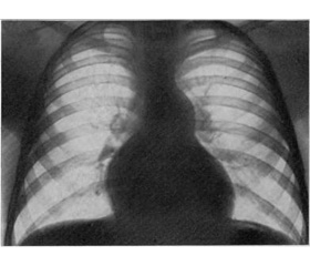
On the X-ray, cardiomegaly with a highly poor pulmonary circulation is found. The electrocardiogram shows dextrogram, signs of the right ventricle and right atrium hypertrophy. On the echocardiogram there is a stenosis on the valve level or of the subvalvular space, myocardium hypertrophy of the anterior wall of the right ventricle and of the interventricular septum, increase of the right atrium cavity, etc.
Differential diagnosis must be lead with the defect of the interatrial, interventricular septi, Fallot’s tetrad, Ebstein’s anomaly.
Treatment. Patients with a slight and moderate stenosis don’t need the implementation of the drug correction. It’s forbidden to prescribe digoxin to them. Antibacterial prophylaxy of the infectional endocarditis is recommended in all the cases. With the development of the right ventricle insufficiency with cyanosis, the oxygenotherapy and the infusion of the E1 prostaglandin are prescribed, until the surgical correction is done.
The early school age is optimal for the surgery. With the emergency, cases of the highly marked stenosis are operated in the first months of children’ life.
Prognosis. Children with a successfully done ballon valvuloplasty have a promising life prognosis. Patients with a severe stenosis are marked to have a progression of the defect with time. Average lifespan with the natural flow of the defect is 25 years. A remote favorable result after a surgery is fixed with 84,3 % of the patients. Restenosis and pulmonary artery valvular insufficiency are possible after the surgery.