Резюме
Актуальність. Визначення походження еритроцитурії в практиці педіатра є важливим завданням, воно дозволяє сформувати правильний діагноз. Розглянуто можливість застосування фазово-контрастної мікроскопії (ФКМ) для виявлення гломерулярної та негломерулярної еритроцитурії. Метою роботи було визначення морфологічної характеристики еритроцитів сечі в дітей з еритроцитурією для покращення якості диференціальної діагностики. Матеріали та методи. Дослідження морфологічної характеристики еритроцитів сечі методом ФКМ проведено 73 пацієнтам віком від 1 до 18 років, із них 45 (61,6 %) хворим на гематуричну форму гломерулонефриту, 23 (31,5 %) хворим на спадковий нефрит та 5 (6,8 %) хворим на дисметаболічну нефропатію. Результати. У дітей із клінічно встановленим діагнозом «гломерулонефрит, гематурична форма» виявлено незмінені еритроцити в 44,4 % пацієнтів, у 42,2 % випадків еритроцити були дисморфними, і в 13,4 % випадків виявлено акантоцити. Серед дітей з еритроцитурією, яким було клінічно встановлено діагноз «спадковий нефрит», переважали пацієнти з дисморфними еритроцитами — 52,2 %, у 26,1 % хворих еритроцити не були зміненими, і в 21,7 % виявлено акантоцити. У дітей з еритроцитурією, хворих на дисметаболічну нефропатію, еритроцити осаду сечі були незмінені й простежувались кристали солей. Висновки. Метод ФКМ можна використовувати в сукупності із загальноклінічними методами в рутинній практиці для визначення подальшої тактики обстеження та лікування дітей з еритроцитурією
Актуальность. Определение происхождения эритроцитурии в практике педиатра является важной задачей, оно позволяет формировать правильный диагноз. Рассматривается возможность применения фазово-контрастной микроскопии (ФКМ) для выявления гломерулярной и негломерулярной эритроцитурии. Целью работы было определение морфологической характеристики эритроцитов мочи у детей с эритроцитурией для улучшения качества дифференциальной диагностики. Материалы и методы. Исследование морфологической характеристики эритроцитов мочи методом ФКМ проведено 73 пациентам в возрасте от 1 до 18 лет, из них 45 (61,6 %) больным с гематурической формой гломерулонефрита, 23 (31,5 %) больным с наследственным нефритом, 5 (6,8 %) больным с дисметаболической нефропатией. Результаты. У детей с клиническим диагнозом «гломерулонефрит, гематурическая форма» выявлены измененные эритроциты у 44,4 % пациентов, в 42,2 % случаев эритроциты были дисморфными, и в 13,4 % случаев выявлены акантоциты. Среди детей с эритроцитурией, которым клинически установлен диагноз «наследственный нефрит», преобладали пациенты с дисморфными эритроцитами — 52,2 %, у 26,1 % пациентов эритроциты были неизмененными, и у 21,7 % больных выявлены акантоциты. У детей с эритроцитурией, больных дисметаболической нефропатией, эритроциты мочевого осадка были неизмененными и прослеживались кристаллы солей. Выводы. Метод ФКМ возможно применять в совокупности с общеклиническими методами в рутинной практике для определения дальнейшей тактики обследования и лечения детей с эритроцитурией.
Background. Nephropathies with erythrocyturia make up about 1/3 of all diseases of the kidneys and the urinary system, and they have some difficulties in differential diagnostics. Quite often, erythrocyturia is the only symptom of these diseases. In connection with this, determination of its origin is an important task in forming the correct diagnosis. Erythrocyturia in most diseases of the lower urinary tract is not accompanied by proteinuria or the presence of cylinders in the urine. The presence of proteinuria (more than 0.3 g/l or 1 g protein in urine per day), along with the appearance of erythrocytic cylinder in the urine sediment, raises suspicion in favor of glomerular or tubular diseases. Glomerular erythrocytes may be detected by means of urea concentration factor (UCF) in the urinary sediment as a preliminary test for the determination of the erythrocyturia site. Erythrocytes that pass through the glomerular membrane have a changed form (dysmorphic). Determination of acanthocytes in the urine (ring-shaped erythrocytes with one or several bulges in the form of bubbles of different sizes and types) is a more precise criterion of glomerular nephropathy than the presence of dysmorphic erythrocytes. The purpose of the study was to determine the morphological characteristics of urine erythrocytes in children with erythrocyturia, to improve the quality of differential diagnosis. Materials and methods. Determination of the morphological characteristics of urinary erythrocytes using UCF in 73 patients aged 1 to 18 years, of which 45 (61.6 %) are patients with hematuric form of glomerulonephritis, 23 (31.5 %) — with hereditary nephritis, and 5 (6.8 %) — with dysmetabolic nephropathy. Detection of 50 to 80 % of dysmorphic erythrocytes in the urine sediment and finding in urine of more than 5 % of acanthocytes is a highly sensitive and specific diagnostic criterion for glomerular hematuria. Results. In children with a clinical diagnosis “glomerulonephritis, the hematuric form”, unchanged red blood cells were detected in 44.4 % of patients, in 42.2 % of cases red blood cells were dysmorphic, and in 13.4 % of cases acanthocytes were detected. In children with erythrocyturia, who were clinically diagnosed with hereditary nephritis, dysmorphic erythrocytes prevailed — 52.2 %, in 26.1 % of patients erythrocytes were unchanged, and in 21.7 % of patients acanthocytes were identified. In children with erythrocyturia and dysmetabolic nephropathy, erythrocytes in the urine sediment were unchanged, and salt crystals were detected. Conclusions. The severity of erythrocyturia does not always correspond to the degree of alteration of red blood cell morphology. The most significant erythrocyturia is in children with hereditary nephritis; in this group the percentage of acanthocytes is highest, suggesting the presence of glomerular nephropathy. The presence of high number of dysmorphic erythrocytes is not a purely specific feature of glomerular erythrocyturia. Studying morphological characteristics of erythrocytes in the urine sediment with the help of UCF does not allow establishing the final diagnosis, but gives an opportunity to determine the tactics of further examination of the patient. A further research of the sensitivity and specificity of this method is carried out, the results of the study of morphological characteristics of erythrocytes in the urine sediment are studied in comparison with kidney biopsy.
Вступ
Нефропатії, які перебігають з еритроцитурією, становлять близько 1/3 від усіх захворювань нирок і сечовидільної системи, і в цих випадках мають місце певні труднощі в диференціальній діагностиці. Досить часто еритроцитурія є єдиним симптомом цих захворювань. У зв’язку з цим визначення її походження є важливим завданням, воно необхідне для формування правильного діагнозу. За місцем виникнення еритроцитурія може бути нирковою та позанирковою, у свою чергу, ниркова еритроцитурія поділяється на гломерулярну та тубулярну.
Еритроцитурія при більшості захворювань нижніх сечових шляхів не супроводжується протеїнурією чи наявністю циліндрів у сечі. Наявність протеїнурії (понад 0,3 г/л, або 1 г білка в сечі за добу) разом із появою в осаді сечі еритроцитарних циліндрів підвищує підозру щодо гломерулярних чи тубулярних захворювань. Гломерулярну еритроцитурію можливо встановити за допомогою фазово-конт–растної мікроскопії (ФКМ) сечового осаду як попереднього тесту на визначення топіки еритроцитурії. Уперше визначення морфологічної характеристики еритроцитів сечі було проведене D.F. Birch і K.F. Fairlye в 1979 р. У подальшому праці G. Rizzoni et al., Stapleton переконливо довели, що для гломерулярної еритроцитурії характерний дисморфізм еритроцитів. Еритроцити, що проходять через гломерулярну мембрану, мають змінену форму (дисморфічні) та дегемоглобінезовані на відміну від еритроцитів з нижнього відділу сечового тракту, які не змінюють свого розміру та форми й зберігають достатню кількість гемоглобіну (рис. 1) [6–8].
/678-1.jpg)
Дисморфічні еритроцити можуть спостерігатись і в здорових осіб, а також при негломерулярній патології, тому велике значення має співвідношення нормальних та дисморфних еритроцитів сечі. Виявлення від 50 до 80 % дисморфних еритроцитів у сечовому осаді є діагностичним критерієм гломерулярної еритроцитурії. За даними багатьох авторів, визначення в сечі акантоцитів (кільцеподібних еритроцитів з одним або декількома випинаннями у вигляді бульбашок різного розміру та виду) — більш точний критерій гломерулярної нефропатії, ніж наявність дисморфних еритроцитів.
За умови виявлення більше ніж 5 % акантоцитів серед усіх еритроцитів сечі гломерулярну еритроцитурію можливо діагностувати з чутливістю 52–99 % та специфічністю 98–100 %. Акантоцити, на відміну від інших форм еритроцитів, не виникають в експериментальних умовах in vitro при зміні рН та осмоляльності сечі, підвищенні концентрації білка, діурезу, а також через 24 години зберігання сечового осаду. Акантоцити не виявляються при фізіологічній еритроцитурії в здорових людей, для якої характерні інші види дисморфних еритроцитів (ехіноцити — еритроцити з короткими зубчиками, що знаходяться через рівні проміжки на незміненій поверхні, анулоцити — пласкі еритроцити з щільною мембраною).
Мета роботи: визначення морфологічної характеристики еритроцитів сечі у дітей з еритроцитурією.
Матеріали та методи
Визначення морфологічної характеристики еритроцитів сечі методом ФКМ проведено в умовах нефрологічного відділення дитячої міської клінічної лікарні № 2 м. Дніпро 73 пацієнтам віком від 1 до 18 років: 45 (61,6 %) хворим на гематуричну форму гломерулонефриту, 23 (31,5 %) хворим на спадковий нефрит та 5 (6,8 %) хворим на дисметаболічну нефропатію. Дослідження сечового осаду методом ФКМ проводилося за допомогою фазово-контрастного мікроскопа ФКМ МС 10 за такими критеріями:
— використовувалась ранкова порція сечі, яка знаходилась не більше ніж 2 години в сечовому міхурі;
— підрахунок кількості еритроцитів проводився при збільшенні × 40;
— сечу було центрифуговано при 1500 обертів за хвилину упродовж 5 хв;
— надосадова рідина зливалась;
— сечовий осад досліджувався на предметному склі;
— у процесі оцінювався вміст різноманітних морфологічних форм еритроцитів за гематологічною класифікацією [3, 6, 7].
Виявлення від 50 до 80 % дисморфних еритроцитів у сечовому осаді та знаходження в сечі понад 5 % акантоцитів є високочутливим та специфічним діагностичним критерієм гломерулярної гематурії.
Статистична обробка даних виконана за допомогою прикладних програм: Microsoft Excel 2010, Statistica 9.0. Перевірку відповідності емпіричних розподілів кількісних даних нормальному закону проводили за критеріями Колмогорова — Смирнова і Лілієфорса. Дані наведені як середнє значення (М), стандартна похибка (± m) або стандартне відхилення (SD) для нормального розподілу і як медіана (Ме) з інтерквартильним розкидом (25-й; 75-й перцентилі) в інших випадках. Вірогідність відмінностей кількісних показників оцінювалась за критеріями Стьюдента (t) або Манна — Уїтні (U), якісних — за критерієм хі-квадрат Пірсона (χ2), у тому числі з поправкою Йєтса. Взаємозв’язок між показниками визначали за коефіцієнтом рангової кореляції Спірмена (rs). Рівень значимості вважали вірогідним при р < 0,05.
Результати та обговорення
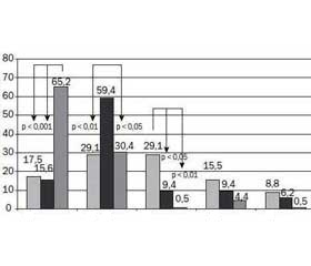
Понад половину дітей, хворих на ГН, мали виражену еритроцитурію — 55/53,4 %, у тому числі макрогематурію — 9/8,8 %, на той час як у більшості пацієнтів, хворих на СН (24/75 %) і ДН (22/95,6 %), еритроцитурія була незначною або помірною (рис. 2).
У результаті проведеного дослідження було виявлено, що в дітей із клінічно встановленим діагнозом ГН переважали незмінені еритроцити — 20/44,4 % випадків, у 19/42,2 % встановлено дисморфний вид еритроцитів (дискоцити, ехіноцити, шизоцити) (рис. 3) та в 6/13,4 % зустрічались акантоцити (рис. 4). У хворих на ГН в осаді сечі було виявлено дисморфні еритроцити та кристали солей (рис. 5).
Серед пацієнтів, яким при обстеженні було клінічно встановлено діагноз СН, найбільшу частку становили особи з дисморфними еритроцитами — 12/52,2 %, у 6/26,1 % еритроцити не були зміненими, і в 5/21,7 % виявлені акантоцити.
У всіх пацієнтів зі встановленим діагнозом ДН еритроцити осаду сечі були незмінені та спостерігались кристали солей (рис. 6).
Порівняльний аналіз морфологічної структури еритроцитів в обстежених дітей із гематурією показав її статистичну порівнянність зі структурою еритроцитів у хворих на ГН і СН (p > 0,05) та відмінність від такої у хворих на ДН (p < 0,05) (рис. 7).
У дітей з еритроцитурією, хворих на ГН, СН, ДН, визначено функціональний стан нирок залежно від морфологічного виду еритроцитів осаду сечі. Встановлено, що у хворих, в осаді сечі яких виявлено акантоцити, була зниженою швидкість клубочкової фільтрації (ШКФ), підвищена протеїнурія та альбумінурія (АУ); у дітей, в осаді сечі яких переважали дисморфні еритроцити, встановлено протеїнурію (p < 0,001), оксалурію. У дітей, в осаді сечі яких встановлено незмінені еритроцити, переважала оксалурія.
Характеристики функціонального стану нирок залежно від встановленого морфологічного виду еритроцитів осаду сечі в дітей з еритроцитурією, хворих на ГН, СН, ДН, та рівень значимості подано в табл. 1.
За даними кореляційного аналізу встановлено наявність оберненого зв’язку між незміненим характером еритроцитів і показниками азотовидільної функції, а також прямого зв’язку з рівнем ШКФ і оксалурії (табл. 2).
Висновки
За результатами аналізу морфологічної структури захворювань з еритроцитурією в дітей, хворих на ГН, СН та ДН, слід відзначити:
— у дітей, хворих на ГН, встановлено виражену еритроцитурію в 53,4 % випадків, у тому числі макрогематурію — в 8,8 % випадків, на той час як у пацієнтів із СН в 75 % та у хворих на ДН — у 95,6 % випадків еритроцитурія була незначною або помірною;
— серед дітей із клінічно встановленим діагнозом ГН виявлено незмінені еритроцити в 44,4 % осіб, у 42,2 % випадків — дисморфні еритроцити, і в 13,4 % випадків — акантоцити. У хворих на ГН в осаді сечі було виявлено кристали солей, які травмують слизову оболонку сечовивідних шляхів та посилюють прояви еритроцитурії;
— серед дітей з еритроцитурією, яким було клінічно встановлено діагноз СН, переважали хворі з дисморфними еритроцитами — 52,2 %; у 26,1 % хворих еритроцити не були зміненими, і в 21,7 % виявлено акантоцити;
— у дітей з еритроцитурією, хворих на ДН, еритроцити осаду сечі були незмінені та спостерігалися кристали солей;
— встановлено, що у хворих, в осаді сечі яких виявлено акантоцити, була зниженою ШКФ, підвищена протеїнурія та АУ;
— у дітей, в осаді сечі яких переважали дисморфні еритроцити, встановлено протеїнурію, оксалурію;
— у дітей, в осаді сечі яких встановлено незмінені еритроцити, переважала оксалурія;
— встановлено наявність кореляційного зв’язку між характером еритроцитів і показниками азотовидільної функції, рівнем ШКФ і оксалурії;
— вираженість еритроцитурії не завжди відповідає ступеню змін морфології еритроцитів. Найбільше виражена еритроцитурія в дітей зі спадковим нефритом, у даній групі найбільш високий відсоток виявлення акантоцитів, що дає підстави припустити наявність гломерулярної нефропатії. Наявність високого відсотка дисморфних еритроцитів не є суто специфічною ознакою гломерулярної еритроцитурії. Вивчення морфологічної характеристики еритроцитів осаду сечі за допомогою ФКМ не дає змогу встановити заключний діагноз, але дає можливість визначитися з тактикою подальшого обстеження пацієнта. Можна використовувати даний метод у хворих з еритроцитурією в рутинній практиці в сукупності із загальноклінічними методами дослідження як простий та неінвазивний. Проводиться подальше дослідження чутливості та специфічності даного методу, вивчаються результати дослідження морфологічної характеристики еритроцитів осаду сечі в зіставленні з даними біопсії нирки.
Конфлікт інтересів. Автори заявляють про відсутність конфлікту інтересів при підготовці даної статті.

/678-1.jpg)
/678-2.jpg)
/679-1.jpg)
/679-2.jpg)
/680-1.jpg)