Інформація призначена тільки для фахівців сфери охорони здоров'я, осіб,
які мають вищу або середню спеціальну медичну освіту.

Підтвердіть, що Ви є фахівцем у сфері охорони здоров'я.

Журнал "Гастроэнтерология" Том 55, №3, 2021

Вернуться к номеру

Поширеність синдрому надлишкового бактеріального росту серед пацієнтів із хронічними запальними захворюваннями кишечника, його вплив на показники нутритивного статусу та клінічні прояви

Авторы: Yu.M. Stepanov, M.V. Titova, N.V. Nedzvetska
SI “Institute of Gastroenterology of the National Academy of Medical Sciences of Ukraine”, Dnipro, Ukraine

Рубрики: Гастроэнтерология

Разделы: Клинические исследования

Версия для печати


Резюме

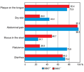
Актуальність. Останніми роками спостерігається висока зацікавленість щодо поширеності синдрому надлишкового бактеріального росту (СНБР) у тонкому кишечнику в різних популяціях. Відомо, що хронічні запальні захворювання кишечника (ХЗЗК) є гетерогенною групою розладів із високим ступенем географічної мінливості щодо симптомів, характеру прогресування, клінічних проявів або зі сполученням з іншими типами патологій. Зважаючи на те що участь мікробіому кишечника відіграє важливу роль в етіопатогенезі запальних захворювань кишечника, поєднання СНБР та ХЗЗК все частіше розглядається та досліджується останнім часом. Оскільки симптоматика обох патологічних станів значно перекликається, а отже і посилюється при поєднанні, прояви мальнутриції стають більш вираженими, що негативно впливає на нутритивний статус пацієнтів із ХЗЗК. Мета дослідження: визначити частоту СНБР в осіб із ХЗЗК залежно від нозологічних форм та дослідити його влив на клініко-лабораторні показники нутритивного статусу та клінічні прояви. Матеріали та методи. Було обстежено 100 пацієнтів із ХЗЗК віком від 19 до 79 років (у середньому (42,54 ± 1,50) року), у тому числі 70 осіб із неспецифічним виразковим колітом (НВК), 30 — із хворобою Крона (ХК). Усім хворим були проведені загальноклінічне обстеження, антропометричні вимірювання, загальний та біохімічний аналізи крові (із визначенням загального білка, альбуміну, преальбуміну). Для характеристики стану мікробіоти тонкого кишечника (наявності СНБР) усім хворим проведений водневий дихальний тест із навантаженням глюкозою з використанням газоаналізатора Gastro+ Gastrolyzer компанії Bedfont Scientific Ltd (Великобританія). Результати. Аналіз частоти виявлення СНБР показав, що зміни у стані мікрофлори тонкої кишки спостерігались у 45 % пацієнтів із ХЗЗК. Поширеність СНБР виявилася вищою в групі пацієнтів із ХК — 53,3 % (16), ніж у групі з НВК — 41,4 % (29). Наявність СНБР у групі хворих на НВК мала статистичну значущість та прямий кореляційний зв’язок із тривалістю захворювання — (9,3 ± 6,2) проти (2,9 ± 3,1) року (р = 0,001, r = 0,55). Спостерігалося зниження ваги та індексу маси тіла в пацієнтів із СНБР, особливо в осіб із ХК: так, середній показник індексу маси тіла в цих хворих становив (19,8 ± 3,5) кг/м2. Виявлено вірогідну різницю між рівнями загального білка у хворих із СНБР та без нього як у загальній групі, так і в групі НВК: (65,8 ± 8,4) проти (70,2 ± 8,2) г/л (р = 0,009, r = –0,232) та (66,5 ± 8,3) проти (70,7 ± 7,4) г/л (р = 0,029) відповідно, а також були знижені рівні альбуміну в обох нозологічних групах. Не виявлено залежності між вираженістю абдомінального болю та наявністю СНБР. При виявленому СНБР прояви метеоризму значно переважали у хворих на НВК — 75,8 % (n = 22), а діарейний синдром у пацієнтів із ХК — 75 % (n = 12). Висновки. Отримані результати свідчать про високу поширеність СНБР у пацієнтів із ХЗЗК. Пацієнти з ХК страждали від СНБР частіше (53,3 %), ніж пацієнти з НВК (41,4 %) (із переважанням хворих із тяжким ступенем вираженості). Виявлено прямий кореляційний зв’язок СНБР із тривалістю захворювання в осіб із НВК (r = 0,55; р < 0,05), що пояснюється порушенням фізіологічних бар’єрів, які попереджають виникнення СНБР, внаслідок більшої кількості епізодів загострення, тривалого прийому медичних препаратів та приєднання супутньої патології з часом. Негативний вплив СНБР на нутритивний статус проявлявся в зниженні маси тіла, індексу маси тіла та інших антропометричних (окружність середньої частини плеча, окружність м’язів плеча, товщина шкірно-жирової складки) та лабораторних (загальний білок, альбумін, преальбумін) параметрів у цих пацієнтів. Найчастішими симптомами в пацієнтів із ХЗЗК із СНБР були абдомінальний біль, діарейний синдром та метеоризм, що явно відображало типову клінічну картину СНБР.

Background. In recent years, there has been high interest in the prevalence of small intestinal bacterial overgrowth (SIBO) syndrome in various populations. Chronic inflammatory bowel disease (IBD) is known to be a heterogeneous group of disorders, with a high degree of geographical variability in terms of symptoms, nature of progression, clinical manifestations, or combination with other types of pathologies. Since the involvement of the intestinal microbiome plays an important role in the etiopathogenesis of inflammatory bowel disease, the combination of SIBO and IBD is increasingly being considered and studied recently. Since the symptoms of both pathological conditions are significantly echoed, and therefore exacerbated by the combination, the manifestations of malnutrition become more pronounced, which negatively affects the nutritional status of patients with IBD. The purpose of the study is to determine the frequency of SIBO in patients with IBD depending on the nosological forms and to investigate its effect on clinical and laboratory indicators of nutritional status and clinical manifestations. Materials and methods. We examined 100 patients with IBD, aged 19 to 79 years, on average (42.54 ± 1.50) years, including 70 patients with ulcerative colitis (UC), and 30 — with Crohn’s disease (CD). All patients underwent general clinical examination, anthropometric measurements, general and biochemical blood tests (with the determination of total protein, albumin, prealbumin). To characterize the state of the small intestine microbiota (presence of SIBO), all patients underwent a hydrogen breath test with glucose loading using a Gastro+ Gastrolyzer gas analyzer from Bedfont Scientific Ltd (UK). Results. The analysis of SIBO frequency showed the changes in the state of the small intestinal microflora in 45 % of patients with IBD. The prevalence of SIBO was higher in the group of patients with CD — 53.3 % (16) than in the group with UC — 41.4 % (29). The presence of SIBO in the group of patients with UC had statistical significance and a direct correlation with the duration of the disease — (9.3 ± 6.2) versus (2.9 ± 3.1) years (p = 0.001, r = 0.55). There was a decrease in weight and body mass index (BMI) in patients with SIBO, especially in patients with Crohn’s disease, and accounted for (19.8 ± 3.5) kg/m2. There was a significant difference between the levels of total protein in patients with SIBO and without it, both in the basic group and in the group of UC: (65.8 ± 8.4) vs. (70.2 ± 8.2) g/l (p = 0.009, r = –0.232) and (66.5 ± 8.3) vs. (70.7 ± 7.4) g/l (p = 0.029), respectively, and albumin levels were reduced in both nosological groups. No relationship was found between the severity of abdominal pain and the presence of SIBO. When SIBO was detected, the manifestations of flatulence significantly prevailed in patients with UC — 75.8 % (n = 22), and diarrheal syndrome in patients with CD — 75 % (n = 12). Conclusions. The obtained results indicate a high prevalence of SIBO in patients with IBD. Patients with CD suffered from SIBO more often (53.3 %) than patients with UC (41.4 %) (with a predominance of patients with severe disease). A direct correlation of SIBO with the disease duration in patients with UC (r = 0.55, р < 0.05) was revealed, which is explained by the violation of physiological barriers that prevent the emergence of SIBO, due to more episodes of exacerbation, long-term use of drugs and concomitant pathology with time. The negative impact of SIBO on nutritional status manifested in weight loss, reduced BMI and other anthropometric (mid-upper arm circumference, mid-arm muscle circumference, triceps skinfold) and laboratory (total protein, albumin, prealbumin) parameters in these patients. The most common symptoms in patients with IBD with SIBO were abdominal pain, diarrhea, and flatulence that reflected the typical clinical picture of SIBO.


Ключевые слова

нутритивний статус; неспецифічний виразковий коліт; хвороба Крона; антропометрія; синдром надлишкового бактеріального росту

nutritional status; ulcerative colitis; Crohn’s disease; anthropometry; small intestinal bacterial overgrowth syndrome

Introduction

Chronic inflammatory bowel disease (IBD), which is represented by ulcerative colitis (UC) and Crohn’s disease (CD), is gaining increasing attention due to the steady increase in morbidity and prevalence worldwide. Underinvestigated etiology and various hypotheses of the pathogenesis of these diseases lead to a deeper study of clinical features and mechanisms of systemic lesions in patients with UC and CD. This will allow the development of new, more accurate and personalized schemes for the diagnosis and treatment of these patients.
It is known that nutritional disorders are a common manifestation in patients with IBD, which significantly affects the course of the disease and worsens its prognosis. In addition to the common causes of nutrient loss in these patients (frequent bowel movements, chronic blood loss, enzymatic insufficiency, dietary restriction due to pain and intoxication), more and more attention has recently been paid to the changes in intestinal microbiota and their effects on inflammation, digestion and absorption [1, 2]. This is due to the fact that the most do-minant etiological hypothesis indicates an immune response to the altered microbiota in a genetically predisposed host [3, 4].
One of the frequent manifestations of intestinal dysbiosis in patients with IBD is the small intestinal bacterial overgrowth (SIBO) syndrome. Some studies report a high predisposition to the development of SIBO in these patients, especially in patients with CD [1, 5–7].
The small intestine is a transition zone between the sto-mach, which contains a small number of microorganisms, and the large intestine with a high level of bacterial colonization. The small intestinal bacterial overgrowth syndrome develops with excessive colonization of bacteria in the small intestine due to opportunistic microflora of colon origin, including Streptococcus, Escherichia coli, Staphylococcus, Micrococcus, etc., and is accompanied by certain signs and symptoms depending on the type of bacteria in the intestine. Under physiological conditions, the proximal part of the small intestine is characterized by a bacterial population corresponding to 103–104 colony-forming units (CFU) per milliliter of aspirate. SIBO is characterized by a population over 105 CFU/ml aspirate [8].
The clinical picture of SIBO can be different: from asymptomatic to severe symptoms, such as constant abdominal pain, excessive flatulence, diarrhea. In severe cases, the symptoms manifested themselves as a consequence of malabsorption: vitamin B12 deficiency or even iron deficiency anemia, deficiency of fat-soluble vitamins, hypoalbuminemia, weight loss, signs of malnutrition, and even severe exhaustion [9]. Often these symptoms in patients with IBD are perceived as an outbreak of the underlying disease and SIBO remains undetected. In turn, the accession of the SIBO to the next exacerbation of IBD significantly affects the severity of symptoms, severity, the possibility of complications, and the overall prognosis [7, 10].
The microflora of patients with SIBO is mainly characterized by the predominance of coliform bacteria and anaerobes, which cause fermentation of carbohydrates, compete with the absorption of vitamins and trace elements and provoke microscopic inflammation of the mucosa, which leads to the above symptoms [8, 11]. Most experts suggest that a culture of small bowel aspirate (with the number of bacteria in colonies ≥ 105 CFU/ml) is the gold standard for the diagnosis of SIBO. However, this method has several disadvantages, the most important of which are the invasiveness of the procedure and the use of special equipment. The glucose hydrogen breath test (GBT) for the diagnosis of SIBO has gained increasing approval and is most commonly used in clinical practice (lactulose breath test and glucose breath test) [12]. The glucose hydrogen breath test is non-invasive and based on determining the concentration of hydrogen in the air exhaled after carbohydrate loading since there is a direct relationship between the degree of bacterial contamination of the small intestine and the concentration of hydrogen in exhaled air. However, it should be noted that the lactulose test is not suitable for the diagnosis of SIBO in patients with IBD, because lactulose is a synthetic disaccharide that does not break down and passes intact through the small intestine to the cecum, where it is metabolized by the colon bacteria to the short intestine, and gases, including hydrogen and/or methane, which are systematically absorbed and eventually exhaled. Due to accelerated intestinal transit in these patients, the test may give false-positive results (peak hydrogen concentration of 90 minutes or more) [12]. Therefore, GBT, due to its simplicity and high specificity, should be included in the list of mandatory studies in patients with IBD to detect SIBO in the early stages of diagnosis. This will allow timely correction of therapy and avoid complications, which in turn will improve the prognosis of the underlying disease.
So, the purpose of the study is to determine the frequency of SIBO in patients with IBD depending on the nosological forms and to investigate its effect on clinical and laboratory indicators of nutritional status and clinical manifestations.

Materials and methods

We examined 100 patients with IBD, aged from 19 to 79 years, on average (42.54 ± 1.50) years, including 70 patients with UC and 30 individuals with CD. In 51 cases with UC and in 11 with CD, there was a moderate severity of the disease; 19 patients with UC and 19 individuals with CD presented with severe disease. All patients underwent a general clinical examination to determine anthropometric parameters as well as a detailed collection of medical history and complaints.
Anthropometric parameters. Body mass index (BMI) was calculated by the formula:
BMI = m/ h2
where m — body weight in kilograms; h — height in meters.
The triceps skinfold (TSF) was measured in millimeters with a caliper. The mid-upper arm circumference (MUAC) was measured in centimeters at the level of the middle third (midway between the acromion and the olecranon processes) of the non-working unstressed arm. The mid-arm muscle circumference (MAMC) was calculated by the formula:
MAMC = MUAC – 3.14 • TSF.
To characterize the condition of the small intestinal microbiota (presence of SIBO), all patients underwent a GBT using a Gastro+ Gastrolyzer gas analyzer from Bedfont Scientific Ltd (UK).
Recommendations for preparing and conducting testing were as follows:
— avoidance of taking:
– antibiotics for 4 weeks before testing,
– bismuth preparations and probiotics for 2–4 weeks before testing,
– prokinetics for 3 half-lives before testing;
— avoidance of colon cleansing for 4 weeks before testing;
— adherence to a diet that does not contain complex carbohydrates (pasta, bread, cereals, beans) in the evening before testing;
— night fasting before testing;
— avoidance of smoking before and during testing.
The study was conducted following the standards of GBT performance [13, 14]. The method is based on measuring the concentration of hydrogen in the air exhaled by the patient after ingestion of glucose solution (50 g per 250 ml of water), at regular intervals (0 min, 15 min, 30 min, 45 min, and 60 min). The GBT begins with the measurement of the basal level of H2. To do this, the patient is asked to hold his breath for 15 seconds and then slowly exhale into the mouthpiece of the device. The hydrogen content is determined in parts per million (ppm), and diagnostically significant is the increase of its level by 12 ppm or more from the initial value.
The test can be started if the basal level of hydrogen in the exhaled air (on an empty stomach) does not exceed 5 ppm. The data obtained during the study are entered into the protocol and a schedule is built. Graphs can be of two types: normal — the glucose does not decompose in the small intestine, and the concentration of H2 does not exceed 10 ppm (Fig. 1); pathological — with bacterial overgrowth, glucose is fermen-ted in the small intestine and the concentration of hydrogen increases earlier (SIBO) (Fig. 2).
It should be noted that the concentration of hydrogen in the exhaled air depends on the position of the body, therefore, each time the patient must be in the same position as in the initial measuring.
The results were processed using the Statistica 6.0 package. Since most of the data had a normal distribution, parametric statistics were used. The probability of the difference between the samples was evaluated by Student’s t-test, the differences were considered probable if p < 0.05. All quantitative indicators are given in the form of M ± m, where M is the arithmetic mean, m is the arithmetic mean error. Correlation analysis was performed using the Pearson correlation coefficient.
All measuring equipment used in the work was metrologically verified in the prescribed manner.

Results and discussion

The analysis of the frequency of SIBO showed that the changes in the state of the small intestinal microflora were observed in 45 % of patients with IBD. The prevalence of SIBO was found to be higher in the group with CD — 53.3 % of patients than in the group with UC — 41.4 % of patients (Fig. 3).
It was also found that in the group of CD with detected SIBO, patients with severe disease prevailed, namely in 11 patients (68.7 %) out of 16, while in the group of UC only 8 patients (27.5 %) out of 29. This may be explained by the fact that most patients with CD had a predominant lesion of the terminal small intestine, and the accession of the SIBO had a negative impact on the severity of the disease. The presence of SIBO in the group of patients with UC had statistical significance and a direct correlation with the disease duration — (9.3 ± 6.2) versus (2.9 ± 3.1) years (p = 0.001, r = 0.55). No relation was found between the positive result of GBT and therapy for the underlying condition, which is confirmed by the literature resources [7].
We analyzed the relationship between the results of GBT and age, anthropometric and some laboratory parameters of patients (Table 1).
The groups of patients with positive and negative GBT did not differ in age or height. The average body weight of patients with a positive result of GBT was less than in those with a negative result: for UC — (64.0 ± 11.6) against (69.8 ± 17.8) kg, for CD — (58.3 ± 13.8) against (63.3 ± 12.5) kg, but the difference was not statistically significant. Body mass index was lower in patients with CD and positive SIBO — (19.8 ± 3.5) kg/m2.
There was a significant difference between the levels of total protein in patients with SIBO and without it, both in the basic group and in the group of UC: (65.8 ± 8.4) vs. (70.2 ± 8.2) g/l (p = 0.009, r = –0.23) and (66.5 ± 8.3) vs. (70.7 ± 7.4) g/l (p = 0.03), respectively. Albumin levels were also reduced in both nosological groups with detected SIBO but were significant only in the UC group — (36.3 ± 9.2) against (42.0 ± 8.5) g/l (p = 0.009, r = –0.35) (Table 2).
This indicates a violation of the processes of digestion and absorption in the small intestine. The correlation analysis in the UC group revealed a reversible relationship between the indicators of MUAC and MAMC and the presence of SIBO (r = –0.240, r = –0.235, respectively).
The data from a detailed survey of the patient’s data on the most common complaints and symptoms were analyzed. The results depending on the presence of SIBO and nosological group are shown in Fig. 4 and 5.
The most common symptom in the groups was abdominal pain. Almost all patients complained about it, regardless of the nosological form or the presence of SIBO. Dry skin was also a common symptom in patients with SIBO in both nosological groups (41.4 % (n = 12) in patients with UC and 56.3 % (n = 9) in patients with CD) and indicated an increase in the general manifestations of malnutrition. In SIBO, however, the indicators were not statistically significant.
Diarrhea is known to be a common clinical manifestation in patients with IBD. However, in some cases, the increase in the frequency of defecation in these patients is associated with the presence of SIBO. In our study, the diarrheal syndrome was observed in almost the same number of patients with UC regardless of the presence of SIBO. Manifestations of flatulence significantly prevailed in patients with UC with detected SIBO — 75.8 versus 26.8 % in patients without SIBO that reflects the typical clinical picture of SIBO.
In contrast, in the group of patients with CD, the diarrheal syndrome significantly prevailed in patients with positive SIBO: 75.0 % (n = 12) versus 28.5 % (n = 4). It can therefore be concluded that SIBO exacerbates the manifestations of this symptom in patients with CD in the active phase. However, it should be borne in mind that SIBO can mimic exacerbations of CD, including increased bowel movements and weight loss, so it should be detected in the early stages of diagnosis and treatment.

Conclusions

Thus, our results indicate a high prevalence of SIBO in patients with IBD — 45 %. The prevalence of SIBO was higher in the group of patients with CD than in patients with UC — 53.3 and 41.4 %, respectively. SIBO was more common in patients with severe CD, namely 68.7 %, while in the group of UC only 27.5 %.
There is a direct correlation between the duration of the disease and the presence of SIBO in patients with UC (r = 0.55, р < 0.05) that is explained by the violation of physiological barriers that prevent the emergence of SIBO, more episodes of exacerbation, prolonged use of drugs, and concomitant pathology in time.
The laboratory study found a significant decrease in total protein and albumin in patients with SIBO. They also showed a tendency to decrease body weight, BMI, and other anthropometric parameters (MUAC, MAMC, TSF) that indicates the negative impact of SIBO on the nutritional status of patients. The most common symptoms in patients with IBD and SIBO were abdominal pain, diarrhea, and flatulence that reflected the typical clinical picture of SIBO. But we should not forget that the clinical manifestations of SIBO and IBD often resonate and can be perceived as an exacerbation of the underlying disease. That is why it is necessary to timely diagnose SIBO in this group of patients, which will allow timely correction of disorders of the intestinal microbiota and avoid the strengthening of basic therapy.
 
Received 01.07.2021
Revised 12.07.2021
Accepted 16.07.2021

Список литературы

  1. Cohen-Mekelburg S., Tafesh Z. et al. Testing and Treating Small Intestinal Bacterial Overgrowth Reduces Symptoms in Patients with Inflammatory Bowel Disease. Dig. Dis. Sci. 2018 Sep. 63 (9). 2439-2444. doi: 10.1007/s10620-018-5109-1. Epub 2018 May 14. PMID: 29761252.
  2. Stepanov Yu.M, Titova M.V., Stoikevich M.V. Nutrytyvnyi status khvorykh na khronichni zapalni zakhvoriuvannia kyshechnyka ta metody yoho otsinky [Nutritional status of patients with chronic inflammatory bowel disease and methods for its assessment]. Gastroenterologia. 2019. 4 (53). 273-281. (in Ukrainian).
  3. Khan I., Ullah N., Zha L. et al. Alteration of Gut Microbiota in Inflammatory Bowel Disease (IBD): Cause or Consequence? IBD Treatment Targeting the Gut Microbiome. Pathogens. 2019 Aug 13. 8 (3). 126. doi: 10.3390/pathogens8030126. PMID: 31412603; PMCID: PMC6789542.
  4. Lo Presti A., Zorzi F., Del Chierico F. et al. Fecal and Mucosal Microbiota Profiling in Irritable Bowel Syndrome and Inflammatory Bowel Disease. Front. Microbiol. 2019 Jul 17. 10. 1655. doi: 10.3389/fmicb.2019.01655. PMID: 31379797; PMCID: PMC6650632.
  5. Shah A., Morrison M., Burger D. et al. Systematic review with meta-analysis: the prevalence of small intestinal bacterial overgrowth in inflammatory bowel disease. Aliment. Pharmacol. Ther. 2019 Mar. 49 (6). 624-635. doi: 10.1111/apt.15133. Epub 2019 Feb 8. PMID: 30735254.
  6. Greco A., Caviglia G.P., Brignolo P. et al. Glucose breath test and Crohn’s disease: Diagnosis of small intestinal bacterial overgrowth and evaluation of therapeutic response. Scand. J. Gastroenterol. 2015. 50 (11). 1376-81. doi: 10.3109/00365521.2015.1050691. Epub 2015 May 19. PMID: 25990116.
  7. Kulygina Yu.A., Osipenko M.F., Lukinov V.L., Lukashova L.V., Pomogaeva A.P. Sindrom izbytochnogo bakterial’nogo rosta i gastrointestinal’nye simptomy u pacientov s vospalitel’nymi zabolevanijami kishechnika [The bacterial overgrowth syndrome in the small intestine and gastrointestinal symptoms in patients with inflammatory bowel diseases depending on the presence of small intestinal bacterial overgrowth syndrome]. Experimental and Clinical Gastroenterology. 2018. 159 (11). 68-74. doi: 10.31146/1682-8658-ecg-159-11-68-74. (in Russian).
  8. Adike A., DiBaise J.K. Small Intestinal Bacterial Overgrowth: Nutritional Implications, Diagnosis, and Management. Gastroenterol. Clin. North Am. 2018 Mar. 47 (1). 193-208. doi: 10.1016/j.gtc.2017.09.008. Epub 2017 Dec 7. PMID: 29413012.
  9. Andrei M., Gologan S., Stoicescu A., Ionescu M., Nicolaie T., Diculescu M. Small Intestinal Bacterial Overgrowth Syndrome Prevalence in Romanian Patients with Inflammatory Bowel Disease. Curr. Health Sci. J. 2016 Apr-Jun. 42 (2). 151-156. doi: 10.12865/CHSJ.42.02.06. Epub 2016 Jun 28. PMID: 30568826; PMCID: PMC6256157.
  10. Danilova N.A., Abdulhakov R.A., Abdulhakov S.R., Odincova A.H., Ramazanova A.H., Sadykova L.R. Sindrom izbytochnogo bacterial’nogo rosta u pacientov s vospalitel’nymi zabolevanijami kishechnika [Syndrome of intestinal bacterial overgrowth in patients with inflammatory bowel diseases]. Prakticheskaja medicina. 2015. 6 (91). 122-126. (in Russian).
  11. Ponziani F.R., Gerardi V., Gasbarrini A. Diagnosis and treatment of small intestinal bacterial overgrowth. Expert Rev. Gastroenterol. Hepatol. 2016. 10 (2). 215-27. doi: 10.1586/17474124.2016.1110017. Epub 2015 Dec 4. PMID: 26636484.
  12. Massey B.T., Wald A. Small Intestinal Bacterial Overgrowth Syndrome: A Guide for the Appropriate Use of Breath Testing. Dig. Dis. Sci. 2021 Feb. 66 (2). 338-347. doi: 10.1007/s10620-020-06623-6. Epub 2020 Oct 10. PMID: 33037967.
  13. Losurdo G., Leandro G., Ierardi E. et al. Breath Tests for the Non-invasive Diagnosis of Small Intestinal Bacterial Overgrowth: A Systematic Review With Meta-analysis. J. Neurogastroenterol. Motil. 2020 Jan 30. 26 (1). 16-28. doi: 10.5056/jnm19113. PMID: 31743632; PMCID: PMC6955189.
  14. Rezaie A., Pimentel M., Rao S.S. How to Test and Treat Small Intestinal Bacterial Overgrowth: an Evidence-Based Approach. Curr. Gastroenterol. Rep. 2016 Feb. 18 (2). 8. doi: 10.1007/s11894-015-0482-9. PMID: 26780631.

Вернуться к номеру