Резюме
Актуальність. Запальні захворювання кишечника, що включають виразковий коліт (ВК) і хворобу Крона, є актуальною проблемою сучасної гастроентерології. Тому виявлення нових лабораторних підходів надасть можливість оцінити ступінь перебігу захворювання. Мета: виявити зв’язки між морфологічними проявами та імунологічними показниками у хворих на ВК. Матеріали та методи. Дослідження проведені на біологічному матеріалі (кров та колонобіоптати) 90 пацієнтів з ВК. Морфологічним та морфометричним шляхом у біоптатах підраховували товщину слизової оболонки (СО), щільність запального інфільтрату та його склад, розміри крипт, їх архітектоніку, наявність крипт-абсцесів, атрофічних та фібротичних змін. Імунологічні дослідження включали визначення рівня В-лімфоцитів, ІЛ-10, TNF-α, вмісту імуноглобулінів (Ig) класів А, М, G. Результати. Гістологічна активність захворювання визначалась збільшеною щільністю запального інфільтрату (14 431,4 ± 483,3 на 1 мм2 строми) і наявністю в ньому великої кількості нейтрофільних гранулоцитів (212,2 ± 20,9 на 1 мм2 строми) та лімфоцитів (2922,8 ± 76,6 на 1 мм2 строми). Також у частини пацієнтів виявлялись крипт-абсцеси (36,7 % від загальної кількості пацієнтів) і порушення цілісності епітелію (54,4 % від загальної кількості пацієнтів). Було встановлено кореляційний зв’язок рівня СD22+ лімфоцитів з окремими морфометричними показниками: шириною крипт (r = 0,27; Р < 0,01) та висотою поверхневого епітелію (r = 0,30; Р < 0,01); між концентрацією IgМ та клітинною щільністю інфільтрату СО (r = 0,29; Р < 0,01), нейтрофілами (r = 0,28; Р < 0,01) та базофілами (r = 0,24; Р < 0,05); рівнем IgА та макрофагами (r = 0,21; Р < 0,05), лімфоцитами (r = 0,24; Р < 0,05), базофілами (r = 0,25; Р < 0,05). Висновки. Показано, що окремі морфологічні та морфометричні показники пов’язані з імунологічними показниками. Встановлено, що підвищений рівень цитокінів корелює з активністю запалення у пацієнтів з ВК. Рівень СD22+ лімфоцитів та зміни окремих морфометричних показників (ширина крипт та висота поверхневого епітелію) безпосередньо пов’язані з посиленням запальних процесів в СО кишечника.
Background. Inflammatory bowel diseases, including ulcerative colitis and Crohn’s disease, are an urgent problem of modern gastroenterology. Therefore, the discovery of new laboratory approaches makes it possible to assess the degree of the disease. Purpose: to reveal the relationship between morphological manifestations and immunological indicators in patients with ulcerative colitis. Materials and methods. The studies were conducted on biological material (blood and colonic biopsy samples) of 90 patients with ulcerative colitis. The thickness of the mucosa, density of the inflammatory infiltrate and its composition, crypt sizes, their architectonics, the presence of crypt abscesses, atrophic and fibrotic changes were calculated in biopsies by morphological and morphometric methods. Immunological studies included the evaluation of mononuclear cells, the levels of B-lymphocytes, interleukin-10, tumor necrosis factor α, immunoglobulins (Ig) A, M, G. Results. The histological activity of the disease was determined by an increased level of inflammatory infiltrate (14,431.4 ± 483.3 per 1 mm2 of stroma) and the presence of many neutrophilic granulocytes (212.2 ± 20.9 per 1 mm2 of stroma) and lymphocytes (2,922.8 ± 76.6 per 1 mm2 of stroma) in it. Also, some patients had crypt abscesses (36.7 % of the total number of patients) and breaches in the epithelial integrity (54.4 % of the total number of patients). A correlation was found between the level of CD22+ lymphocytes and some morphometric parameters: the width of the crypts (r = 0.27; P < 0.01) and the height of the surface epithelium (r = 0.30; P < 0.01); between IgM concentrations and cellular density of mucosal infiltrate (r = 0.29; P < 0.01), neutrophils (r = 0.28; P < 0.01) and basophils (r = 0.24; P < 0.05); level of IgA and macrophages (r = 0.21; P < 0.05), lymphocytes (r = 0.24; P < 0.05), basophils (r = 0.25; P < 0.05). Conclusions. It is shown that some morphological and morphometric indicators are related to immunological parameters. It was found that the elevated level of cytokines correlates with the activity of inflammation in patients with ulcerative colitis. The level of CD22+ lymphocytes and changes in some morphometric indicators (crypt width and surface epithelium height) are directly related to an increase in inflammatory processes in the intestinal mucosa.
Introduction
Inflammatory bowel disease (IBD) is a chronic immune-mediated inflammatory disease that predominantly affects the gastrointestinal tract (GIT) and is represented by the main nosological forms: ulcerative colitis (UC) and Crohn’s disease [10]. As a chronic disease, UC causes inflammatory reactions and an immune response in the colonic mucosa (CM) [27]. The disease is mainly detected in the age of 20–40 years, but it can occur at any age. The most common clinical symptoms are gastrointestinal distress, such as abdominal pain, bloody or mucous diarrhea, nausea, and vomiting; however, general symptoms, inclu–ding fever, weight loss, and anemia, are also common. UC is characterized by alternating periods of clinical relapse and remission [19].
The pathogenesis of UC involves damage of the CM and deterioration of its digestive and absorptive functions. Inflammation of the CM plays a significant role in the pathogenesis of UC, leading to ulceration. The changes observed in the intestinal mucosa are localised in the rectum and extend proximally to other parts of the colon. Inflammation of the CM occurs due to an imbalance between the expression of proinflammatory and anti-inflammatory factors and the migration of neutrophils to the damaged area, which leads to the activation of inflammation. Although UC is constantly being researched, its exact pathogenesis is not fully understood; however, it is known that UC is associated with an excessive immune response to environmental factors or resident microbiota in genetically predisposed subjects, and immune status plays a crucial role in increasing intestinal permeability and impairing barrier function. However, va–rious biological compounds can be detected in patients that cause damage to the GI tract [14, 24].
UC is primarily associated with chronic inflammation, possibly resulting from an activated immune response to the gut microbiota and/or food antigens. In order to analyse the pathogenesis of UC at the molecular level, it is necessary to have a good knowledge of the cellular populations of the CM. The GI tract is lined by a single-layer columnar epithelium with a thin brush border, which is essential for maintai–ning intestinal homeostasis and functions as a physical, biochemical barrier and a focal point for immune defense and crosstalk between bacteria and immune cells [5, 7]. Intestinal stem cells, responsible for the rapid renewal of the intestinal epithelium, are located at the base of these crypts and develop into temporary proliferative cells that differentiate as they pass through the transition zone, where intestinal epithelial cells eventually emerge into the lumen at the top of the crypts [22]. The epithelium consists of various subtypes of specialised intestinal epithelial cells (absorptive, goblet, enteroendocrine, Paneth, M cells), which all differentiate from epithelial stems, which are functionally different in nature and are necessary for maintaining intestinal homeostasis [2, 21]. A reliable histological criterion and indicator of colitis activity is the detection of neutrophils (not single ones) in the crypt epithelium (crypts). The appearance of crypt abscesses indicates the beginning of erosion formation and can be interpreted as moderate inflammation activity, and the detection of erosions and ulcers will be considered as a manifestation of its severe activity [3, 16].
It is assumed that one of the key defects of the immune system that leads to the development of UC is a disruption of the process of recognition of common infectious molecular patterns by dendritic cells, which in turn triggers the activation of cellular and humoral immunity reactions and hyperactivation of proinflammatory signaling pathways [29]. These factors determine the development of a systemic inflammatory process with a predominant localisation in the CM. Prolonged activation of the cellular-humoral link, antigenic aggression lead to a breakdown of immunological tolerance and the formation of autoimmune inflammation, in the development of which immunoregulatory disorders play a significant role [9].
An understanding of the clinical and endoscopic features of UC is crucial for making a timely diagnosis. The full range of available tools should be used to accurately diagnose IBD, including medical history, clinical presentation, non-invasive and invasive imaging (endoscopy), and histological interpretation. The diagnosis of IBD is consi–dered reliable only with histological confirmation. The main significance of histological features is that they are crucial in assessing the activity and seve–rity of the disease and, therefore, have great clinical and prognostic value. Many studies have been conducted on immunological disorders in UC and leave no doubt that the immunopathogenesis of this disease is formed with the participation of all parts of the immune system [8, 25, 26, 28].
The purpose of our studies was to identify the relationship between the morphological changes and the immunological profile of the CM in patients with UC.
Мaterials and methods
Ninety patients with ulcerative colitis who were in the inpatient department of intestinal diseases of the State Institution “Institute of Gastroenterology of the National Academy of Medical Sciences of Ukraine” were examined. Diagnosis was established on the basis of endoscopic examination of the colon. Endoscopy was performed in all patients according to generally accepted methods using Olympus EVIS EXERA III equipment (Japan).
Morphological and morphometric evaluation were carried out in the pathomorphology laboratory of the Institute of Gastroenterology of the National Academy of Medical Sciences of Ukraine. The biopsied material was fixed in a 10.0% solution of neutral formalin, dehydrated in alcohols of increasing concentration and embedded in paraffin. To study the general histological structure of the mucous membrane, sections of biopsies with a thickness of 5–7 μm were cut with the help of a rotary microtome PM60-EKA. Staining of the samples was carried out according to the standard method with hematoxylin-eosin.
For morphometric research, sections were photographed using an XSZ-21 light microscope (Micro Med, Ukraine) and measured using ImageJ 1.45S software (National Institutes of Health, USA). Morphometric studies of histological preparations included: measurement of the depth of the crypts (μm), height of the surface epithelium (μm), height of the epithelium of the crypts (μm), the number of goblet cells in the epithelium of the crypts per 100 cells, the density of the cellular infiltrate and the number of inflammatory cells (per 1 mm2 of stroma).
The material for the study of immunological parameters was venous blood, which was taken from the ulnar vein in a volume of 10 ml on an empty stomach. Mononuclear cells were isolated from the patients’ peripheral venous blood in a density gradient of 1.077 g/cm. The level of B-lymphocytes was determined using monoclonal antibodies to CD22+ (Sorbent).
Determination of the quantitative serum content of Ig of classes A, M, G was performed by ELISA using test systems Granum Ltd. (Ukraine) according to the manufacturer’s recommendations. Serum levels of IL-10 and TNF-α were determined by ELISA using test systems of Wuhan Fine Biotech Co., Ltd (China). ELISA was performed using a Stat Fax 303 Plus (USA).
Statistical processing of the results was performed using the Statistica 10.0 software package. The statistical analysis of the data included the calculation of the mean (M) and standard error (m) of indicators with a normal distribution of data, the median (Me) and the lower and upper quartiles (Q1; Q3) for data that did not have a normal distribution. The Shapiro-Wilk test was used to test the normality of the distribution. The comparison of mean values of variables was carried out using the non-parametric Mann-Whitney U test. Statistical significance was assessed at a level not lower than 95.0 % (p < 0.05). Correlation between indicators were assessed using significant Spearman correlation coefficients (r).
Results
In the majority of patients (49 patients), microscopic examination of colon biopsies showed a violation of the integrity of the epithelium: ulcers were 4.7 times more frequent than erosion (3 patients) (Table 1).
/58.jpg)
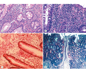
In 22 patients with UC (24.4 %), crypt architecture distortion was noted (shortening, deformation, branching, decrease in the number and/or uneven distribution of goblet cells). Such phenomena are typical for this disease. The histological activity of UC was determined by the presence of the epithelium damage mediated by neutrophils, which, in some cases, took the form of neutrophilic infiltration of the epithelium of the crypts (cryptitis), accumulation of neutrophils in the lumen of the crypts (crypt abscesses) or infiltration of the surface epithelium with mucosal ulcers formation. The chronic nature of UC was determined by changes in the architecture of the crypts and basal lymphoplasmacytosis of the CM (lymphoplasmacytic infiltrate between the bases of the crypts). The architectural change took the form of shortened (by the presence of space between the bottom of the crypts and the upper edge of the muscular shell) and branched crypts (Fig. 1).
/59.jpg)
Attention is drawn to the fact that individual structural disorders (loss of glands, decrease in the number of goblet cells) can be interpreted as an analogue of the histological activity of UC.
Moderately expressed inflammatory infiltration (focal or diffuse) of the lamina propria of the mucous membrane was noted in more than half of the patients. Increased infiltration of the CM in UC was present in a third of patients. Mild infiltration was less common in patients (13.3 % of cases). Inflammatory infiltration was detected together with the presence of crypt abscesses. Crypt abscesses, as a typical sign of UC, were found in 55.6 % of cases, and single crypt abscesses were found almost twice as often as multiple ones. Atrophic changes of the mucous membrane appeared 3.1 times more often than fibrous changes, and dysplasia was diagnosed in 9 patients (10.0 %).
From a morphological point of view, morphometric parameters are the most informative signs for an objective evaluation of the diagnosis of UC. Morphometry characteri–zes the course of inflammatory and reparative processes, restoration of the morphofunctional state and local immunity of the CM. Microscopic examination of colon biopsies from patients with UC revealed inflammatory cell infiltration both in the lamina propria and in the lumen of the crypts, which indicates the development of cryptitis and the formation of crypt abscesses. The density of the cellular infiltrate in UC according to the morphometry was 1.35 times higher than the density of the cellular infiltrate according to morphological staining. Inflammatory infiltration was represented by neutrophilic and eosinophilic leukocytes, macrophages, basophils, fibroblasts and lymphocytes (Table 2).
/59_2.jpg)
/59_2.jpg)
It should be noted that neutrophilic leukocytes are not found in a healthy intestine. These cells are the first links of the body’s immune defense and are the first to be found in the focus of inflammation. With the death and degranulation of neutrophils in a pathological cell, an excessive amount of leukocyte elastase enters the blood, which is characterized by a powerful proteolytic and destructive potential and plays an important role in the pathogenesis of inflammation. Normally, eosinophils are present in the mucous membrane of the digestive organs except the esophagus. In the healthy intestine, eosinophils are found no more than 3–5 per field of view. In IBD, the presence of a high number of eosinophilic leukocytes in the mucosa indicates increased inflammatory processes in the intestinal tissues [4].
The increase in UC activity leads to an increase in inflammatory cell infiltration of the CM, as evidenced by an increase in the density of the cell infiltrate; the severity of inflammatory changes in the crypts and an increase in the number of crypt abscesses; decrease in the number of cases with focal infiltration in the lamina propria and increase in the number of cases with diffuse infiltration; the spread of inflammatory cell infiltration from the surface parts of the lamina propria to its deep parts with subsequent involvement of its thickness; an increase in the cell infiltration, infiltration by plasma cells, T-lymphocytes, macrophages, and neutrophilic leukocytes. Macrophage cells and basophils were characterized by a diffuse or focal location (in areas of ulcers, erosions, and damaged surface epithelium). Damage to the epithelial barrier and tissues causes an immune response, which leads to chronic inflammation and excessive synthesis of the extracellular matrix components and intestinal fibrosis, which is confirmed by the presence of a large number of fibroblasts in the CM.
The median level of the relative content of B-lymphocytes in the examined patients did not differ significantly from the control values (Table 3). At the same time, a significant decrease in the number of IgM (2.0 times, p < 0.05) and IgG (1.4 times, p < 0.05) was found.
The concentration of TNF-α in patients with UC was significantly higher by 9.9 times (p < 0.05) compared to the control group. Meanwhile, the median level of IL-10 in these patients (5.4 times, p < 0.05) was significantly reduced compared with the control group.
According to the results of the correlation analysis, the level of CD22+ lymphocytes was associated with such morphometric parameters as crypt width (r = 0.27; P < 0.01) and height of the surface epithelium (r = 0.30; P < 0.01) (Fig. 2).
There was also a correlation between the concentration of IgM and the cellular density of the CM infiltrate (r = 0.29; P < 0.01), neutrophils (r = 0.28; P < 0.01) and basophils (r = 0.24; P < 0.05); IgA levels and macrophages (r = 0.21; P < 0.05), lymphocytes (r = 0.24; P < 0.05) and basophils (r = 0.25; P < 0.05).
Discussion
From a clinical-morphological point of view, to provide an objective assessment of the establishment of a histological diagnosis, the most informative are the morphometric parameters that most accurately characterize the course of the inflammatory state and reparative processes, the restoration of the morpho-functional state of the intestinal mucosa [3].
The general histological pattern identified in UC is a change in the architecture of the intestinal epithelium, cha–racterized by shortening and reduced branching of the crypts. Our studies showed that in some patients with UC, the crypts have branched, shortened crypts, which indicates atrophic changes during the development of the disease. When microscopically examining the colon biopsies of patients with UC, inflammatory cell infiltration both in the lamina propria and in the lumen of the crypts indicates the development of cryptitis and the formation of crypt abscesses, which were also found in some histological preparations. Such morphometric indicators as the height and width of the crypts can be considered as signs of atrophy of the intestinal glands [22].
Attention is drawn to the fact that during a recurrent course, the maximum infiltration of the CM was determined mainly due to neutrophilic and eosinophilic granulocytes, which determined the activity of inflammation [6, 11]. The development of fibrotic changes is regulated by macrophages, since macrophages are always located near fibroblasts that produce collagen and regulate fibrosis regardless of direct interaction with myofibroblasts [7].
The precise mechanisms that contribute to the development of UC remain unclear, but significant progress has been made recently and it has been shown that the immune response plays a major role in the pathogenesis of UC. Inflammation promotes lymphocyte activation through increased activity of antigen-presenting cells (macrophages and dendritic cells) in IBD [12, 15]. Th17-cells play a dual role: on the one hand, they secrete proinflammatory cytokines (IL-17, TNF-α), and on the other hand, cytokines that protect the intestinal epithelium (IL-10, IL-22). Therefore, the correlations of IL-10 with inflammatory cells number do not contradict the literature [13, 18].
B-lymphocytes play an important role alongside T-cells. B-cells are responsible for the synthesis of antibodies, presentation of antigen to T-cells, and adaptation of the inflammatory response by secreting IL-2, IL-4, interferon-gamma (IFN-γ), transforming growth factor-beta (TGF-β), and granulocyte-macrophage colony-stimulating factor (GM-CSF). Plasma cells secrete Ig A, which inhibits the infiltration of pathogens and helps maintain a homeostatic balance between the host and the commensal microbiota [11, 20, 25]. Immunoglobulins are secreted by plasma cells as a result of the complex interaction of T- and B-cells, antigens, antigen-presenting cells. Immunoglobulin deficiency, primary or secondary, is a type of humoral immune system disorder due to the quantitative or qualitative inability of plasma cells to secrete immunoglobulins. Serum IgM levels reflect the immune response to inflammatory processes in UC [6, 12].
TNF-α is one of the most important cytokines mediating intestinal inflammation, and its increased expression correlates with inflammation activity in patients with UC [14], which was confirmed in our studies. Namely, in patients with UC, TNF-α content was found to be associated with a high degree of infiltration (r = 0.23; P < 0.05), edema (r = 0.36; P < 0.01), histologic activity (r = 0.240; P = 0.026) and erosions (r = 0.293; P = 0.006).
IL-10 can be produced by a variety of immune cells, including macrophages, mast cells, natural killer cells, eosinophils, neutrophils, CD4+, CD8+ T-cells, and B-cells [23, 26]. In addition to its anti-inflammatory role, IL-10 enhan–ces immune response, such as immunoglobulin production by B-cells, cytotoxicity of natural killer cells and CD8+ T-cells, and thymocyte proliferation [16, 26]. Elevated levels of IL-10 indicate upregulation during the healing of intestinal CO, which is confirmed with the correlation between IL-10 levels and inflammatory infiltrate cells [1, 28]. In the UC patients we examined, elevated levels of proinflammatory cytokines (TNF-α) in the blood do not induce the secretion of anti-inflammatory cytokines (IL-10), which leads to excessive activation of macrophages, maintenance of the inflammatory process, and disease progression.
Conclusions
1. The detected changes in the cellular composition of the CM, especially the significant granulocytic (neutrophilic and eosinophilic) infiltration of its surface and deep layers, reflect the significant severity of the inflammation. Morphometric examination revealed a decreased crypt depth, crypt epithelium height and the number of goblet cells with a simultaneous increase in the height of the surface epithelium, which confirms the deep destructive changes in the CM.
2. Correlations between the level of IgM and the total cell density of the CM (r = 0.29; P < 0.05), the level of neutrophils (r = 0.28; P < 0.01), basophils (r = 0.24; P < 0.05); immunoglobulin IgA and macrophages (r = 0.21; P < 0.05), lymphocytes (r = 0.24; P < 0.05) and basophils (r = 0.25; P < 0.05) was found. The established links between humoral immunity parameters and morphological changes in the CM indicate the involvement of B-lymphocytes and immunoglobulins in the development and progression of UC.
3. There were correlations between TNF-α levels and a high degree of infiltration (r = 0.23; P < 0.05), UC edema (r = 0.36; P < 0.01) and the presence of UC erosions (r = 0.29; P < 0.01).
Received 23.03.2023
Revised 07.04.2023
Accepted 12.04.2023

/59.jpg)
/60.jpg)
/60_2.jpg)