Резюме
Мета дослідження: стратифікація факторів, що призводять до хронізації запальних захворювань сечовидільної системи в дітей, а також створення математичної моделі прогнозування їх перебігу. Матеріали та методи. Групу дослідження становили 97 дітей (середній вік — 10,0 ± 1,3 року). Основну групу розділили на підгрупи: першу — 43 дитини з гострими інфекціями сечовивідних шляхів (ІСШ) та другу — 34 пацієнти з хронічними ІСШ. Контрольну групу становили 20 умовно здорових дітей. Рівні 1,25(OH)2D3, вітамін D-зв’язуючого білка, індукованої синтази оксиду азоту (NOS2), цистатину С, кателіцидину, гепсидину, лактоферину, інтерлейкінів 6, 15 дослідили методом імуноферментного аналізу. За допомогою факторного та кластерного аналізів вивчали вплив факторних ознак на процес хронізації ІСШ. Для прогнозування ймовірності розвитку хронічних ІСШ використовували рівняння логістичної регресії. Якість побудованої моделі оцінювали за її чутливістю та специфічністю, також використовувався ROC-аналіз. Результати. Установлено, що найбільшу значущість мали 6 факторів: фактор функціональних розладів сечовивідних шляхів, фактор коморбідних станів, захисний фактор, імунний фактор, хронічні вогнища захворювання та фактор NOS2. За результатами проведеної логістичної регресії модель прогнозу ймовірності розвитку хронічних ІСШ у дітей мала вигляд рівняння, що включало в себе 6 змінних (рання маніфестація захворювання, рівень вітаміну D, міхурово-сечовідний рефлюкс, дисметаболічна нефропатія, нейрогенний сечовий міхур, ІСШ у матері в дитинстві). Класифікаційна здатність моделі визначалася за даними навчальної вибірки і становила 75,0 %. Чутливість моделі — 78,3 %, специфічність — 76,5 %. Площа ROC-кривої, що відповідала нашій математичній моделі, дорівнювала 0,776. Індекс Gini становив 55,2 %, що відповідає добрій якості. Висновки. Процес хронізації запального процесу в сечовидільній системі в дітей відбувається в умовах взаємодії цілої низки патологічних факторів. Провідними факторами ризику хронізації запального процесу виступають наявність функціональних розладів сечовивідних шляхів, рання маніфестація захворювання, рівень вітаміну D, дисфункція кишечника, наявність ІСШ у матері в дитинстві.
Background. The purpose was stratification of factors that lead to the chronicity of inflammatory diseases of the urinary system in children, as well as creation of a mathematical model for predicting their course. Materials and methods. The research group consisted of 97 children (average age — 10.0 ± 1.3 years). The main group was divided into subgroups: the first one — 43 children with acute urinary tract infections (UTIs), the second one — 34 patients with chronic UTIs. The control group consisted of 20 conditionally healthy children. The content of 1,25(OH)2D3, vitamin D-binding protein, inducible nitric oxide synthase (NOS2), cystatin C, cathelicidin, hepcidin, lactoferrin, interleukins 6, 15 was investigated by immunoenzymatic analysis. The impact of factor characteristics on the process of UTI chronicity was evaluated using factor and cluster analyses. A logistic regression equation was used to predict the probability of developing chronic UTIs. The quality of the constructed model was assessed by its sensitivity and specificity, and receiver operator characteristic (ROC) analysis was also used. Results. It was found that 6 factors had the greatest significance: the factor of functional disorders of the urinary tract, the factor of comorbid conditions, the protective factor, the immune factor, chronic foci of the disease, and the NOS2 factor. According to the results of logistic regression, the model for predicting the probability of developing chronic UTI in children had the form of an equation that included 6 variables (early manifestation of the disease, vitamin D level, vesicoureteral reflux, dysmetabolic nephropathy, neurogenic bladder, UTI in the mother in childhood). The classification ability of the model was determined based on the data of the training sample and was 75.0 %. The sensitivity of the model was 78.3 %, and the specificity was 76.5 %. The area under the ROC curve that corresponded to our mathematical model was equal to 0.776. The Gini index was 55.2 %, which corresponds to the good quality of the model. Conclusions. The process of chronicity of the inflammatory process in the urinary system in children occurs under the conditions of the interaction of some pathological factors. The leading risk factors for the chronicity of the inflammatory process are the presence of functional disorders of the urinary tract, early manifestation of the disease, the level of vitamin D, intestinal dysfunction, and the presence of UTI in the mother in childhood.
Introduction
According to the data of the Ministry of Health of Ukraine, namely the Center of Medical Statistics of the Ministry of Health of Ukraine, the negative dynamics of the primary morbidity of the population is noted, particularly due to genitourinary diseases [1]. Among children aged 0–18 years, the most common microbial and inflammatory diseases of the genitourinary system are urinary tract infections (UTIs) [2]. More than 1 million children with a diagnosis of UTI visit the hospital annually, 500,000 visit the emergency department, and more than 50,000 children are hospitalized [3]. The frequency of UTI recurrence ranges from 8 to 30 % among children in high-income countries [4]. Risk factors for UTI can be behavioral, anatomical, or genetic and depend on both the population and the form of disease. Understanding the risk factors associated with the chronicity of UTIs can help physicians adapt preventive strategies to effectively reduce the development of UTIs [5].
The purpose of the research was stratification of factors that lead to the chronicity of inflammatory diseases of the urinary system in children, as well as the creation of a mathe–matical model for predicting their course.
Materials and methods
We examined 97 children aged 6 to 14 (on average 10.0 ± 1.3) years who were receiving inpatient treatment at the Zaporizhzhia Regional Clinical Children’s Hospital of the Zaporizhzhia Regional Council in 2018–2020. The main study group included 77 children with primary urinary tract infections. Patients with urinary tract abnormalities, as well as those who received antibacterial therapy prior to the experiment, were excluded from the study. The children were divided into groups taking into account the classification and criteria for the diagnosis of UTI, according to the 2021 European Association of Urology guidelines (levels of evidence I, II) [6] and the order of the Ministry of Health of Ukraine No. 627 dated 03.11.2008 [7].
The main group was divided into two subgroups. The first one included 43 children with acute inflammatory di–seases of the urinary system: 17 with acute pyelonephritis, 16 with acute cystitis, and 10 with unspecified urinary system infections. The second subgroup included 34 patients with chronic inflammatory diseases of the urinary system: 27 children were diagnosed with chronic pyelonephritis, and 7 children were diagnosed with chronic cystitis. The control group consisted of 20 conditionally healthy children without signs of inflammation of the urinary system.
The serum inducible nitric oxide synthase (NOS2), cystatin C, cathelicidin, hepcidin, lactoferrin, interleukin (IL) 6, IL-15, vitamin D-binding protein (DBP), as well as 1,25-dihydroxyvitamin D (1,25(OH)2D3) concentrations were evaluated by enzyme-linked immunosorbent assay (ELISA) using commercial kits: ELISA Kit for Nitric Oxide Synthase 2, Inducible (NOS2) (Cloud-Clone Corp., USA); Cystatin C Human ELISA (BioVendor, Czech Republic); LL-37, Human, ELISA kit (Hycult Biotech, Netherlands); Hepcidin-25 (human) Enzyme Immunoassay Kit (H-sr, pl): Extraction Free (USA); Human LTF/LF (Lactoferrin) ELISA Kit (Elabscience, USA); Human IL-6 (Interleukin 6) ELISA Kit (Elabscience, USA); Human IL-15 (Interleukin 15) ELISA Kit (Elabscience, USA); 1,25-Dihydroxy Vitamin D EIA (Immunodiagnostic Systems, UK); Human DBP (Vitamin D Binding Protein) ELISA Kit (Elabscience, USA).
Mathematical analysis and statistical processing of the data were performed using the Statistica for Windows 13.0 (JPZ8041382130ARCN10-J) and IBM SPSS Statistics 23 licensed program. We used correlation analysis with determination of Spearman’s rank correlation coefficient. The method of factor analysis was used to identify the signs mostly associated with the development of urinary system infections. Spearman’s correlation matrix was basis of mo–deling for the selection of factor complexes, followed by the determination of the factor loading of the studied indicators. The Varimax raw orthogonal rotation method was used to select indicators with a high factor loading on the complex (over 0.6). Factor analysis using Varimax raw rotation was performed taking into account the results of the initial analysis and using the principal components to describe the dispersion of the data array. Major factors were determined using Kaiser test and Cattell scree plot. To identify stable groups of factors characterized by the commonality of the studied parameters for the entire sample, a cluster analysis was conducted. The object classification procedure was carried with hierarchical clustering by the method of centroid-based clustering whose graphic representation was de–monstrated by constructing a dendrogram. The Euclidean distance was used as a measure of the distance between the formed clusters. The method of genetic algorithm was applied to identify the factors mostly associated with the risk of developing chronic inflammatory diseases of the urinary system [8]. The logistic regression equation was used to predict the probability of developing chronic UTIs:
р = 1 / (1 + exp(–z)),
where z = a0 + a1 · x1 + a2 · x2 + ... + an · xn, where x1, ..., xn are independent variables, and a0, ..., an are regression coefficients.
If the calculated value of p ≥ 0.5, then this patient should be classified to the risk group for chronic inflammatory di–seases of the urinary system. If the calculated value of p < 0.5, then the probability of chronicity of the inflammatory process is quite low.
The quality of the constructed model was assessed by its sensitivity and specificity [9]. To determine the quality of the obtained predictive model, receiver operator characteristic (ROC) analysis was used, and the area under the ROC curve (AUC) was calculated. Value from 0.9 to 1 corresponds to excellent model quality, 0.8–0.9 is very good, 0.7–0.8 is good, 0.6–0.7 is average, 0.5–0.6 — unsatisfactory. To assess the discriminative ability of the model, the Gini index was calculated according to the formula:
Gini = 2 · (AUC – 0.5) · 100.
Gini index > 40 % corresponds to the acceptable qua–lity of the method, Gini > 60 % is an excellent quality analysis.
All human studies complied with the ethical standards of the Institutional and National Research Committee and the 1964 Declaration of Helsinki and its subsequent amendments or comparable ethical standards. Informed consent was obtained from all individual participants included in the study. A complete set of data on children, their parents and physicians confirming the results of this study was not publicly available due to limited initial ethics approvals.
Results
In order to study the aspects that determined the development of the chronic process in the urinary system, a factor analysis was conducted with the selection of the principal components that can cause the chronicity of the inflammation in the urinary tract. Potentially significant factors were singled out, namely patient’s gender, UTI episodes in mother during pregnancy, frequent UTIs in mo–ther in childhood, the presence of vulvitis, bowel dysfunction, neurogenic bladder, dysmetabolic nephropathy, vesicoureteral reflux, chronic foci of the disease, the manifestation of the disease, erythrocyte sedimentation rate, leukocytes in blood and urine, creatinine, urea, the etiology of the pathogen and the levels of cystatin C, cathelicidin (LL-37), lactoferrin, NOS2, IL-6, IL-15, hepcidin, DBP, as well as vitamin D3.
According to the results of the factor analysis based on the Kaiser test and the Cattell scree plot, 6 factors with eigenvalues more than 1 were determined. The obtained data are shown in Table 1.
As can be seen from Table 1, these factors described 71.34 % of the total variance of the variables under study. It is worth noting that the first 4 factors described the greater part (54.926 %) of the total load, which indicated that they determine the main pathogenetic aspects for the development of the chronic inflammatory process in the urinary tract of the examined patients.
Later, using the method of principal components, we created a matrix of factor loadings. The results are shown in Table 2.
According to the data in Table 1, the most significant was factor 1, which described 19.342 % of the total variance, and, according to the data in Table 2, included 3 variables with the leading factor loading: neurogenic bladder (factor loading 0.646), vesicoureteral reflux (factor loading –0.726), early disease manifestation (factor loa–ding 0.795). Conditionally, we have designated this factor as the factor of functional disorders of the urinary tract. Early manifestation of the disease meant that the first episode of pyelonephritis occurred between the ages of 3 and 6 years.
The second factor accounted for 13.403 % of the total variance and included vulvitis (factor loading 0.759), intestinal dysfunction (factor loading 0.770), and dysmetabolic nephropathy (factor loading 0.682). We tentatively called this factor the factor of comorbid conditions.
Factor 3 accounted for 12.269 % of the total variance and included cystatin C (factor loading 0.740) and 1,25(OH)2D3 level (factor loading 0.632). This factor was conventionally designated as a protective factor.
The fourth factor described 9.912 %. It included the following indicators: IL-6 (factor loading –0.829) and DBP content (factor loading 0.775). Conventionally, this factor was designated as immune factor.
The fifth factor described 8.595 % of the total variance and included the presence of chronic disease foci (factor loading –0.826). This indicates that untimely sanitation of chronic foci of diseases can be one of the predictors for chronicity of urinary tract infections.
The last sixth factor described 7.821 % of the total variance and included the level of inducible NO synthase (factor loading 0.825).
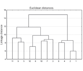
Taking into account the obtained data of the factor analysis, a cluster analysis was performed. And, based on the results of the hierarchical cluster analysis, the interaction of factors for the development of the chronic inflammatory process in the urinary system of children was determined (Fig. 1).
According to Fig. 1, 2, main clusters were formed for children with chronic UTI. The first cluster indicates the correlation between vulvitis and intestinal dysfunction and dysmetabolic nephropathy and the presence of chronic di–sease foci. As you can see, cluster 1 describes the relationship between concomitant pathologies in children with chronic inflammatory diseases of the urinary system.
Hierarchical cluster analysis data show that there is also an associative relationship between DBP, NOS2 and –IL-6, which is quite logical, because they are all linked to the body’s immune response. Further, we observe an associative relationship between urinary dysfunction (neurogenic bladder, vesicoureteral reflux), early manifestation of the disease, and levels of vitamin D and cystatin C (cluster 2).
The next stage of the work was the creation of a mathe–matical model for predicting the development of chronic inflammatory diseases of the urinary system in children using the binary logistic regression. To identify the factors mostly associated with the risk of developing chronic UTIs in children, the most significant features were selected –using the genetic algorithm method. The obtained results are presented in Table 3.
According to the results of the logistic regression, the model for predicting the probability of developing chronic inflammatory diseases of the urinary system in children had the form of an equation:
Z = 1 / (1 + еxp(–8.053 + 0.365X1 + 0.280X2 + 1.579X3 + 1.243X4 + 1.118X5 – 0.894Х6).
The classification ability of the model was determined based on the data of the training sample and was 75.0 % (Table 4). The sensitivity of the model was 78.3 %, and the specificity was 76.5 %.
The diagnostic significance of the obtained mathematical model was determined by ROC analysis (Fig. 2). The logistic regression equation is represented by the AUC. The AUC that corresponded to our mathematical model was equal to 0.776. The Gini index was 55.2 %, which indicated the good quality of the model.
Discussion
The results of the study showed that the factor of functional disorders of the urinary tract, the factor of comorbid conditions, the protective factor, the immune factor, as well as the presence of chronic foci of diseases and the level of NOS2 played a certain role in the chronicity of inflammatory diseases of the urinary system.
The presence of abnormalities in the urinary system was ruled out in all children under observation. Accor–dingly, the early manifestation of the disease, namely the development of the first case of pyelonephritis at the age of 3 to 6 years, had other causes, for example, functional disorders of the urinary tract, disruption of intracellular signaling cascades of innate immunity, etc. The innate immune system plays an important role in the prevention of recurrent UTIs, because it generates a faster response to invading pathogens [10]. According to Godaly G. et al. (2015), susceptibility to UTI is determined by the efficiency of the host organism’s defense, and the weakening of innate immunity initially leads to asymptomatic bacteriuria [11]. Rudaitis S. et al. (2009) demonstrated that a first urinary tract infection at the age of ≤ 6.5 years is a risk factor for recurrent UTIs (odds ratio 0.9; 95% confidence interval 0.85–0.98) [12]. In our work, we showed that the development of an acute UTI at the age of 3 to 6 years is a predictor of a chronic inflammatory process in the urinary system, which, apparently, reflected an inadequate immune response in these children.
The relevance of the factor of functional disorders of the urinary tract was confirmed by the works of se–veral researchers. So, Chase J. et al. (2010) indicated that the inability to empty the bladder often leads to urinary retention, urinary stasis, and ineffective elimination of uropathogens [13], thereby increasing the risk of recurrent UTIs. Becknell V. et al. (2015) showed that increased bladder pressure due to poor emptying can also cause a secondary vesicoureteral reflux, which increases the potential risk of kidney damage, namely the development of pyelonephritis [3].
According to the literature, concomitant pathologies can contribute to the development of UTI, complicate its course and act as a background for the chronicity of the process [14]. This is also confirmed in our research. Becknell V. et al (2015) noted in their work that with intestinal dysfunction, a large intestine filled with feces can make it difficult to empty the bladder and increases the risk of developing both primary UTI and recurrence [3]. In addition, Abaturov O.E., Vakulenko L.I. (2019) showed that dysmetabolic nephropathy was a concomitant pathology in chronic pyelonephritis in more than half of children [15]. During our factor analysis, the presence of dysmetabolic nephropathy was also considered the comorbid conditions factor.
Commenting on the role of the protective factor to which we attributed the levels of cystatin C and vitamin D3, it is important to emphasize that cystatin C plays a fundamental role in many biological processes, such as protein metabolism, regulation of phagocytosis of innate immune cells, activation of progenitor proteins, mediated antigen presentation, and apoptosis [16]. Vitamin D3 is an important antimicrobial peptide that takes an active part in the body’s defense due to its antimicrobial activity against a wide range of pathogens [17], and is also capable of controlling immune function at various levels by stimulating autophagy, media–ting the expression of the antimicrobial peptide LL-37 [18].
According to the data obtained in our factor analysis, the immune factor included the levels of DBP and IL-6. Pursuant to literature data, DBP, in addition to transporting vitamin D metabolites, performs other, no less important functions, including influence on the functioning of the immune system due to macrophage activation, participation in chemotaxis, etc. [19]. For its part, IL-6 is a multifunctional cytokine that regulates numerous body functions, such as the response of the acute phase of inflammation [20]. Also, Bikle D.D. and Schwartz J. (2019) noted in their study that certain cytokines, such as IL-6, increase the production of DBP [21].
According to the literature, inducible NO synthase, albeit in small amounts, is also present in the blood under normal physiological conditions, but is expressed in response to the invasion of pathogens and/or inflammatory cytokines. Once induced, NOS2 produces large, sustained amounts of NO, which in turn can limit growth or be lethal to pathogens [22]. In our study, NOS2 was singled out as a separate factor, which emphasizes its importance in the process of UTI chronicity.
Conclusions
1. The chronicity of the inflammatory process in the urinary system of children occurs under the interaction of several pathological factors. Based on the analysis, a model was created to predict the occurrence of chronic inflammatory diseases of the urinary system in children with acute UTI.
2. It has been found that a significant contribution to the development of chronic inflammatory process in the urinary system is made by the presence of functional disorders of the urinary tract in combination with an inadequate, non-specific immune response, the mediated symptom of which is the early manifestation of the acute process. Vitamin D level, intestinal dysfunction, and the presence of UTI in the mother in childhood are also leading risk factors for the chronicity of the inflammatory process.
3. The determined risk factors and the results of prognostic modeling should be used in groups at high risk of developing a chronic inflammatory process in the urinary system.
Received 01.04.2023
Revised 11.04.2023
Accepted 18.04.2023

/31.jpg)
/32.jpg)
/33.jpg)
/32_2.jpg)
/33_2.jpg)