
Журнал "Гастроэнтерология" Том 57, №3, 2023
Вернуться к номеру
Структура печінки та маркери фіброзу при моделюванні алкогольного ураження печінки й корекції виявлених порушень
Авторы: V.I. Didenko, I.A. Klenina, O.I. Hrabovska, Yu.A. Gaidar, O.O. Halinskyi, V.A. Karachynova, D.F. Mylostуva
State Institution “Institute of Gastroenterology of the National Academy of Medical Sciences of Ukraine”, Dnipro, Ukraine
Рубрики: Гастроэнтерология
Разделы: Клинические исследования
Версия для печати
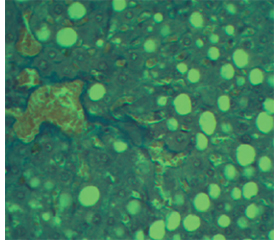
Актуальність. Хронічне вживання алкоголю призводить до алкогольного фіброзу печінки. На сьогодні багато наукових досліджень спрямовано на вивчення патометаболічних механізмів розвитку та формування фіброзу печінки у тваринних моделях. Мета дослідження: вивчення структурних змін та жорсткості печінки, біохімічних маркерів фіброзу при моделюванні хронічного алкогольного ураження печінки (ХАУП) у щурів та оцінка змін цих параметрів при різних типах корекції. Матеріали та методи. Вісімдесят дев’ять щурів були поділені на експериментальні групи залежно від терміну впливу алкоголю (4 й 12 тижнів) і виду корекції (метадоксин та пребіотик). У роботі використовували морфологічні методи, зсувнохвильову еластографію, біохімічні та статистичні методи. Результати. При моделюванні ХАУП на 4 тижні морфологічні дослідження виявили помірний крупнокрапельний жировий гепатоз та легкий фіброз у ділянці центральної венули печінкових часток. Після 12 тижнів примусової алкоголізації, коли загальна інтоксикація є більш вираженою, гепатоцити мають дистрофічні зміни, що виражаються появою в паренхімі поодиноких або зібраних в групи дистрофічних клітин. Частіше спостерігалося поєднання білкової та жирової дистрофії. Еластографія дозволила виявити структурні зміни в печінці на ранніх стадіях формування фіброзу при моделюванні ХАУП упродовж 12 тижнів. Також змінювалися рівні біохімічних показників: вільного й білковозв’язаного гідроксипроліну, глікозаміногліканів. Після корекції ХАУП, як коротко-, так і довгострокового, уміст маркерів фіброзу в гомогенаті печінки щурів нормалізувався після введення метадоксину й пребіотика. Після корекції пребіотиком на 12 тижні алкоголізації спостерігалося зниження жорсткості паренхіми печінки на 12 % у групі моделювання ХАУП, на 19 % (p < 0,05) у групі плацебо-контролю. Після корекції метадоксином виявлено збільшення рівня вільного гідроксипроліну в гомогенаті печінки щурів в 1,5 раза на 12 тижні та збільшення глікозаміногліканів в 1,2 раза (p < 0,05) на 4 тижні порівняно з групою моделювання ХАУП. Висновки. Довгострокова алкоголізація тварин призвела до розвитку дистрофічних змін у гепатоцитах, білкової та жирової дистрофії, збільшення кількості капілярів. На цьому фоні змінювалися жорсткість печінки та біохімічні параметри. Після корекції метадоксином і пребіотиком зміни у показниках жорсткості печінки та рівнях маркерів фіброзу спостерігалися на 12 тижні моделювання ХАУП.
Background. Chronic alcohol use leads to alcoholic liver fibrosis. Today, a sufficient number of scientific studies are focused on the pathometabolic mechanisms of liver fibrosis development and formation in animal models. The purpose of our study was to investigate structural changes and liver stiffness, biochemical markers of fibrosis in rats with chronic alcoholic liver injury (CALI) modeling and to evaluate the changes of these parameters with different types of treatment. Materials and methods. Eighty-nine rats were divided into experimental groups depending on the duration of alcohol exposure (4 and 12 weeks) and the corresponding type of correction (metadoxine and prebiotic). Results. When modeling CALI at week 4, morphological studies revealed moderate large-droplet fatty hepatosis and mild fibrosis in the central venule of the liver lobes. After 12 weeks of forced alcoholization, with more pronounced general intoxication, hepatocytes have dystrophic changes such as appearance of single or grouped dystrophic cells in the parenchyma. A combination of protein and fatty dystrophy was more common. Elastography allowed to detect structural changes in the liver at the early stages of fibrosis formation when modeling CALI for 12 weeks. There were also changes in the levels of biochemical parameters: free and protein-bound hydroxyproline, glycosaminoglycans. According to the results of elastography, liver stiffness in rats increased maximally after prebiotic correction in all approaches compared to the controls. After correction of CALI, both early- and long-term, fibrosis markers normalized in rat liver homogenate after administration of metadoxine and prebiotic. After prebiotic correction at week 12 of alcoholization, we observed a 12% decrease in liver parenchymal stiffness in the CALI modeling group and a 19% decrease (p < 0.05) in the placebo group. After correction with metadoxine, there was a 1.5-fold increase in free hydroxyproline levels in rat liver homogenate at week 12 and a 1.2-fold increase in glycosaminoglycans (p < 0.05) at week 4 compared to the CALI modeling group. Conclusions. Long-term alcoholization of animals led to the development of dystrophic changes in hepatocytes, protein and fatty degeneration, and an increase in the number of capillaries. Against this background, liver stiffness and biochemical parameters changed. After correction with metadoxine and prebiotic, changes in the liver stiffness and fibrosis markers were observed at week 12 of CALI modeling.
фіброз; хронічне ураження печінки; гідроксипролін; глікозаміноглікани; жорсткість печінки; морфологічні дослідження
fibrosis; chronic liver damage; hydroxyproline; glycosaminoglycans; liver stiffness; morphological studies
Introduction
Materials and methods
Results and discussion
/33.jpg)
/34.jpg)
/34_2.jpg)
/34_3.jpg)
/35.jpg)
/35_2.jpg)
Conclusions
- Жорсткість паренхіми печінки щурів при моделюванні стеатозу аліментарного генезу та його корекції / В.І. Діденко та ін. Проблеми екології та медицини. 2019. № 5–6. С. 36-41. doi: 10.31718/mep.2019.23.5-6.06.
- Методи визначення глікопротеїнів та їх компонентів з діагностичною метою у змішаній слині у дітей з гастродуоденальної патологією: методичні рекомендації. Київ, 2015. 19 с.
- Взаємозв’язок між морфологічними змінами печінки та жорсткістю її паренхіми при моделюванні алкогольного та токсичного гепатиту / Ю.М. Степанов та ін. Журнал Національної Академії Медичних Наук України. 2017. № 23(3–4). С. 196-204.
- Glycosaminoglycans: Carriers and Targets for Tailored Anti-Cancer Therapy / A. Berdiaki et al. Biomolecules. 2021. Vol. 11(3). P. 395. doi: 10.3390/biom11030395.
- Bertola A. Rodent models of fatty liver diseases. Liver Research. 2018. Vol. 2(1). P. 3-13. doi: 10.1016/j.livres.2018.03.001.
- Identification of key metabolic changes during liver fibrosis progression in rats using urine and serum metabolomics approach / H. Chang et al. Springer Science and Business Media LLC. 2017. Vol. 7 (1). P. 11433. doi: 10.1038/s41598-017-11759-z.
- Di Miceli M., Gronier B. Pharmacology, Systematic Review and Recent Clinical Trials of Metadoxine. Rev Recent Clin Trials. 2018. Vol. 13(2). P. 114-125. doi: 10.2174/1574887113666180227100217.
- Gabr S.A., Alghadir A.H., Sherif Y.E., Ghfar A.A. Hydroxyproline as a Biomarker in Liver Disease. Biomarkers in Disease. Dordrecht; 2017. P. 471-491.
- Ghadie N.M., St-Pierre J.P., Labrosse M.R. The Contribution of Glycosaminoglycans/Proteoglycans to Aortic Mechanics in Health and Disease: A Critical Review. IEEE Trans Biomed Eng. 2021 Dec. Vol. 68(12). P. 3491-3500. doi: 10.1109/TBME.2021. 3074053.
- Rodent Models of Alcoholic Liver Disease: Role of Binge Etha–nol Administration / S. Ghosh Dastidar et al. Biomolecules. 2018. Vol. 8(1). Р. 3. doi: 10.3390/biom8010003.
- Habash N.W., Sehrawat T.S., Shah V.H., Cao S. Epigenetics of alcohol-related liver diseases. JHEP Rep. 2022. Vol. 4(5). P. 100466. doi: 10.1016/j.jhepr.2022.100466.
- Han K., Zhang Y., Yang Z. Cilostazol protects rats against alcohol-induced hepatic fibrosis via suppression of TGF-β1/CTGF activation and the cAMP/Epac1 pathway. Experimental and Therapeutic Medicine. 2019. Vol. 17. P. 2381-2388. doi: 10.3892/etm.2019.7207.
- Hofman K., Hall B., Cleaver H., Marshall S. High-throughput quantification of hydroxyproline for determination of collagen. Analit Biochem. 2011. Vol. 417(2). P. 289-91. doi: 10.1016/j.ab.2011.06.019.
- Kim J., Seki E. Hyaluronan in liver fibrosis: basic mechanisms, clinical implications, and therapeutic targets. Hepatol Commun. 2023 Mar. Vol. 7(4). P. e0083. doi: 10.1097/HC9.0000000000000083.
- Pathogenesis, Early Diagnosis, and Therapeutic Management of Alcoholic Liver Disease / L.Z. Kong et al. Int J Mol Sci. 2019. Vol. 20(11). P. 2712. doi: 10.3390/ijms20112712.
- Alcoholic liver disease: Utility of animal models / A. Lamas-Paz et al. World J Gastroenterol. 2018. Vol. 24(45). P. 5063-5075. doi: 10.3748/wjg.v24.i45.5063.
- Liu S.Y., Tsai I.T., Hsu Y.C. Alcohol-Related Liver Disease: Basic Mechanisms and Clinical Perspectives. Int J Mol Sci. 2021. Vol. 22(10). P. 5170. doi: 10.3390/ijms22105170.
- Alcohol Consumption and Risk of Liver Fibrosis in People Living With HIV: A Systematic Review and Meta-Analysis / H. Lyu et al. Front Immunol. 2022. Vol. 18(13). P. 841314. doi: 10.3389/fimmu.2022.841314.
- Compositional Analysis of Glycosaminoglycans in Different Lung Cancer Types — A Pilot Study / D. Pál et al. Int J Mol Sci. 2023. Vol. 24(8). P. 7050. doi: 10.3390/ijms24087050.
- Sehrawat T.S., Liu M., Shah V.H. The knowns and unknowns of treatment for alcoholic hepatitis. Lancet Gastroenterol Hepatol. 2020. Vol. 5(5). P. 494-506. doi: 10.1016/S2468-1253(19)30326-7.
- Shouman M.M., Abdelsalam R.M., Tawfick M.M., Kenawy S.A., El-Naa M.M. Antisense Tissue Factor Oligodeoxynucleotides Protected Diethyl Nitrosamine/Carbon Tetrachloride-Induced Liver Fibrosis Through Toll Like Receptor4-Tissue Factor-Protease Activated Receptor1 Pathway. Front Pharmacol. 2021. Vol. 12. P. 676608. doi: 10.3389/fphar.2021.676608.
- Natural Recovery by the Liver and Other Organs after Chronic Alcohol Use / P.G. Thomes et al. Alcohol Res. 2021. Vol. 41(1). P. 5. doi: 10.35946/arcr.v41.1.05.
- Wang Q., Chi L. The Alterations and Roles of Glycosaminoglycans in Human Diseases. Polymers (Basel). 2022. Vol. 4(22). P. 5014. doi: 10.3390/polym14225014.
- Yang Y.M., Cho Y.E., Hwang S. Crosstalk between Oxidative Stress and Inflammatory Liver Injury in the Pathogenesis of Alcoholic Liver Disease. Int J Mol Sci. 2022. Vol. 23(2). P. 774. doi: 10.3390/ijms23020774.
- Dynamic changes of key metabolites during liver fibrosis in rats / J. Yu et al. World J Gastroenterol. 2019. Vol. 25(8). P. 941-954. doi: 10.3748/wjg.v25.i8.941.