Інформація призначена тільки для фахівців сфери охорони здоров'я, осіб,
які мають вищу або середню спеціальну медичну освіту.

Підтвердіть, що Ви є фахівцем у сфері охорони здоров'я.

Журнал "Гастроэнтерология" Том 58, №1, 2024

Вернуться к номеру

Оцінка функціонального стану мікробіоти кишечника в пацієнтів із метаболічно-асоційованою жировою хворобою печінки в поєднанні з цукровим діабетом 2 типу

Авторы: O.K. Didyk
Bogomolets National Medical University, Kyiv, Ukraine

Рубрики: Гастроэнтерология

Разделы: Клинические исследования

Версия для печати


Резюме

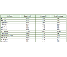
Актуальність. Метою дослідження було оцінити функціональний стан мікробіоти кишечника в осiб із метаболічно-асоційованою жировою хворобою печінки (МАЖХП) у поєднанні з цукровим діабетом (ЦД) 2 типу. Матеріали та методи. У дослідженні взяв участь 71 пацієнт із МАЖХП, поєд­наною з ЦД 2 типу, яких було обстежено та розділено на 2 групи. До першої групи ввійшли 39 осіб із МАЖХП у поєднанні з ЦД 2 типу без синдрому надмірного бактеріального росту (СНБР), до другої — 32 пацієнти з МАЖХП, поєднаною з ЦД 2 типу та СНБР. Групу контролю становили 25 практично здорових осіб. Уміст зонуліну в сироватці крові визначали методом імуноферментного аналізу (ІФА) з використанням тест-систем IDK Zonulin ELISA, KR5601. Концентрацію інтерлейкіну (ІЛ) 6 та ІЛ-10 у сироватці крові оцінювали за допомогою ІФА та тест-систем Human Interleukin 6 and 10 ELISA Kit. Рівень коротколанцюгових жирних кислот у калі визначали методом газової хроматографії з мас-спектрометрією на газовому хроматографі PerkinElmer Clarus 680 GC. Результати. У хворих першої та другої груп установлено підвищення печінкових амінотрансфераз, загального холестерину, тригліцеридів, глюкози крові на­тще, інсуліну, індексу HOMA-IR, глікозильованого гемо­глобіну, зонуліну сироватки крові, лептину, ІЛ-6 і коефіцієнта затухання ультразвуку та зниження рівня ліпопротеїнів високої щільності. За результатами стеатометрії зафіксовано тяжкий ступінь стеатозу (S3) у пацієнтів із МАЖХП у поєднанні з ЦД 2 типу й СНБР та без СНБР. При кількісному дослідженні рівня масляної кислоти у фекаліях встановлено його вірогідне зменшення в 2,3 раза в першій групі та в 3,4 раза в другій порівняно з групою контролю (p < 0,001). У хворих другої групи вміст масляної кислоти був знижений в 1,4 раза порівняно з пацієнтами першої групи (p < 0,001). При порівнянні рівня оцтової кислоти виявлено його вірогідне підвищення в 1,2 раза в першій групі та в 1,3 раза в другій порівняно з контрольною групою (p < 0,001). Установлено збільшення концентрації пропіонової кислоти в 1,3 раза в першій групі та в 1,5 раза в другій порівняно з групою контролю (p < 0,05). У хворих першої та другої груп спостерігалося вірогідне підвищення рівня оцтової кислоти в 1,3 раза відносно контрольної групи (p < 0,001). При порівнянні концентрацій пропіонової та оцтової кислоти в другій групі спостерігалося їх збільшення в 1,1 раза порівняно з першою (p < 0,05). Аналіз коефіцієнтів кореляції в осіб із МАЖХП у поєднанні з ЦД 2 типу й СНБР та без СНБР виявив прямий пропорційний сильний зв’язок між рівнями масляної, оцтової та пропіонової кислот і сироваткового зонуліну, ІЛ-6, ІЛ-10. При вивченні кореляцій установлено прямий помірний зв’язок між умістом коротколанцюгових жирних кислот і лептину, показниками ліпідного (загальний холестерин, тригліцериди), вуглеводного обміну (глюкоза крові, HOMA-IR, HbA1c) і зворотну слабку кореляцію з рівнем ліпопротеїнів високої щільності. Зафіксовано прямий помірний кореляційний зв’язок між рівнями коротколанцюгових жирних кислот та показниками функціональної активності печінки (аланінамінотрансфераза, аспартатамінотрансфераза), ступенем стеатозу в осіб із МАЖХП у поєд­нанні з ЦД 2 типу й СНБР та без СНБР. Висновки. У пацієн­тів із МАЖХП, поєднаною з ЦД 2 типу й СНБР та без СНБР, виявили підвищену кишкову проникність та гіперлептинемію. При дослідженні функціонального стану мікробіоти кишечника встановлено підвищення рівнів пропіонової та оцтової кислот та зниження масляної, що свідчить про збільшення кількості Bacteroidetes у кишечнику та зменшення Firmicutes. У пацієнтів із МАЖХП у поєднанні з ЦД 2 типу й СНБР та без СНБР виявлено прямий пропорційний сильний кореляційний зв’язок між рівнями масляної, оцтової та пропіонової кислот, зонуліну в сироватці крові та IЛ-6, IЛ-10, що асоціюється з прозапальними і протизапальними процесами в слизовій оболонці кишечника й порушенням проникності кишкового бар’єра. Установлено, що концентрації коротколанцюгових жирних кислот корелюють з показниками функціональної активності печінки (аланінамінотрансфераза, аспартатамінотрансфераза), рівнем лептину, показниками вуглеводного та ліпідного обміну, ступенем стеатозу печінки.

Background. The purpose of the study was to assess the functional state of gut microbiota in patients with metabolic-associated fatty liver disease (MAFLD) combined with type 2 diabetes mellitus (T2DM). Materials and methods. The prospective interventional randomized study included 71 patients with MAFLD in combination with T2DM, who were examined and divided into the 2 groups. The first group included 39 people with MAFLD and T2DM without small intestinal bacterial overgrowth (SIBO). The second group consisted of 32 patients with MAFLD in combination with T2DM and SIBO. The control group included 25 practically healthy patients. The content of serum zonulin was determined by enzyme-linked immunosorbent assay (ELISA) using test systems IDK Zonulin ELISA, KR5601. Serum concentration of interleukin (IL) 6 and IL-10 was assessed by the ELISA method using the Human Interleukin 6 and 10 ELISA Kit test systems. The content of short-chain fatty acids in feces was determined by gas chromatography with mass spectrometry in the PerkinElmer Clarus 680 GC Gas Chromatograph. Results. Patients of the first and second groups had an increase in hepatic aminotransferases, total cholesterol, triglycerides, fasting blood glucose, insulin, HOMA-IR, glycosylated hemoglobin, serum zonulin, leptin, IL-6 and liver attenuation coefficient and a decrease in high-density lipoprotein. The results of steatometry revealed a severe degree of hepatic steatosis (S3) in patients with MAFLD combined with T2DM and SIBO and without SIBO. During the quantitative study of the level of butyric acid in feces, it was found to be significantly reduced, by 2.3 times in the first group and by 3.4 times in the second one, compared to the controls (p < 0.001). The content of butyric acid was decreased by 1.4 times in the second group compared to the first one (p < 0.001). When evaluating the level of acetic acid, it was found to be significantly increased compared to the controls, by 1.2 times in the first group and by 1.3 times in the second one (p < 0.001). The concentration of propionic acid was increased by 1.3 times in the first group and by 1.5 times in the second one compared the control group (p < 0.05). When comparing the level of acetic acid, a significant increase by 1.3 times was observed in patients of the first and second groups compared to patients of the control group (p < 0.001). The content of propionic and acetic acids was increased by 1.1 times in the second group compared to patients of the first group (p < 0.05). When analyzing the correlation coefficients in patients with MAFLD combined with T2DM and SIBO and without SIBO, a direct proportional strong correlation was revealed between the levels of butyric, acetic and propionic acids and serum zonulin, IL-6, IL-10. A direct moderate correlation was found between the content of short-chain fatty acids and leptin, indicators of lipid metabolism (total cholesterol, triglycerides), carbohydrate metabolism (fasting blood glucose, HOMA-IR, HbA1c) and inverse weak correlation with high-density lipoprotein. A direct moderate correlation was revealed between levels of short-chain fatty acids and indicators of functional activity of the liver (alanine aminotransferase, aspartate aminotransferase), liver attenuation coefficient in patients with MAFLD combined with T2DM and SIBO and without SIBO. Conclusions. Increased intestinal permeability and hyperleptinemia have been found in patients with МAFLD combined with T2DM and SIBO and without SIBO. When studying the functional state of the gut microbiota, an increase was found in propionic and acetic acids and a decrease in butyric acid, which indicates an increase in the number of Bacteroidetes in the intestine and a decrease in Firmicutes. Patients with MAFLD combined with Т2DM and SIBO and without SIBO had a direct proportional strong correlation between the levels of butyric, acetic and propionic acids and serum zonulin, IL-6, IL-10, which is associated with pro-inflammatory and anti-inflammatory processes in the intestinal mucosa and a violation of the permeability of the intestinal barrier. It was found that concentrations of short-chain fatty acids correlate with indicators of functional activity of the liver (alanine aminotransferase, aspartate aminotransferase), leptin, indicators of carbohydrate and lipid metabolism, degree of hepatic steatosis.


Ключевые слова

кишкова проникність; цукровий діабет 2 типу; лептин; коротколанцюгові жирні кислоти; зонулін; метаболічно-асоційована жирова хвороба печінки

intestinal permeability; type 2 diabetes mellitus; leptin; short-chain fatty acids; zonulin; metabolic-associated fatty liver disease

Introduction

The microbiome plays a key role in the development and functioning of innate and adaptive immune responses [1]. Violation of the permeability of the intestinal barrier due to intestinal dysbiosis is an important pathogenetic aspect for the development and progression of metabolic-associated fatty liver disease (MAFLD) [2]. It is extremely important that oxidative stress in the liver, as one of the key mechanisms of the development of MAFLD, can be caused by high concentrations of endotoxin due to a violation of the permeability of the intestinal barrier [3].
Increased permeability is associated with translocation of bacteria and their cell wall components and elevated bacterial lipopolysaccharide, which triggers a TLR4-mediated pro-inflammatory cascade in immune cells (monocytes, macrophages and Kupffer cells) leading to increased synthesis of zonulin, a protein that is one of the regulators of intestinal permeability [4].
Short-chain fatty acids (SCFAs) play an important role in regulating the integrity of the epithelial barrier through coordinated regulation of tight junction proteins, which themselves regulate the intracellular molecular pathway between the lumen and hepatic portal system [5].
SCFAs are monocarboxylic acids with a chain length of up to 6 carbon atoms. SCFAs, mainly acetate, propionate, and butyrate, are organic fatty acids synthesized from non-digestible proteins and fibers via anaerobic fermentation by the gut microbiota [6]. There are numerous studies demonstrating that SCFAs activate the G-protein-coupled receptors GPR41 and GPR43 in the surface of the gut enteroendocrine L cells. Activation of GPR41 and GPR43 on the surface of the L cells increases secretion of glucagon-like peptide-1 [7]. Glucagon-like peptide-1 leads to reduction of glucagon secretion and improves insulin sensitivity, a decrease in oxidative stress and enhances lipid oxidation in the liver, which contributes to reduced steatosis [7].
Further work is urgently needed in human models to determine whether SCFAs play an important role in mucosal maintenance and integrity.
The aim of the investigation was to assess the functional state of gut microbiota in patients with MAFLD combined with type 2 diabetes mellitus (T2DM).

Materials and methods

The study was approved by the commission on biotic expertise and ethics of scientific research (protocol No. 150 dated October 18, 2021) at Bogomolets National Medical University and performed at the clinical base of the Department of Internal Medicine 1 from 2021 to 2023. All patients who participated in the study signed an informed consent.
The prospective interventional randomized study inclu–ded 71 patients with MAFLD combined with T2DM, who were examined and divided into the 2 groups. The first group included 39 patients with MAFLD and T2DM without small intestinal bacterial overgrowth (SIBO). The second group consisted of 32 people with MAFLD combined with T2DM and SIBO. The control group included 25 practically healthy patients.
The inclusion criteria were men and women aged 25–78 years, patients with МAFLD in combination with type 2 diabetes, whose diagnosis was established by determining the degree of steatosis based on the results of ultrasound steatometry (Ultrasign Soneus P7 device with a 1–6 MHz convex sensor) performed on the scale of ultrasound attenuation (ultrasound attenuation coefficient ≥ 2.2 dB/cm) and diagnostic criteria of carbohydrate metabolism disorders according to the 2023 American Diabetes Association guidelines [8].
The exclusion criteria were viral hepatitis, alcoholic liver disease, autoimmune hepatitis, drug-induced liver damage, Wilson-Konovalov disease, type 1 diabetes, decompensated type 2 diabetes mellitus, cancer, pregnancy, refusal to participate in the study.
Blood samples were collected to evaluate indicators of liver functional activity (alanine aminotransferase (ALT), aspartate aminotransferase (AST)), carbohydrate (fasting blood glucose (FBG), insulin, HOMA-IR, glycosylated hemoglobin (HbA1c)) and lipid (total cholesterol (TC), triglycerides (TG), high-density lipoprotein (HDL)) metabolism. Analyses were performed using a biochemistry analyzer Cobas 6000 with appropriate reagent kits (Roche Diagnostics, Switzerland). The content of serum zonulin was determined by enzyme-linked immunosorbent assay (ELISA) using test systems IDK Zonulin ELISA, KR5601 (Immundiagnostik AG, Germany). ELISA method was used to quantify serum leptin.
The concentration of human interleukin (IL) 6 and IL-10 in serum was assessed by the ELISA method using the Human Interleukin 6 and 10 ELISA Kit test systems (Elabscience, USA). The content of short-chain fatty acids in feces was determined by gas chromatography with mass spectrometry on the PerkinElmer Clarus 680 GC Gas Chromatograph (USA).
The lactulose hydrogen breath test was used to diagnose SIBO. It was performed using the Advanced Hydrogen Breath Testing (Micro H2 Meter) device.
The Microsoft Office 2016 software package, MedStat version 5.2. and EZR version 3.4.1 (the R Foundation for Statistical Computing) were used for statistical processing of the obtained results. When checking the distribution of the obtained data for normality, the Shapiro-Wilk test was used. With a normal distribution, quantitative variables were described by the arithmetic mean value with a standard deviation (mean ± SD); with a deviation from the normal distribution, by the median with the first and third quartiles (median (Q1-Q3)). To check the differences between 3 groups when the data is normally distributed, the method of multiple comparisons ANOVA was used, when it was different from the normal, the Kruskal-Wallis test. Spearman’s rank correlation coefficient (r) was calculated for correlation analysis between variables. Differences between groups were considered likely to be significant when the value of р < 0.05 was reached.

Results

The clinical and diagnostic characteristics of patients are shown in Table 1. In patients of the first and second groups, an increase in hepatic aminotransferases, total choleste–rol, triglycerides, fasting blood glucose, insulin, HOMA-IR, glycosylated hemoglobin, serum zonulin (Fig. 1), leptin (Fig. 2), IL-6 and liver attenuation coefficient (LAC) was found and a decrease in high-density lipoprotein.
The results of steatometry revealed a severe degree of hepatic steatosis (S3) in patients with MAFLD combined with T2DM and SIBO and without SIBO.
During the quantitative study of the level of butyric acid in feces, it was found to be significantly reduced, by 2.3 times in the first group and by 3.4 times in the second group, compared to the controls (р < 0.001). The content of butyric acid was decreased by 1.4 times in the second group compared to the first one (р < 0.001) (Тable 1, Fig. 3).
When comparing the level of acetic acid, it was found to be significantly increased, by 1.2 times in the first group and by 1.3 times in the second group, compared to the controls (р < 0.001). During the study, the concentration of propio–nic acid was increased by 1.3 times in the first group and by 1.5 times in the second group compared to the controls (р < 0.05) (Тable 1, Fig. 3). When comparing the level of acetic acid, a significant increase by 1.3 times was observed in patients of the first and second groups compared to the control group (р < 0.001). The content of propionic and acetic acid increased by 1.1 times in the second group compared to the first one (р < 0.05) (Тable 1, Fig. 3).
Tables 2 and 3 show the results of the correlation analy–sis. A direct proportional strong correlation was revealed between the levels of butyric, acetic, and propionic acids and serum zonulin, IL-6, IL-10 in patients with MAFLD combined with T2DM and SIBO and without SIBO.
When studying relationships, a direct moderate correlation was found between the content of SCFAs and leptin, indicators of lipid metabolism (ТС, ТG), carbohydrate meta–bolism (FBG, HOMA-IR, HbA1c) and inverse weak correlation with HDL.
A direct moderate correlation was revealed between the levels of SCFAs and indicators of functional activity of the liver (ALT, AST), liver attenuation coefficient (LAC) in patients with MAFLD combined with T2DM and SIBO and without SIBO.

Discussion

Determination of a specific type of SCFAs allows to carry out comprehensive assessment of the functional state of the gut microbiota.
Among SCFAs produced in the colon, butyrate appears to be the most important regulator of tight junction proteins and has been shown to enhance intestinal barrier function through increased expression of claudin-1 and zonula occludens-1 and occludin redistribution, proteins, which are critical components of the tight junction assembly [9]. Butyrate has been shown to reverse the aberrant expression of zonula occludens-1 and decrease lipopolysaccharide translocation leading to inhibition of macrophage activation, pro-inflammatory cytokine production and neutrophil infiltration resulting in reduced hepatic liver injury in rats [10].
Butyrate and propionate reduce the expression of tumor necrosis factor and nitric oxide synthase in lipopolysaccharide-induced monocytes. In macrophages, butyrate exerts anti-inflammatory effects by reducing the production of induced nitric oxide synthase, monocyte chemoattractant protein-1, tumor necrosis factor α, and IL-6 through free fatty acid receptor 3 [11].
Recent literature data indicate that acetіс and propionіс acids participate in gluconeogenesis and lipogenesis in the liver. Butyrate induces expression of genes participated in gluconeogenesis in the intestine [12]. SCFAs can stimulate the secretion of glucagon-like peptide-1 by activating free fatty acid receptor 2, which indirectly regulates blood glucose levels by increasing insulin secretion and decreasing pancreatic glucagon secretion [13]. In addition to glucagon and insulin, leptin also plays an important role in blood glucose balance. Leptin resistance causes hyperinsulinemia and insulin resistance, which play a key role in the development of obesity, MAFLD and T2DM [14].
The study has demonstrated increased serum zonulin and leptin levels, suggesting increased intestinal permeability and hyperleptinemia in patients with MAFLD combined with T2DM and SIBO and without SIBO. An increase in the level of IL-6 in patients with MAFLD, type 2 diabetes and SIBO and without SIBO indicates chronic subclinical inflammation.
When studying the functional state of the intestinal microbiota, a decrease in butyric acid was detected, which indicates a decrease in butyrate-producing bacteria and, as a result, butyrate, which is important for modulating the immune system, increasing the antibacterial activity of macrophages, promoting protective and reparative processes in the intestine, regulating the integrity and functions of the intestinal barrier. It may also indicate a decrease in the number of Firmicutes in the gut that produce butyrate.
An increased concentration of acetic and propionic acids was revealed, which contributed to pro-inflammatory processes in the intestinal mucosa. This suggests that acetic and propionic acids are involved in the pro-inflammatory process of chronic subclinical inflammation. It can also indicate the presence of an increased number of Bacteroidetes in the intestine, which mainly produce acetate and propionate.
The obtained results of the correlation analysis give reason to assert that an increase in the concentration of propio–nic, acetic acids and a decrease in butyric acid are associa–ted with pro-inflammatory and anti-inflammatory processes in the intestinal mucosa and a violation of the permeability of the intestinal barrier. This is evidenced by a strong correlation between the level of SCFAs, serum zonulin and interleukins 6 and 10.
It was found that concentrations of short-chain fatty acids correlate with indicators of functional activity of the liver (ALT, AST), leptin, parameters of carbohydrate and lipid metabolism, degree of hepatic steatosis.

Conclusions

1. Increased intestinal permeability and hyperleptinemia have been found in patients with МAFLD in combination with T2DM and SIBO and without SIBO.
2. When studying the functional state of the gut microbiota, an increase in propionic and acetic acid was found and a decrease in butyric acid, which indicates an increase in the number of Bacteroidetes in the intestine and a decrease in Firmicutes.
3. Іn patients with MAFLD combined with T2DM and SIBO and without SIBO, a direct proportional strong correlation was revealed between the levels of butyric, acetic, and propionic acids and serum zonulin, IL-6, IL-10. This is associated with pro-inflammatory and anti-inflammatory processes in the intestinal mucosa and a violation of the permeability of the intestinal barrier.
4. It was found that concentrations of short-chain fatty acids correlate with indicators of functional activity of the liver (ALT, AST), leptin, indicators of carbohydrate and lipid metabolism, degree of hepatic steatosis.
 
Received 02.02.2024
Revised 13.02.2024
Accepted 24.02.2024

Список литературы

  1. Leung C. et al. The role of the gut microbiota in NAFLD. Nature Reviews. Gastroenterology & Hepatology. 2016. Vol. 13. No. 7. P. 412-425. doi: 10.1038/nrgastro.2016.85.
  2. Augustyn M. et al. Small intestinal bacterial overgrowth and nonalcoholic fatty liver disease. Clinical and Experimental Hepatology. 2019. Vol. 5. No. 1. P. 1-10. doi: 10.5114/ceh.2019.83151.
  3. Kessoku T. et al. Endotoxins and Non-Alcoholic Fatty Liver Disease. Frontiers in Endocrinology. 2021. Vol. 12. P. 770-786. doi: 10.3389/fendo.2021.770986.
  4. Ferolla S.M. et al. The role of intestinal bacteria overgrowth in obesity-related nonalcoholic fatty liver disease. Nutrients. 2014. Vol. 6. No. 12. P. 5583-99. doi: 10.3390/nu6125583.
  5. Silva Y.P. et al. The Role of Short-Chain Fatty Acids from Gut Microbiota in Gut-Brain Communication. Frontiers in Endocrinology. 2020. Vol. 11. No. 25. doi: 10.3389/fendo.2020.00025.
  6. Wolever T.M., Brighenti F., Royall D., Jenkins A.L., Jenkins D.J. Effect of rectal infusion of short chain fatty acids in human subjects. Am J Gastroenterol. 1989. Vol. 84. No. 9. Р. 1027-33.
  7. Tolhurst G. et al. Short-chain fatty acids stimulate glucagon-like peptide-1 secretion via the G-protein-coupled receptor FFAR2. Diabetes. 2012. Vol. 61. No. 2. P. 364-71. doi: 10.2337/db11-1019.
  8. ElSayed N.A., Aleppo G., Aroda V.R. et al. Erratum. 2. Classification and diagnosis of diabetes: Standards of Care in Diabetes-2023. Diabetes Care 2023;46(Suppl. 1):S19-S40. Diabetes Care. 2023. Vol. 46. No. 5. P. 1106. doi: 10.2337/dc23-er05.
  9. Vallianou N. et al. Understanding the Role of the Gut Micro–biome and Microbial Metabolites in Non-Alcoholic Fatty Liver Di–sease: Current Evidence and Perspectives. Biomolecules. 2021. Vol. 12. No. 1. P. 56. doi: 10.3390/biom12010056.
  10. Gao Zh. et al. Butyrate improves insulin sensitivity and increases energy expenditure in mice. Diabetes. 2009. Vol. 58. No. 7. P. 1509-17. doi: 10.2337/db08-1637.
  11. Vinolo M.A.R. et al. Regulation of inflammation by short chain fatty acids. Nutrients. 2011. Vol. 3. No. 10. P. 858-76. doi: 10.3390/nu3100858.
  12. Den Besten G. et al. Short-Chain Fatty Acids Protect Against High-Fat Diet-Induced Obesity via a PPARγ-Dependent Switch from Lipogenesis to Fat Oxidation. Diabetes. 2015. Vol. 64. No. 7. P. 2398-408. doi: 10.2337/db14-1213.
  13. Miele L. et al. Increased intestinal permeability and tight junction alterations in nonalcoholic fatty liver disease. Hepatology (Baltimore, Md). 2009. Vol. 49. No. 6. P. 1877-87. doi: 10.1002/hep.22848.
  14. Minokoshi Y. et al. Leptin stimulates fatty-acid oxidation by activating AMP-activated protein kinase. Nature. 2002. Vol. 415. No. 6869. P. 339-43. doi: 10.1038/415339a.

Вернуться к номеру