
Журнал «Здоровье ребенка» 3 (54) 2014
Вернуться к номеру
Ultrasonic density of kidneys at healthy and children with pyelonephritis
Авторы: Vakulenko L.I., Kondratiyev V.A., Vakulenko A.V. - SI« Dnepropetrovsk medical academy of MH of Ukraine»; Andreychenko I.I. - CE «Dnepropetrovsk regional children''s clinical hospital», Ukraine
Рубрики: Педиатрия/Неонатология
Разделы: Клинические исследования
Версия для печати
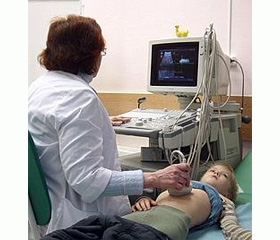
Introduction. Ultrasonic investigation helps to carry out diagnostics of most renal diseases which are accompanied by macrostructural morphological changes, which allowes to estimate echogenesity (ultrasonic density) of renal parenchyma for determination of structural changes, which includes the diffuse changes of ultrasonic denbsity, and also presence of focal changes.
It is accepted that an ultrasonic density represents the degree of renal morphological disorders (sclerosis, fibrosis, cellular infiltration, atrophy of canaliculi). There are two variants of changes of echostructure of renal parenchima: first, at which the ultrasonic density of renal cortical substanse inrceases with the preservation of cortical-medullary differentiation (at glomerulonephritis, vasculitis, interstitial nephritis, nephrosclerosis, acute canalicular necrosis); second, at which process has widespread character and the changes between cortical layer and pyramids disappears, so an ultrasonic density becomes similar. Such variant can be seen at polycistosis, chronic glomerulonephritis, acute and chronic pyelonephritis. However, determination of the presence and the degree of expressiveness of pathological changes (inflammation, fibrosis, nephrosclerosis) of kidney structures is often difficulty performed owing to technical circumstances. Due to this there is a problem of morphometric standardization of renal ultrasonic investigations. The research was performed owing to insufficient knowledge of the discussed question, the purpose of the work was the development of quantitative normative indexes of ultrasonic densiry of kidneys at healthy children for determination of changes and consequences at the inflammatory renal diseases.
A purpose of work is the development of quantitative normative indexes of ultrasonic density of the kidney at healthy children and determination of its changes at the pyelonephritis.
Materials and methods.
For the development of normative indexes 90 healthy children were inspected aged from 1 to 16 years. For determination of indexes of ultrasonic density of kidneys at its inflammation 40 children with pyelonephritis were inspected. 1th group was composed from 18 patients with acute PN, 2th group - 22 patients with chronic PN. The computer assessment of ultrasonic skanograms with the measurement of ultrasonic density of kidneys and its structures was carried out by computer program. Ultrasonic density of the standardized areas of renal parenchyma, its capsule, canaliculi, renal pelvis were determined by the coefficient of ultrasonic density, that is automatically calculated by the formula. Measuring the coefficient of ultrasonic density in kidneys was carried out in 8 standard areas: upper and lower pole of renal capsule, external and internal edge of renal capsule, cortical matter of parenchyma, medullar matter of parenchyma, renal pelvis, canaliculi. Next according to the size of sigmal deviations of the coefficient of ultrasonic density from the normal parameters the degree of increase (decline) of ultrasonic density of the kidney and its structures were determined.
Results and discussion.
The analysis of age-specific features of ultrasonic density of kidney revealed that healthy children older than 6 years had a tendency to the decline of ultrasonic density of renal capsule and pelvis, that was accompanied by reliable increase of ultrasonic density of cortical-medullar substance (p < 0,05) and by insignificant increase of renal canaliculi ultrasonic density at the age of 6-12 years (p > 0,05). Healthy children older 12 years didn`t have significant changes of the ultrasonic density of the mentioned structures.
There was performed renal ultrasonic investigation of 40 children aged 1-16 years who had acute and chronic PN. The renal capsule at acute and chronic PN had a reliable moderate increase of ultrasonic density compared to healthy children, on the average on 13%, although at chronic PN this increase was more considerable without substantial difference between groups. While investigating renal cortical substance at acute and chronic PN as compared to healthy children there was found the reliable moderate decline of ultrasonic density, on the average on 20,1 and 24,6 % accordingly. At acute PN the decline of ultrasonic closeness was registered more frequently than at chronic PN (86,4 and 50 % cases, p < 0,01). Ultrasonic density of renal medullar substance at chronic PN in most cases (68,2 %) didn`t differ from the normal parameters, but at acute PN on the average on a group its moderate reliable decline was registered on the average on 21% below than norm in 61,1 % cases. Ultrasonic density of renal canaliculi both at acute and at chronic PN was on the average for certain below as compared to the normal parameters. Such decline, though insignificant (6,7-6,8 % from a norm) it appeared in the most inspected patients on PN (72,2 and 81,8 % cases, accordingly). From the side of renal pelvis at acute PN for all patients the reliable sharp increase of ultrasonic density was registered, on the average on 57,2 % in a group. At chronic PN an ultrasonic density was moderatly enhanceable, on the average on 32,7 % as compared to a norm, also in most – 86,4 % cases patients.
Thus, the conducted researches allowed to define the age-specific tendencies of changes of renal ultrasonic density for healthy children and rotined the high sensitiveness (from 82 to 90 %) of indexes of ultrasonic density for diagnostics of acute and chronic PN for children, here specificity of indexes of ultrasonic closeness hesitated scope from 56 to 62 %.
Conclusions.
Healthy children with age have a tendency to the decline of ultrasonic density of connective tissue structures – renal capsule and pelvis. From the side of renal parenchyma there is an opposite tendency – ultrasonic density of cortical-medullar substance and renal canaliculi increases in age from 6 to 12 years, whereupon, to 17 years substantial changes from one side these structures are not observed. Children with acute PN in 44,4 % cases from ultrasonic data have a moderate increase of ultrasonic density of capsule of the affected kidney and sharp increase of ultrasonic density of renal pelvis in 100 % cases. For children with acute PN characteristic is the decline of ultrasonic density of cortical substance (86,4 % cases) and medullar substance (61,1 % cases) of the affected kidney. The ultrasonic density of canaliculi here goes down insignificantly, but in most patients (72 % cases). At chronic PN there are the same changes in the period of exacerbation: characteristic is a moderate increase of ultrasonic density of renal capsule and pelvis, and also moderate decline of ultrasonic density of cortical-medullar substanse. Measuring of ultrasonic density of renal structures acute and chronic PN for children in the terms of the making this process objective by computer evaluation of ultrasonic skanogram gives additional diagnostic information on the presence and the degree of renal inflammatory process.
1. Vakulenko LI, Kondrat`yev VO, Vakulenko AV, Andrejchenko II. Method of ultrasonic diagnostics of density of kidney and its structures. Patent on an useful model №71388 Promy`slova vlasnist`.-2012.-Byul.№13.
2. Hendlyn HE, Еttynher OA, Reznyk EV et all. Ultrasonic investigation of kidneys: possibilities and limitations of the method. Klynycheskaya nefrolohyya.2009; 2:17-25.
3. Kondrat'yev VO, Kulikova HV. Ultrasonic diagnostics of affection of heart membranes at children .Ukrayins'kyy radiolohichnyy zhurnal.2005.Vоl.XIII;4:539-542.
4. Kondrat'yev YeV, Kondrat'yev VO, Kulikova HV. Computer program Echodd. Certificate to registration of copyright on work ¹15142 from 23.12.2005
5. Ol'khova EB. Ultrasonic diagnostics of kidney and urinary tract diseases at children. Indications. Methods. Echographic anatomy. Variants of structure (Clinical lecture) Radyolohyya Praktyka.2008; 5: 28-42.
6. Petrov VY, Nehoda VY. Evidence-based medicine.— M.: HЭOTAR¬Medya. 2009: 142 р.
7. Physiology of growth and development of children and teenagers (theoretical and clinical questions). Editor AA Baranov, LA Shcheplyahynа. M. 2000.- Р.321-322.
8. Yang H, Wang Q, Luo J, Li Q, Wang L, Li CC, Zhang G, Xu Z, Tao H, Fan Z. Ultrasound of urinary system and urinary screening in 14 256 asymptomatic children in China. Nephrology (Carlton). 2010 Apr;15(3):362-7. doi: 10.1111/j.1440-1797.2009.01262.x.
9. Glodny B, Unterholzner V, Taferner B, Hofmann KJ, Rehder P, Strasak A, Petersen J. Normal kidney size and its influencing factors - a 64-slice MDCT study of 1.040 asymptomatic patients. BMC Urol. 2009 Dec 23;9:19. doi: 10.1186/1471-2490-9-19.
10. Kim JH, Kim MJ, Lim SH, Kim J, Lee MJ. Length and volume of morphologically normal kidneys in korean children: ultrasound measurement and estimation using body size. Korean J Radiol. 2013 Jul-Aug;14(4):677-82. doi: 10.3348/kjr.2013.14.4.677.
11. Michael Riccabona, Fred Efraim Avni, Maria Beatrice Damasio, Lil-Sofi. ESPR Uroradiology Task Force and ESUR Paediatric Working Group—Imaging recommendations in paediatric uroradiology, Part V: childhood cystic kidney disease, childhood renal transplantation and contrast-enhanced ultrasonography in children. Pediatr Radiol.- May 2012.- 11 p. doi: 10.1007/s00247-012-2436-9