
Журнал «Травма» Том 15, №6, 2014
Вернуться к номеру
Experience of operative treatment of the proximal tibia metaepiphysis in patients with polytrauma
Авторы: Fil A., Fil U., Kozopas V., Seniuk Y., Jajechnyk O. - Lvivsskyy National Medical University named after Danylo Galician
Рубрики: Травматология и ортопедия
Разделы: Клинические исследования
Версия для печати
Entry. Intra-articular fractures of the proximal tibia account for 12 % of all fractures locations and up to 9% of all fractures of the tibia. In 64 % of fractures involving violation of congruence of the articular surfaces, while 80 % of damaged lateral grown-up [1,2]. In 50 % of cases multiple trauma injuries are caused by high energy trauma. [4]. Fracture of the proximal tibia metaepiphysis are some of the problematic areas of injuries and the severity of their treatment depends on the anatomic abnormality. High impact injuries and damage to soft tissues result in difficulties for surgical and conservative treatment especially in the field of trauma. Most of these fractures occur as a result of traffic accidents and cycling. [1, 3, 6].
Aim: To improve the results of treatment of intra-articular fractures of the proximal tibia metaepiphysis in patients with polytrauma.
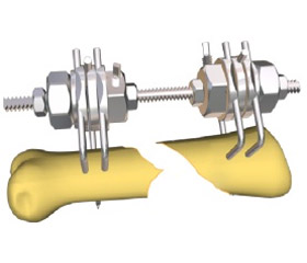
Materials and Methods. From 2009-2013 52 patients were treated in our clinic for polytrauma and fractures of the proximal tibia metaepiphysis. 40% of them were type C classification AO / ASIF of fractures. The age of patients ranged from 18 to 73 years. 39 patients were men and 13 women. The 29 patients had two anatomical damage and malfunction, 18 - three in four - four and one-five. Most fractures of the proximal tibia were related to traumatic brain injury and chest trauma. The RTS anatomical scale is a quick and easy method for assessment of patients with polytrauma. According to the RTSscale of severity of damage , 21 patients fell in the second grade, 16 patients fell in the third grade and 11 fell in the 4th grade. Patients with polytrauma first degree on the RTS scale in the sample were not included. All the injured patients were in a state of shock, and -15 degrees, -28 II, III and -7 in a terminal state 2. In our study, we classified fractures of the proximal tibia according to the Shatskera and AO / ASIFclassification. Clinical examination methods included the assessment and status of soft tissue for the presence of abrasions and wounds intensity edema. Urgent X-ray examination was carried out in two projections to identify the type of fracture and decide the methods of stabilizing rod or hybrid external fixation device. 2 and 3 D computer tomography reconstruction of the proximal mataepiphysisi was done after correcting life treathening damage of trauma patietnts and stabilizing the general condition of the patient, and assessment of the extent of fracture displacement and the extent of the fracture depression. Doppler sonography was used in trauma with dubious viable soft tissue and vascular neurological clinical symptoms. Most patients with injuries of the proximal tibia metaepiphysis with severe polytrauma are (PTS III-VI degree) treated with external fixation devices. One patient did not agree to surgery and so was treated with skeletal system of extraction and plaster of paris. Open reposition and internal fixation was performed on 25 patients with injury of the Second Degree for PTS: 17 males and 8 females, 10-14 days after injury. In our practice, depending on the condition of the patient and the type of fracture of the tibia, we used three options for fixation, external fixtation with external fixation device (RCF), open reposition (OR) internal fixation (IF) plate carts and two landmark treatment with BP and minimally invasive fixation of periarticular malleoli screws or Kirschner wires supplemented RCF, due to suspected inviable tissue. When stabilizing a patient with a simple fracture, the type of fixation is not very important, it will suffice to stabilize the fracture with external fixation apparatus for fractures, and then type C method which remains controversial.
Results. The primary aim during the inspection was to assess the general condition. Patients with unstable hemodynamics were immediately transported to the operating room, both methods of antishock measures, puncture of pleural cavity, diagnostic peritoneal lavage and radiological examination screening method were used for establishing the primary damage and main source of bleeding. So in patients with fractures of the proximal tibia metaepiphysis we applied external fixing device type hip-shin, reduction and fixing of the knee. When the hemodynamics is affected, a complete analysis of the extent of surgery is carried out, then a favourable prognosis of the traumatic disease can be made. In patients with polytrauma and tibia fracture metaepiphysis which resulted in the damage of soft tissues and later resolved we choose a method of treatment of the fracture. The likelihood of surgery with intra-articular fractures were determined based on the severity of the state of the patient, the nature and extent of fracture . In the case of isolated fractures the timing and method of treatment depends on local changes,the nature of the fracture cannot be a determining factor. Indications for surgical treatment were unstable fractures in all cases. The choice of method of osteosynthesis affects the general condition of the patient and the quality of the soft tissues. Osteosynthesis of fractures are often performed by posterior-medial and anterior-lateral approaches. Treatment of fractures 41C1- 41C3 types are divided into several stages - on admission restoration of length and elimination of rotation by fixation: the second stage involved the anatomical reconstruction of the articular surfaces and the treatment of bone depression and defects by auto-allografts and plates. Open reposition and internal fixation,were performed using minimally invasive anatomical approaches, precise method with minimal traumatization of soft tissue and the use of low-profile plates. On the operating table the patient was in the supine position with the knee placed on a roller. Surgery was performed using the turnstile with bleeding limbs to "dry joint". An important aspect for the success of surgical intervention in our opinion was the duration of surgery. There was a sharp increase of soft tissue swelling after 2-hours, which further complicated wound healing. The Recommended duration of surgery is 55 min 10. In our clinic, the average duration of surgery was 70 min + -10 min. In contrast, treatment of isolated lesions of the proximal segment of the metaepiphysis, terms and techniques to assist patients with polytrauma have their own characteristics. The primary objective was to preserve the mobility of the patient, there is no alternative to operative treatment. Cannulated screws spongious are used for more accurate positioning and execution capabilities, minimally invasive fixation. In open fractures and extensive destruction of articulating segments using modular units system "hip-tibia." To improve the quality of care and achieve good functional results in patients with polytrauma and fractures of the proximal tibia metaepiphysis, our clinic developed a simple and practical algorithm of the operating team for a quick adoption of treatment. Consolidation of all patients occurred during the period from 2 to 6 months. Active rehabilitation of the operated joint started the day after surgery. Most patients noted a satisfactory outcome. Patients examined 2-3 years later showed osteoarthritis of the knee joints. Two patients had complications - one patient developed necrosis of the superficial layers of the skin 2 weeks after surgery. Necrosis of the deep layers and osteomyelitis occurred in the second patient - D / s: Bruising of the brain severe, bruised chest, rib fractures, fracture of the tibia (41C3). Due to the severity of the general condition, the operation was performed after 23 days from the date of injury, without the reconstruction of the articular surface. The development of the above complications was a severe and lasted more than 2.5 hours after the reconstruction of the site. We also recorded two deaths due to critical condition of patients in the early stage of hospitalization.
Conclusions.
1. The most common cause of fracture in multiple trauma is a high-impact trauma, the result of which most of them were severe intra-articular fractures according the Shatsker types V and VI classification.
2. In patients with polytrauma priority is to treat life-dangerous abdominal injuries and stabilization of fractures of the pelvis and the use of hip external fixation device. Fracture limb preference should be given to temporary immobilization devices external fixation.
3. Treatment of intra-articular fractures of the proximal tibia in patients with polytrauma should be for a period based on two stages in severely injured patients we need to perform external fixation with fixation devices, if possible with fixation of the articular fragments minimally invasive cannulated screws.
4. To achieve satisfactory treatment of the proximal tibia metaepiphysis, reconstruction of articular surfaces recommended not more than 10-14 days, to avoid problems with insufficient reposition of the articular fragments.
5. State of the soft tissues in the fracture is an indicator of choice of the surgical intervention.
6. To plan a method of surgical treatment of fractures of the metapysis of the distal tibia, restore anatomic length of the legs, analysis of the fracture with spiral computed tomography in 2 and 3D.
7. The combination of using modern low profile carts plates and use minimally invasive approaches provides a good functional outcome in patients with fractures of the proximal tibia metaepiphysis.